. 2016 Feb 15:7:10660.
doi: 10.1038/ncomms10660.

A short G1 phase imposes constitutive replication stress and fork remodelling in mouse embryonic stem cells

Affiliations

A short G1 phase imposes constitutive replication stress and fork remodelling in mouse embryonic stem cells

Akshay K Ahuja et al. Nat Commun. .

Abstract

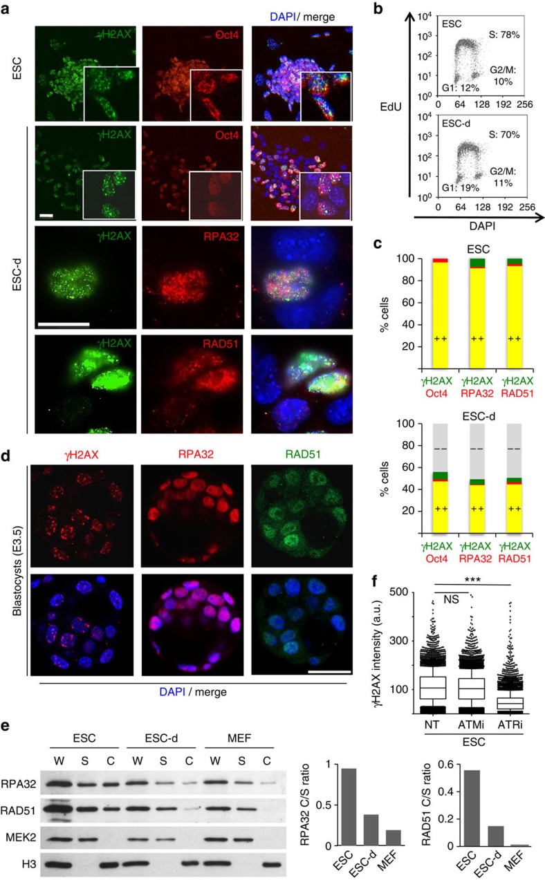
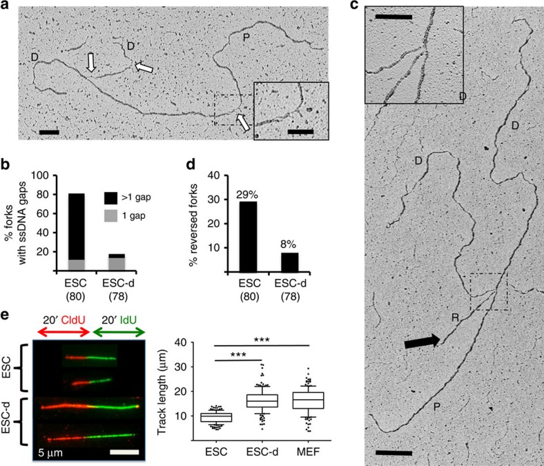
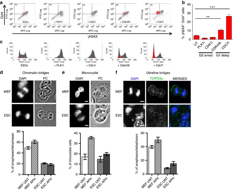
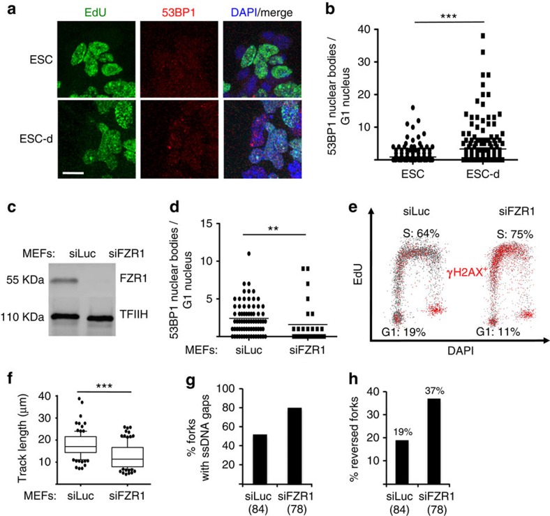
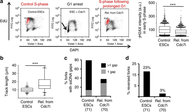
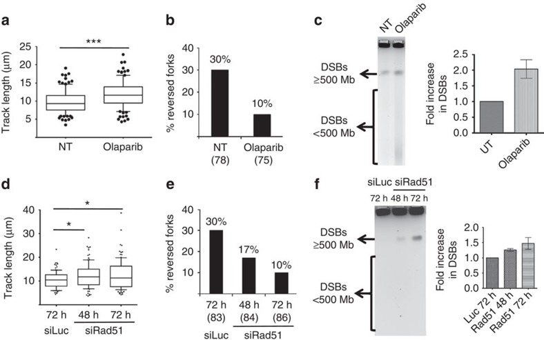
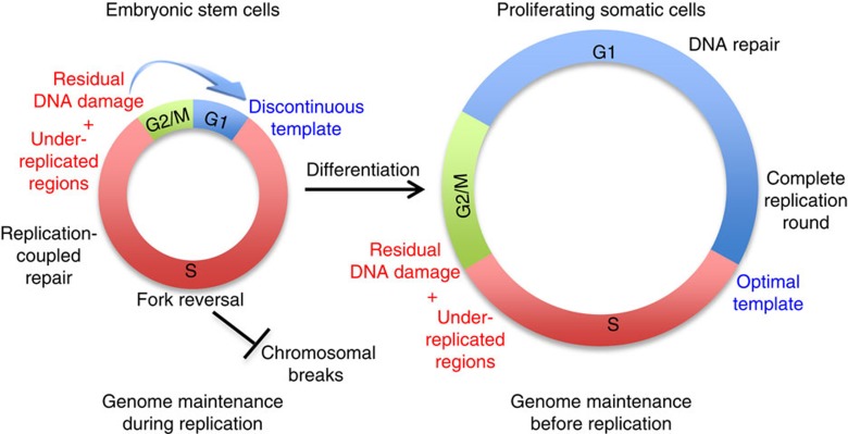
Embryonic stem cells (ESCs) represent a transient biological state, where pluripotency is coupled with fast proliferation. ESCs display a constitutively active DNA damage response (DDR), but its molecular determinants have remained elusive. Here we show in cultured ESCs and mouse embryos that H2AX phosphorylation is dependent on Ataxia telangiectasia and Rad3 related (ATR) and is associated with chromatin loading of the ssDNA-binding proteins RPA and RAD51. Single-molecule analysis of replication intermediates reveals massive ssDNA gap accumulation, reduced fork speed and frequent fork reversal. All these marks of replication stress do not impair the mitotic process and are rapidly lost at differentiation onset. Delaying the G1/S transition in ESCs allows formation of 53BP1 nuclear bodies and suppresses ssDNA accumulation, fork slowing and reversal in the following S-phase. Genetic inactivation of fork slowing and reversal leads to chromosomal breakage in unperturbed ESCs. We propose that rapid cell cycle progression makes ESCs dependent on effective replication-coupled mechanisms to protect genome integrity.


Figures

Figure 1
Figure 1. Replication stress markers are present in vitro in ESCs and in vivo in ICM cells.
(a) Immunofluorescence (IF) staining of an embryonic stem cell (ESC) colony and partially differentiated ESCs (ESC-d; 3d from LIF removal) for the stem cell marker Oct4, the DNA damage marker γH2AX and chromatin-bound ssDNA-binding proteins RPA32 and RAD51. Scale bars, 25 μm. (b) FACS analysis of EdU incorporation and DNA content (DAPI) in ESC and ESC-d. The percentage of cells in the different cell cycle phases is indicated. (c) Quantification of double IF-stainings displayed in a) in ESC and ESC-d. A minimum of 150 cells were scored in each double staining. (d) IF staining for γH2AX, RPA32 and RAD51 of E3.5 blastocysts. Number of embryos analysed per staining was 12, 11 and 9, respectively. Representative images are shown (see also Supplementary Figs 2 and 3). Scale bar, 25 μm. (e) Immunoblot detection of the indicated proteins after biochemical fractionation performed on ESC, ESC-d and mouse embryonic fibroblasts (MEFs). Cytosolic kinase MEK2 and chromatin-bound H3 serve as fractionation controls. Original films were scanned at high resolution and the intensity of immunoblot signals was quantified with ImageJ. C, chromatin-enriched fraction; S, soluble fraction (includes cytosol and abundant nucleosoluble proteins); W, whole-cell extract. Histograms represent chromatin/soluble (C/S) ratios for RPA32 (left) and RAD51 (right). (f) FACS-based quantification of γH2AX staining in ESCs on mock, ATM inhibitor (ATMi) or ATR inhibitor (ATRi) treatment. All these experiments were performed in duplicate. a.u., arbitrary units.
Figure 2
Figure 2. ESCs display massive accumulation of ssDNA gaps, reduced fork speed and frequent fork reversal.
(a,c) Electron micrographs of representative replication forks from ESCs, with indicated parental (P) and daughter (D) duplexes. White arrows indicate ssDNA gaps; black arrow points to the regressed arm (R) of a reversed fork. Insets: (a), a magnified ssDNA gap; (c), the four-way junction at the reversed fork. Scale bar, 500 bp (=217 nm), 200 bp in inset. (b) Frequency of replication forks isolated from ESCs and differentiating ESCs (ESC-d) with the indicated number of ssDNA gaps. (d) Frequency of reversed replication forks isolated from ESC and ESC-d. The number of replication intermediates analysed is indicated in parentheses. Similar results were obtained in an independent experiment. (e) DNA fibre spreading of ESC, ESC-d and MEF. CldU/IdU-containing tracts were immunostained in red and green, respectively. Two representative fibres are shown for ESCs and ‘Differ. ESCs'. The IdU replicated track length was computed using Mann–Whitney test.
Figure 3
Figure 3. Delaying the G1/S and not the G2/M transition suppresses H2AX phosphorylation in ESCs.
(a) FACS analysis of Oct4 and γH2AX in ESCs on the indicated treatments. PLK1i (BI-6727) and CDK1i (RO-3306) treatments delay progression through G2/M, while CDK4/6i (LY-2835219) and CDC7i (PHA-767491) treatments delay S-phase entry. The top-left quadrant identifies the subpopulation of stem cells (Oct4+) that display negative γH2AX staining. (b) Student's t-test of the subpopulation of Oct4+ γH2AX cells (red) identified in a, across three independent experiments. Error bars indicate s.d. **P<0.005, ***P<0.0005. (c) Cell cycle distribution, assessed by FACS-based DNA content (DAPI), of the samples in a. The total population is displayed in grey and the subpopulation of Oct4+ γH2AX in red. Where present, Oct4+ γH2AX cells are invariably detected in G1. (df) Representative images and graphical distribution of untreated (UNT) and aphidicolin (APH)-treated MEFs and ESCs displaying different mitotic defects (d, chromatin bridges; e, micronuclei; f, ultrafine bridges). Scale bars, 10 μm. The histograms indicate mean and standard deviations from three independent experiments.
Figure 4
Figure 4. A shortened G1 phase impairs 53BP1 nuclear body formation and causes replication stress in the following S-phase.
(a) Representative IF images of ESC and ESC-d stained with EdU and 53BP1. ESC differentiation detectably increase 53BP1 nuclear signals in EdU-negative cells. Scale bar, 25 μm. (b) Microscopically discernible 53BP1 nuclear bodies (NBs) in G1 nuclei (as identified by low DAPI and low EdU signals) were quantified in asynchronous populations of ESC and ESC-d by software-assisted segmentation and feature extraction (Supplementary Fig. 6f). Comparable results were observed in two additional, independent experiments. (c) Western blot analysis of FZR1 levels in mock-depleted (siLuc) and FZR1-depleted (siFZR1) MEFs, 96 h after siRNA transfection. TFIIH is used as loading control. (d) Microscopically discernible 53BP1 NBs specifically in G1 nuclei of asynchronous populations of mock- and FZR1-depleted MEFs were quantified as in b. (b,d) **P<0.005, ***P<0.0005 (Mann–Whitney test) (e) FACS analysis for DNA synthesis (EdU incorporation), DNA content (DAPI) and DDR activation (γH2AX) in mock-depleted and FZR1-depleted MEFs. Plots depict EdU incorporation versus DAPI in both populations. γH2AX positive cells are depicted in red. Cell populations in G1 and S-phase are also depicted. (f) DNA fibre analysis to assess fork progression in mock-depleted and FZR1-depleted MEFs. The IdU replicated track length was computed using Mann–Whitney test. (g,h) EM-based assessment of the percentage of replication intermediates containing ssDNA gaps (g) and the percentage of reversed forks (h) in genomic DNA extracted from mock-depleted and FZR1-depleted MEFs.
Figure 5
Figure 5. A prolonged G1 phase in ESCs suppresses replication stress in the following S-phase.
(a) FACS analysis of EdU incorporation and DNA content (DAPI) in control ESCs, ESCs treated with CDC7i, and ESCs on release from a transient CDC7i arrest (PHA-767491, 10 μM, 8 h). The gated (red) subpopulation of cells in early S-phase was analysed quantitatively for γH2AX staining (a.u., arbitrary units). (b) IdU replicated track length, computed using Mann–Whitney test, in control ESCs and cells released from a transient CDC7i arrest. (c) Frequency of replication forks isolated from control ESCs and on release from a transient CDC7i arrest, displaying the indicated number of ssDNA gaps. (d) Frequency of reversed replication forks isolated from control ESCs and on release from a transient CDC7i arrest.
Figure 6
Figure 6. PARP and Rad51 are essential for replication fork protection in ESCs.
(a,d) DNA fibre analysis of mock-treated ESCs, ESCs treated with Olaparib (a), mock-transfected ESCs and ESCs transfected with siRad51 for 48 and 72 h (d). The graphs show the statistical analysis of IdU replicated track length. (b,e) Frequency of reversed replication forks isolated from mock-treated ESCs and ESCs treated with Olaparib (b), mock-transfected ESCs and ESCs transfected with siRad51 for 48 and 72 h (e). (c,f) Pulse field gel electrophoresis (PFGE) to assess accumulation of DNA double-strand breaks (DSBs) in mock-treated ESCs, ESCs treated with Olaparib (c), mock-transfected ESCs and ESCs transfected with siRad51 for 48 and 72 h (f). DSBs equal to and above 500 Mb compact into a single band and DSBs smaller than 500 Mb can be detected as a smear as indicated alongside the gel. DSB (band plus smear) intensity from three independent experiments was quantified and plotted.
Figure 7
Figure 7. Model depicting differential control of genome stability in ESCs and proliferating somatic cells.
Under-replicated regions and residual DNA damage are unavoidably present at the end of each S-phase in both ESCs and somatic cells. However, owing to the brief gap phases, ESCs channel a high number of these lesions in the following S-phase and protect genome integrity by extensive fork reversal and replication-coupled repair. Conversely, differentiated cells have prolonged gap phases, assemble 53BP1 NBs and repair most of these lesions before S-phase entry (see also Supplementary Fig. 7d).

References

    1. Lanner F. Lineage specification in the early mouse embryo. Exp. Cell. Res. 321, 32–39 (2014). - PubMed
    1. Aladjem M. I. et al. ES cells do not activate p53-dependent stress responses and undergo p53-independent apoptosis in response to DNA damage. Curr. Biol. 8, 145–155 (1998). - PubMed
    1. Hong Y. & Stambrook P. J. Restoration of an absent G1 arrest and protection from apoptosis in embryonic stem cells after ionizing radiation. Proc. Natl Acad. Sci. USA 101, 14443–14448 (2004). - PMC - PubMed
    1. Tichy E. D. & Stambrook P. J. DNA repair in murine embryonic stem cells and differentiated cells. Exp. Cell. Res. 314, 1929–1936 (2008). - PMC - PubMed
    1. van der Laan S., Tsanov N., Crozet C. & Maiorano D. High Dub3 expression in mouse ESCs couples the G1/S checkpoint to pluripotency. Mol. Cell. 52, 366–379 (2013). - PubMed

Publication types

MeSH terms