

Inhibition of mTOR by apigenin in UVB-irradiated keratinocytes: A new implication of skin cancer prevention
- PMID: 26876613
- PMCID: PMC4788564
- DOI: 10.1016/j.cellsig.2016.02.008
Inhibition of mTOR by apigenin in UVB-irradiated keratinocytes: A new implication of skin cancer prevention
Abstract
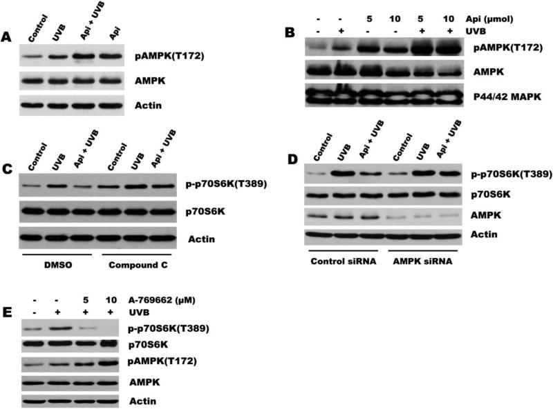
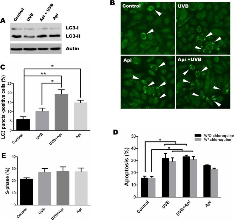
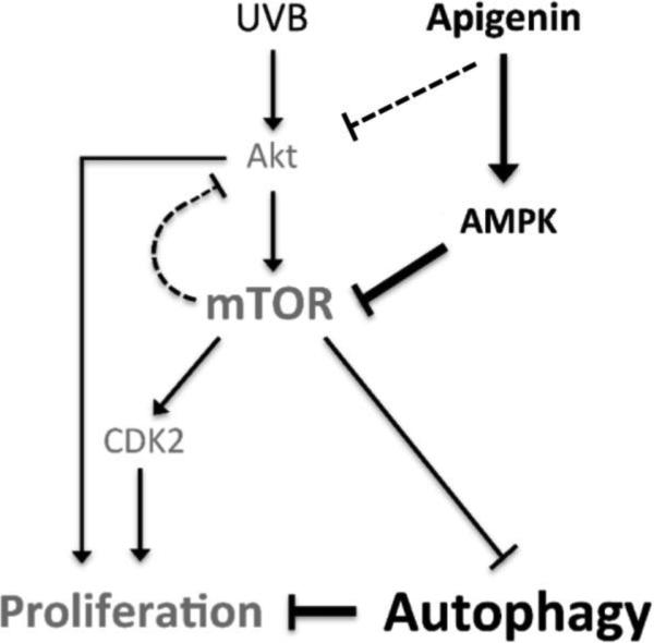
Ultraviolet B (UVB) radiation is the major environmental risk factor for developing skin cancer, the most common cancer worldwide, which is characterized by aberrant activation of Akt/mTOR (mammalian target of rapamycin). Importantly, the link between UV irradiation and mTOR signaling has not been fully established. Apigenin is a naturally occurring flavonoid that has been shown to inhibit UV-induced skin cancer. Previously, we have demonstrated that apigenin activates AMP-activated protein kinase (AMPK), which leads to suppression of basal mTOR activity in cultured keratinocytes. Here, we demonstrated that apigenin inhibited UVB-induced mTOR activation, cell proliferation and cell cycle progression in mouse skin and in mouse epidermal keratinocytes. Interestingly, UVB induced mTOR signaling via PI3K/Akt pathway, however, the inhibition of UVB-induced mTOR signaling by apigenin was not Akt-dependent. Instead, it was driven by AMPK activation. In addition, mTOR inhibition by apigenin in keratinocytes enhanced autophagy, which was responsible, at least in part, for the decreased proliferation in keratinocytes. In contrast, apigenin did not alter UVB-induced apoptosis. Taken together, our results indicate the important role of mTOR inhibition in UVB protection by apigenin, and provide a new target and strategy for better prevention of UV-induced skin cancer.
Keywords: AMPK; Akt; Apigenin; Autophagy; UVB; mTOR.
Copyright © 2016 Elsevier Inc. All rights reserved.
Figures
Similar articles
-
Apigenin, a chemopreventive bioflavonoid, induces AMP-activated protein kinase activation in human keratinocytes.Mol Carcinog. 2012 Mar;51(3):268-79. doi: 10.1002/mc.20793. Epub 2011 May 2. Mol Carcinog. 2012. PMID: 21538580
-
Inhibition of PI3K/Akt/mTOR pathway by apigenin induces apoptosis and autophagy in hepatocellular carcinoma cells.Biomed Pharmacother. 2018 Jul;103:699-707. doi: 10.1016/j.biopha.2018.04.072. Epub 2018 Apr 24. Biomed Pharmacother. 2018. PMID: 29680738
-
Impact on Autophagy and Ultraviolet B Induced Responses of Treatment with the MTOR Inhibitors Rapamycin, Everolimus, Torin 1, and pp242 in Human Keratinocytes.Oxid Med Cell Longev. 2017;2017:5930639. doi: 10.1155/2017/5930639. Epub 2017 Mar 16. Oxid Med Cell Longev. 2017. PMID: 28400912 Free PMC article.
-
Targeting the PI3K/Akt/mTOR axis by apigenin for cancer prevention.Anticancer Agents Med Chem. 2013 Sep;13(7):971-8. doi: 10.2174/18715206113139990119. Anticancer Agents Med Chem. 2013. PMID: 23272913 Free PMC article. Review.
-
Pharmacological Properties of 4', 5, 7-Trihydroxyflavone (Apigenin) and Its Impact on Cell Signaling Pathways.Molecules. 2022 Jul 4;27(13):4304. doi: 10.3390/molecules27134304. Molecules. 2022. PMID: 35807549 Free PMC article. Review.
Cited by
-
Skin cancer: understanding the journey of transformation from conventional to advanced treatment approaches.Mol Cancer. 2023 Oct 6;22(1):168. doi: 10.1186/s12943-023-01854-3. Mol Cancer. 2023. PMID: 37803407 Free PMC article. Review.
-
Apigenin inhibits renal cell carcinoma cell proliferation.Oncotarget. 2017 Mar 21;8(12):19834-19842. doi: 10.18632/oncotarget.15771. Oncotarget. 2017. PMID: 28423637 Free PMC article.
-
A recent update on the connection between dietary phytochemicals and skin cancer: emerging understanding of the molecular mechanism.Ann Med Surg (Lond). 2024 Aug 7;86(10):5877-5913. doi: 10.1097/MS9.0000000000002392. eCollection 2024 Oct. Ann Med Surg (Lond). 2024. PMID: 39359831 Free PMC article. Review.
-
Role of Polyphenols in Dermatological Diseases: Exploring Pharmacotherapeutic Mechanisms and Clinical Implications.Pharmaceuticals (Basel). 2025 Feb 12;18(2):247. doi: 10.3390/ph18020247. Pharmaceuticals (Basel). 2025. PMID: 40006060 Free PMC article. Review.
-
Apigenin Alleviates Intervertebral Disc Degeneration via Restoring Autophagy Flux in Nucleus Pulposus Cells.Front Cell Dev Biol. 2022 Jan 14;9:787278. doi: 10.3389/fcell.2021.787278. eCollection 2021. Front Cell Dev Biol. 2022. PMID: 35096819 Free PMC article.
References
-
- Miller DL, Weinstock MA. J Am Acad Dermatol. 1994;30:774–778. - PubMed
-
- Bode AM, Dong Z. Sci STKE. 2003;2003:RE2. - PubMed
-
- Bowden GT. Nat Rev Cancer. 2004;4:23–35. - PubMed
-
- Ross JA, Kasum CM. Annu Rev Nutr. 2002;22:19–34. - PubMed
-
- Wei H, Tye L, Bresnick E, Birt DF. Cancer Res. 1990;50:499–502. - PubMed
Publication types
MeSH terms
Substances
Grants and funding
LinkOut - more resources
Full Text Sources
Other Literature Sources
Miscellaneous
