

Optical Coherence Tomography Angiography of Asymptomatic Neovascularization in Intermediate Age-Related Macular Degeneration
- PMID: 26876696
- PMCID: PMC5120960
- DOI: 10.1016/j.ophtha.2016.01.044
Optical Coherence Tomography Angiography of Asymptomatic Neovascularization in Intermediate Age-Related Macular Degeneration
Abstract
Purpose: To determine whether angiography with swept-source (SS) optical coherence tomography (OCT) identifies subclinical type 1 neovascularization in asymptomatic eyes with intermediate age-related macular degeneration (iAMD).
Design: Prospective, observational, consecutive case series.
Participants: Patients with asymptomatic iAMD in one eye and neovascular age-related macular degeneration (AMD) in their fellow eye.
Methods: The patients underwent SS OCT angiography (OCTA), fluorescein angiography (FA), and indocyanine green angiography (ICGA), and the images from these 3 angiographic techniques were compared.
Main outcome measures: Identification of subclinical type 1 neovascularization with SS OCTA in asymptomatic eyes with iAMD.
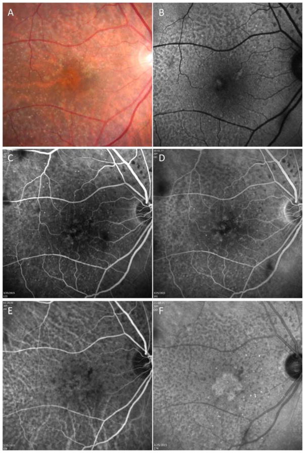
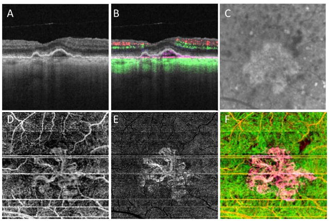
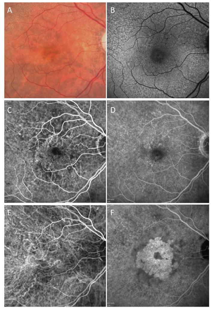
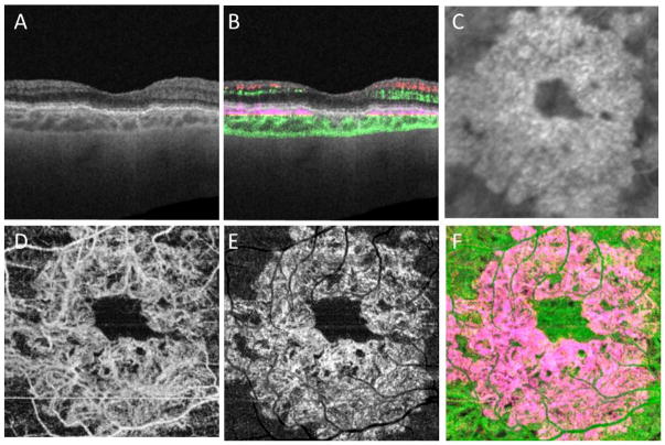
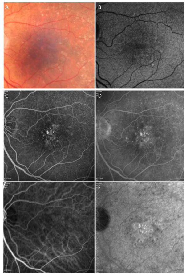
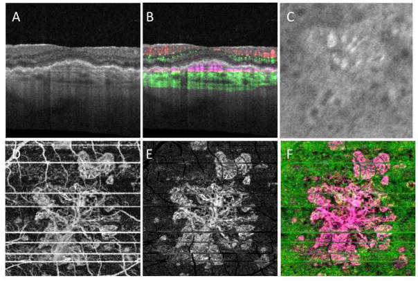
Results: Eleven consecutive patients with iAMD in one eye and neovascular AMD in their fellow eye were imaged with FA, ICGA, and SS OCTA between August 2014 and September 2015. Clinical examination of the 11 eyes revealed drusen and pigmentary abnormalities in the central macula and no evidence of macular fluid on routine OCT imaging. Ten of the 11 eyes had no evidence of leakage on FA and 1 eye had questionable fluorescein leakage. Indocyanine green angiography revealed the presence of central macular plaques in 3 of the 11 asymptomatic eyes with iAMD, and SS OCTA revealed unambiguous type 1 neovascularization corresponding to the plaques in all 3 eyes. Optical coherence tomography angiography did not identify neovascularization in the remaining 8 eyes.
Conclusions: Swept-source OCTA identified type 1 neovascularization corresponding to ICGA plaques in asymptomatic eyes with iAMD. The ability of OCTA to provide noninvasive, fast, detailed, depth-resolved identification of nonexudative neovascular lesions in eyes with iAMD suggests the need for a new classification system that distinguishes between neovascular and nonneovascular iAMD.
Copyright © 2016 American Academy of Ophthalmology. Published by Elsevier Inc. All rights reserved.
Figures
References
-
- Freund KB, Zweifel SA, Engelbert M. Do we need a new classification for choroidal neovascularization in age-related macular degeneration? Retina. 2010;30:1333–49. - PubMed
-
- Brown DM, Kaiser PK, Michels M, Soubrane G, Heier JS, Kim RY, et al. Ranibizumab versus verteporfin for neovascular age-related macular degeneration. The New England journal of medicine. 2006;355:1432–44. - PubMed
-
- Rosenfeld PJ, Brown DM, Heier JS, Boyer DS, Kaiser PK, Chung CY, et al. Ranibizumab for neovascular age-related macular degeneration. The New England journal of medicine. 2006;355:1419–31. - PubMed
-
- Yannuzzi LA, Freund KB, Takahashi BS. Review of retinal angiomatous proliferation or type 3 neovascularization. Retina. 2008;28:375–84. - PubMed
Publication types
MeSH terms
Substances
Grants and funding
LinkOut - more resources
Full Text Sources
Other Literature Sources
