

Understanding Spatial Genome Organization: Methods and Insights
- PMID: 26876719
- PMCID: PMC4792841
- DOI: 10.1016/j.gpb.2016.01.002
Understanding Spatial Genome Organization: Methods and Insights
Abstract
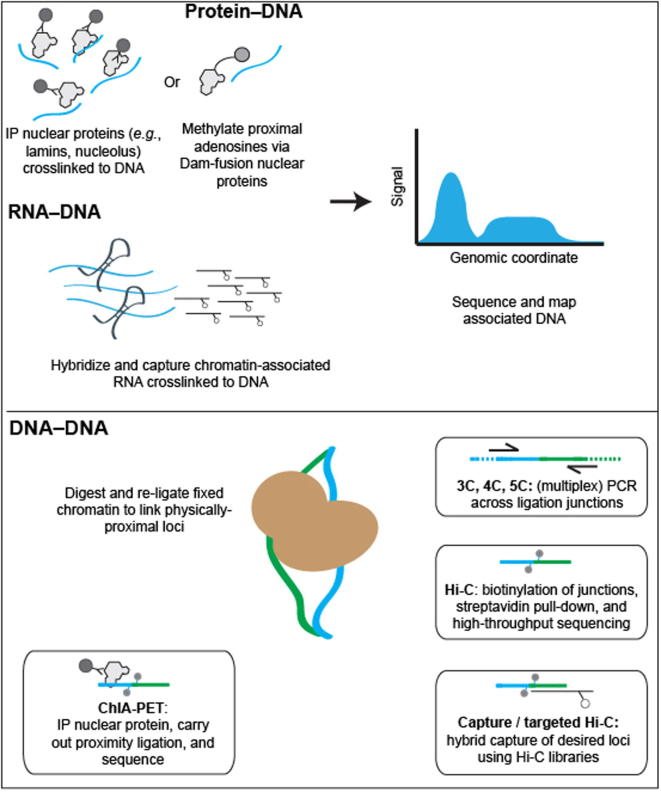
The manner by which eukaryotic genomes are packaged into nuclei while maintaining crucial nuclear functions remains one of the fundamental mysteries in biology. Over the last ten years, we have witnessed rapid advances in both microscopic and nucleic acid-based approaches to map genome architecture, and the application of these approaches to the dissection of higher-order chromosomal structures has yielded much new information. It is becoming increasingly clear, for example, that interphase chromosomes form stable, multilevel hierarchical structures. Among them, self-associating domains like so-called topologically associating domains (TADs) appear to be building blocks for large-scale genomic organization. This review describes features of these broadly-defined hierarchical structures, insights into the mechanisms underlying their formation, our current understanding of how interactions in the nuclear space are linked to gene regulation, and important future directions for the field.
Keywords: 4D nucleome; Chromatin; Chromosome; Epigenomics; Hi-C.
Copyright © 2016 The Authors. Production and hosting by Elsevier Ltd.. All rights reserved.
Figures
References
-
- Felsenfeld G., Groudine M. Controlling the double helix. Nature. 2003;421:448–453. - PubMed
-
- Zhou V.W., Goren A., Bernstein B.E. Charting histone modifications and the functional organization of mammalian genomes. Nat Rev Genet. 2011;12:7–18. - PubMed
-
- Talbert P.B., Henikoff S. Histone variants–ancient wrap artists of the epigenome. Nat Rev Mol Cell Biol. 2010;11:264–275. - PubMed
-
- Oudet P., Gross-Bellard M., Chambon P. Electron microscopic and biochemical evidence that chromatin structure is a repeating unit. Cell. 1975;4:281–300. - PubMed
Publication types
MeSH terms
Substances
Grants and funding
LinkOut - more resources
Full Text Sources
Other Literature Sources
