. 2016 Apr 15:35:166-84.
doi: 10.1016/j.actbio.2016.02.017. Epub 2016 Feb 12.

Decellularized skin/adipose tissue flap matrix for engineering vascularized composite soft tissue flaps

Affiliations

Decellularized skin/adipose tissue flap matrix for engineering vascularized composite soft tissue flaps

Qixu Zhang et al. Acta Biomater. .

Abstract

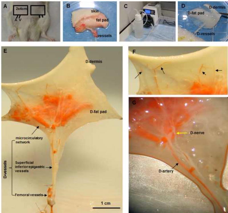
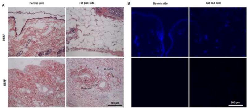
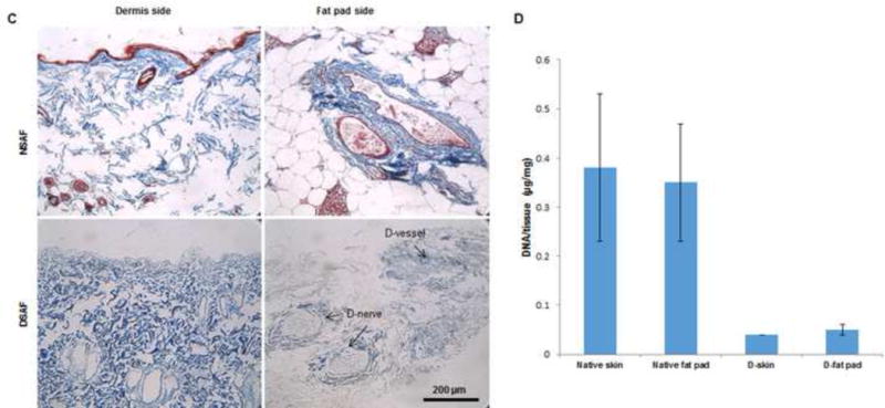
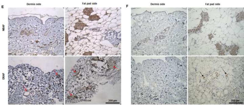
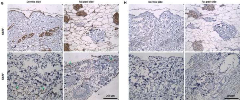
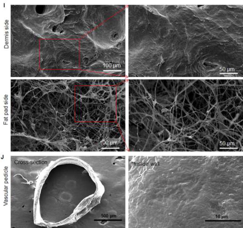
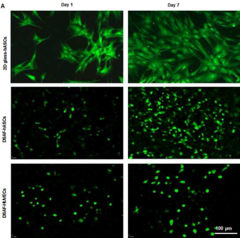
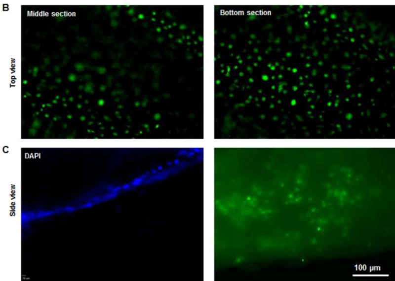
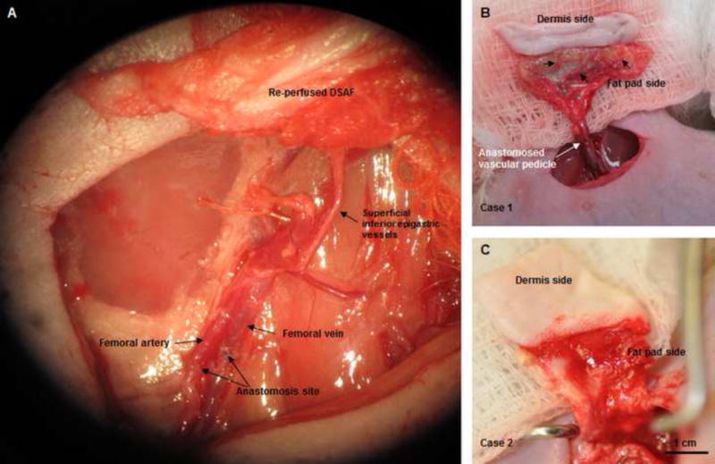
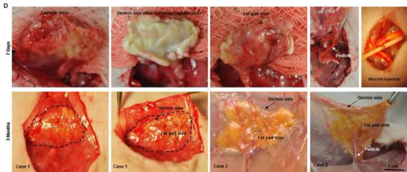
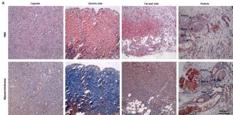
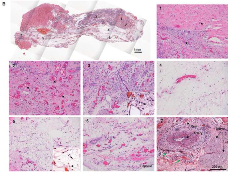
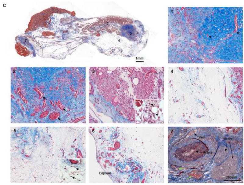
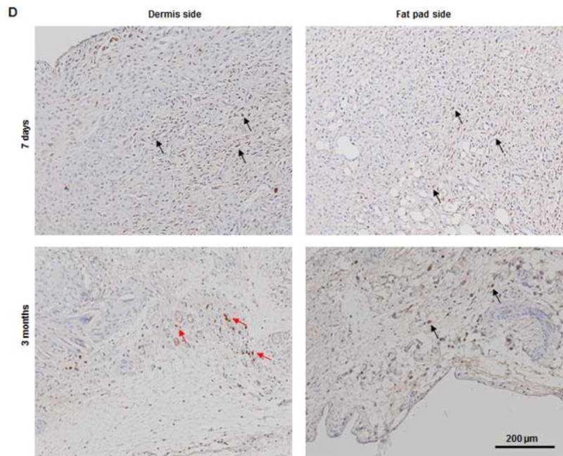
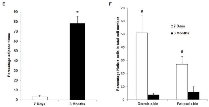
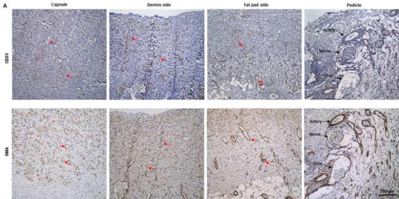
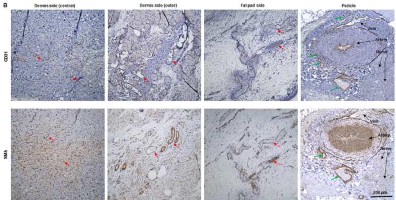
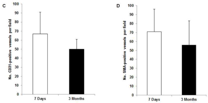
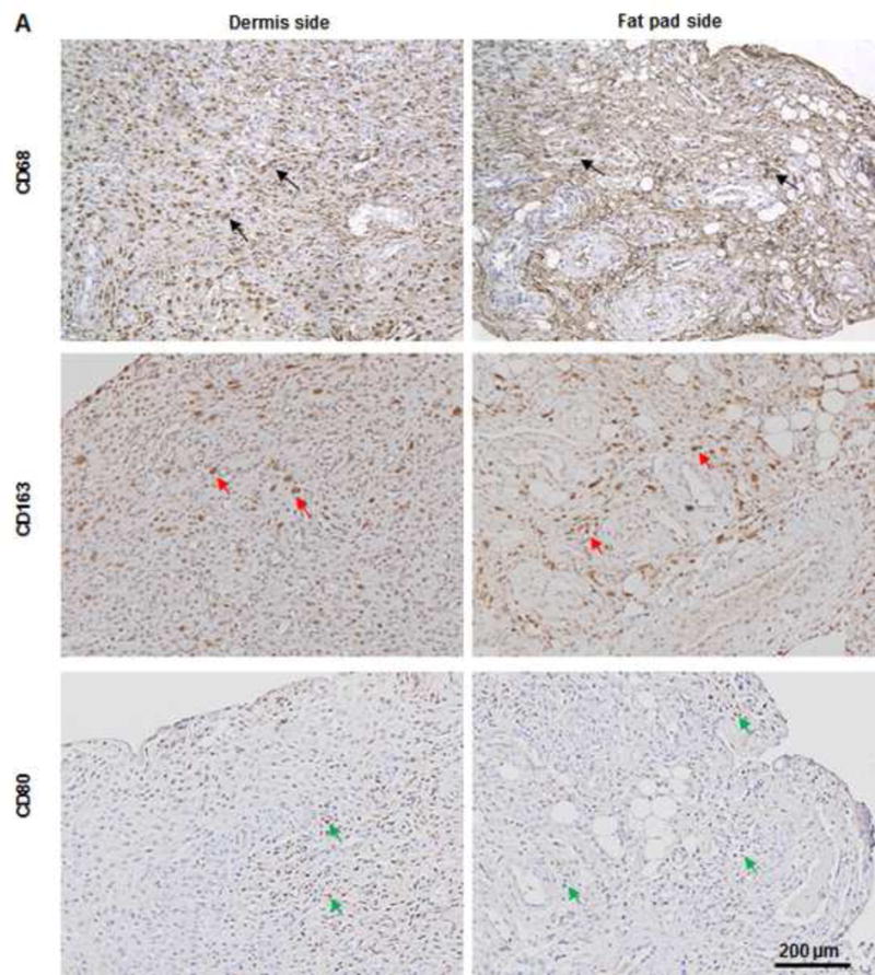
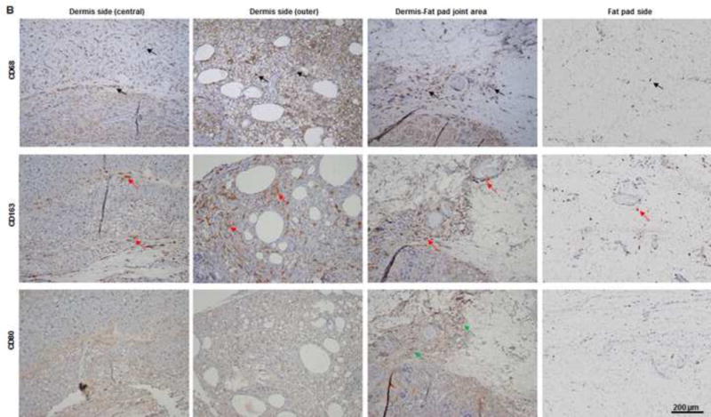
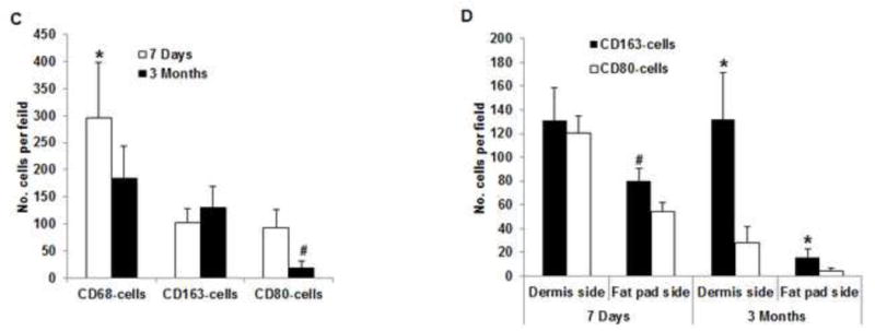
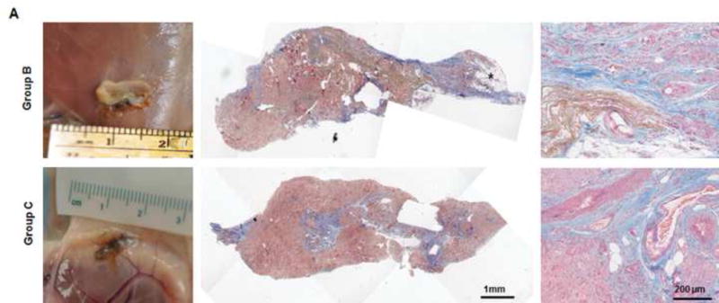
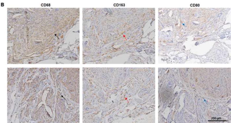
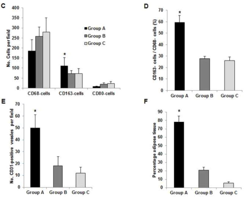
Using a perfusion decellularization protocol, we developed a decellularized skin/adipose tissue flap (DSAF) comprising extracellular matrix (ECM) and intact vasculature. Our DSAF had a dominant vascular pedicle, microcirculatory vascularity, and a sensory nerve network and retained three-dimensional (3D) nanofibrous structures well. DSAF, which was composed of collagen and laminin with well-preserved growth factors (e.g., vascular endothelial growth factor, basic fibroblast growth factor), was successfully repopulated with human adipose-derived stem cells (hASCs) and human umbilical vein endothelial cells (HUVECs), which integrated with DSAF and formed 3D aggregates and vessel-like structures in vitro. We used microsurgery techniques to re-anastomose the recellularized DSAF into nude rats. In vivo, the engineered flap construct underwent neovascularization and constructive remodeling, which was characterized by the predominant infiltration of M2 macrophages and significant adipose tissue formation at 3months postoperatively. Our results indicate that DSAF co-cultured with hASCs and HUVECs is a promising platform for vascularized soft tissue flap engineering. This platform is not limited by the flap size, as the entire construct can be immediately perfused by the recellularized vascular network following simple re-integration into the host using conventional microsurgical techniques.

Statement of significance: Significant soft tissue loss resulting from traumatic injury or tumor resection often requires surgical reconstruction using autologous soft tissue flaps. However, the limited availability of qualitative autologous flaps as well as the donor site morbidity significantly limits this approach. Engineered soft tissue flap grafts may offer a clinically relevant alternative to the autologous flap tissue. In this study, we engineered vascularized soft tissue free flap by using skin/adipose flap extracellular matrix scaffold (DSAF) in combination with multiple types of human cells. Following vascular reanastomosis in the recipient site, the engineered products successful regenerated large-scale fat tissue in vivo. This approach may provide a translatable platform for composite soft tissue free flap engineering for microsurgical reconstruction.

Keywords: Adipose tissue engineering; Decellularization; Extracellular matrix scaffold; Skin/adipose tissue flap matrix; Soft tissue flap engineering; Vascularized composite tissue flap engineering.


Figures

Fig. 1
Fig. 1. Perfusion decellularization and flap matrix angiography
(A, B) Groin skin/adipose tissue flaps with vascular pedicles (2 cm × 4 cm) were harvested from Fischer 344 rats. (C, D) Transparent DSAF matrix was achieved using a bump perfusion system combined with agitation decellularization. (“D-” indicates “decellularized” in this and other figures.) (E) Microfil-117 angiography showed that DSAF retained femoral vessels, superficial inferior epigastric vessels, and microcirculatory vessels. (F) Artery perforators penetrated into the dermis (black arrows). (G) An acellular sensory nerve (yellow arrow) and artery (black arrow) are shown.
Fig. 2
Fig. 2. Characterization of DSAF
(A) H&E staining showed that blood vessels and nerve structures were well maintained in DSAF. Cell nuclei were present in NSAF but absent in DSAF. (B) DAPI staining revealed the absence of nuclei DNA in DSAF. (C) Masson trichrome staining showed that collagen was a major component of DSAF. (D) The DNA content in the decellularized skin and fat pad was significantly lower than that in the native skin and fat pad (P<0.05). (E) IHC analysis indicated that laminin was distributed in vessels, nerves, and nanofibrous structures in DSAF (red arrows). (F) bFGF was present in the glandular and nanofibrous structures of DSAF (black arrows). (G) VEGF was present in the vessels and nerve structures of DSAF (green arrows). (H) The absence of MHC-I indicated the removal of alloantigenicity from DSAF. (I) SEM images confirmed that cells were absent in DSAF, leaving 3D porous structures in the fat pad side. Nanofibrous structures of ECM were well maintained in DSAF. (J) SEM images showed that the decellularized femoral artery remained elastic and patent.
Fig. 3
Fig. 3. Recellularization of DSAF
(A) Confocal microscopy images showed hASCs and HUVECs on DSAF. Cells were stained with Calcein AM (green) and EthD-1 (red) on days 1 and 7 after seeding. (B) Confocal microscopy images of HUVECs integrated with the DSAF pedicle (top view). (C) Confocal (DAPI staining) and fluorescence (Calcein AM and EthD-1 staining) microscopy images of HUVECs integrated with the DSAF pedicle (side view).
Fig. 4
Fig. 4. Implantation and explantation of the engineered DSAF-cells construct in group A
(A) The vascular pedicle was re-integrated into the host using conventional microsurgical anastomosis techniques. (B, C) Blood immediately perfused the engineered flap construct. (D) At 7 days (top row), the implant was encapsulated in swollen tissue (first image). Removing the swollen tissue capsule from the fat pad resulted in bleeding (second and third images). Mircofil-117 went through the vascular pedicle (fourth image) when it was injected through the contralateral femoral artery at day 7 (fifth image). At 3 months (bottom row), the implant was remodeled. Adipose tissue formed on both the dermis side and fat pad side. Numerous visible blood vessels penetrated into the newly formed soft tissue, indicating that it was highly vascularized.
Fig. 5
Fig. 5. Histological analysis of explants in group A
(A) H&E and Masson trichrome staining revealed an inflammation reaction characterized by cellular and vascular infiltration in the tissue at day 7. Lots of intravascular red blood cells were within the large-diameter blood vessels in the pedicle area. (B, C) H&E and Masson trichrome staining showed constructive remodeling in the implant at 3 months. Most dermis tissue was degraded and remodeled as adipose fascial tissue, and a small portion of dense collagen remained. The fat pad side of the implanted flap construct was fully remodeled, showing mature adipose tissue formed by adipocyte cells. (The numbered areas in the top images are magnified in the correspondingly numbered bottom images). The center of the dermis (images 1&2) was undergoing constructive remodeling, with dense collagen, highly vascular infiltration (black arrows), and numerous adipocytes. The outer layer of the dermis (image 3) showed a high level of remodeling, with numerous proliferating adipocytes (black arrows), blood vessel formation, and collagen that was less dense than that in the center of the dermis. The fat pad side of the construct (images 4–6) was completely remodeled, showing mature adipose tissue (black arrows), loose fascial tissue, a high number of blood vessels, and no inflammatory cells. In the pedicle area of the construct (image 7), myointimal hyperplasia of the main artery was present, the vein was patent, a number of functional vessels grew (green arrows), and the intact sensory nerve structure was recellularized. (D) The presence of donor cells was evaluated by immunohistochemical staining for an anti-HuNu antibody. HuNu-positive cells were distinguishable and persistent at 7 days and 3 months. Donor cells were closely located around the vascular structures (red arrow) and adipose structures (black arrow) at 3 months. (E) The area of adipose tissue at 3 months was significantly greater than that at 7 days. *: P<0.05 as compared to 7 days. (F) There were significantly fewer HuNu-positive cells at 3 months than at 7 days. The percentage of donor cells at 3 months was significantly lower than that at 7 days. #: P<0.05 as compared to 3 months.
Fig. 6
Fig. 6. IHC analysis of explants in group A
(A) Abundant CD31- and SMA-positive blood vessels were distributed in the capsule, dermis, and fat pad area of the explant (red arrows) at day 7. (B) The adipose tissue was highly vascularized, showing numerous CD31- and SMA-positive vessels (red arrows) throughout the explant at 3 months; lots of red blood cells were present in the functional vessels. Although the main pedicle artery showed myointimal hyperplasia, lots of vessels grew in the adventitia area (green arrows). (C&D) The numbers of CD31-positive vessels and SMA-positive vessels at 7 days and 3 months were both large and did not differ significantly.
Fig. 7
Fig. 7. IHC analysis of explants in group A
(A) As predominant inflammatory cells, many CD68-positive macrophages (black arrows) infiltrated the construct at day 7. Among these cells, both CD80-positive macrophages (M1 macrophages; green arrows) and CD163-positive macrophages (M2 macrophages; red arrows) were found throughout the sample tissue. (B) There were significantly fewer CD68-positive macrophages (black arrows) and CD80-positive macrophages (green arrows) at 3 months than at 7 days. Most macrophages were CD163-positive M2 macrophages (red arrows), which predominantly infiltrated the dermis side. Much fewer inflammatory macrophages were present in the fat pad side, indicating that the area was fully remodeled. (C) There were significantly fewer CD68- and CD80-positive macrophages at 3 the fat pad side at 7 days and in both the dermis and fat pad side at 3 months. *: P<0.05 as compared to CD80; #: P<0.05 as compared to CD80.
Fig. 8
Fig. 8. IHC analysis of explants in groups B and C at 3 months
(A) Explanted samples of group B and C were relatively hard with lots of undegraded dermis. Masson trichrome staining showed fibrotic remodeling in most areas of the explants. There were only a few vessels located in the vascular pedicle area (third images). The much lower vascularization resulted in less adipose regeneration in the tissues of both groups. *: adipose tissue. (B) Compared to CD80-positive M1 cells (blue arrow), relatively high numbers of CD163-positive M2 cells (red arrow) were distributed in the tissues. Numerous CD68-positive macrophages (black arrow) infiltrated the tissues in both groups. (C) At 3 months, there were significantly more CD163-positive macrophages in the tissue of group A than in the tissues of groups B or C. *: P<0.05 as compared to groups B and C. (D) The ratio of CD163-positive cells to CD68-positive cells in group A was significantly higher than those in groups B and C at 3 months. *: P<0.05 as compared to groups B and C. (E) There were significantly more CD31-positive vessels in the tissue of group A than in the tissues of groups B and C at 3 months. *: P<0.05 as compared to groups B and C. (F) At 3 months, the area of adipose tissue in group A was significantly greater than that in groups B and C. *: P<0.05 as compared to groups B and C.

References

    1. Shores JT, Brandacher G, Lee WP. Hand and upper extremity transplantation: an update of outcomes in the worldwide experience. Plast Reconstr Surg. 2015;135:351e–60e. - PubMed
    1. Kueckelhaus M, Fischer S, Seyda M, Bueno EM, Aycart MA, Alhefzi M, ElKhal A, Pomahac B, Tullius SG. Vascularized composite allotransplantation: current standards and novel approaches to prevent acute rejection and chronic allograft deterioration. Transpl Int. 2015 Aug 12; doi: 10.1111/tri.12652. [Epub ahead of print] - DOI - PMC - PubMed
    1. Siemionow M, Nasir S. Chimerism and bone marrow based therapies in transplantation. Microsurgery. 2007;27:510–21. - PubMed
    1. Siemionow M, Ortak T, Izycki D, Oke R, Cunningham B, Prajapati R, Zins JE. Induction of tolerance in composite-tissue allografts. Transplantation. 2002;74:1211–7. - PubMed
    1. Ott HC, Matthiesen TS, Goh SK, Black LD, Kren SM, Netoff TI, Taylor DA. Perfusion-decellularized matrix: using nature’s platform to engineer a bioartificial heart. Nat Med. 2008;14:213–21. - PubMed

Publication types