. 2016 Feb 15:7:10555.
doi: 10.1038/ncomms10555.

Neutrophil P2X7 receptors mediate NLRP3 inflammasome-dependent IL-1β secretion in response to ATP

Affiliations

Neutrophil P2X7 receptors mediate NLRP3 inflammasome-dependent IL-1β secretion in response to ATP

Mausita Karmakar et al. Nat Commun. .

Abstract

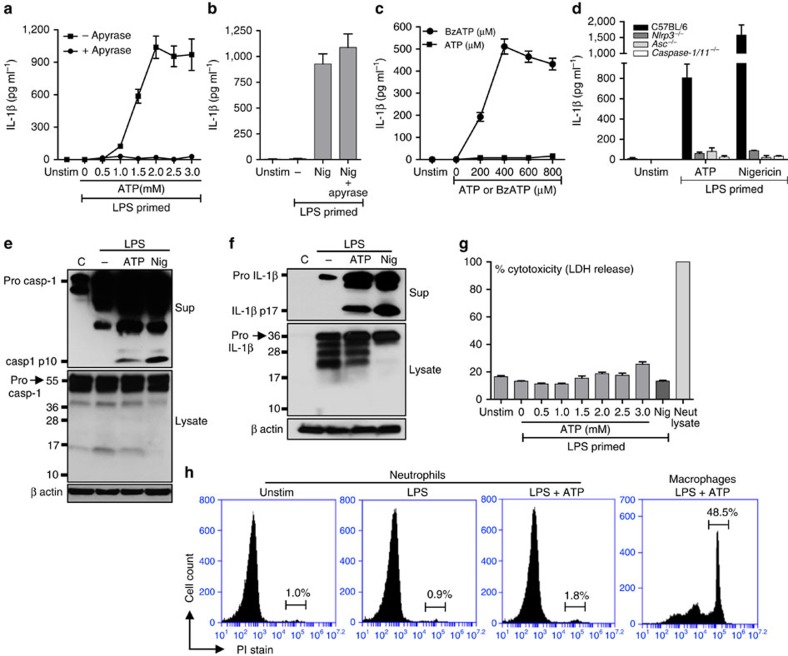
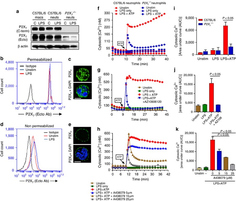
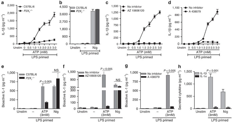
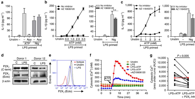
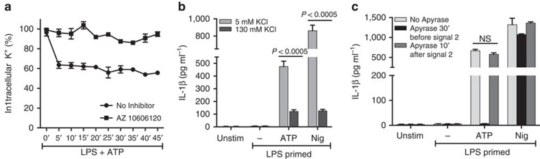
Although extracellular ATP is abundant at sites of inflammation, its role in activating inflammasome signalling in neutrophils is not well characterized. In the current study, we demonstrate that human and murine neutrophils express functional cell-surface P2X7R, which leads to ATP-induced loss of intracellular K(+), NLRP3 inflammasome activation and IL-1β secretion. ATP-induced P2X7R activation caused a sustained increase in intracellular [Ca(2+)], which is indicative of P2X7R channel opening. Although there are multiple polymorphic variants of P2X7R, we found that neutrophils from multiple donors express P2X7R, but with differential efficacies in ATP-induced increase in cytosolic [Ca(2+)]. Neutrophils were also the predominant P2X7R-expressing cells during Streptococcus pneumoniae corneal infection, and P2X7R was required for bacterial clearance. Given the ubiquitous presence of neutrophils and extracellular ATP in multiple inflammatory conditions, ATP-induced P2X7R activation and IL-1β secretion by neutrophils likely has a significant, wide ranging clinical impact.


Figures

Figure 1
Figure 1. ATP—induced IL-1β secretion by murine bone marrow neutrophils.
(a,b). IL-1β in the cell supernatants of C57BL/6 bone-marrow-derived neutrophils primed 3 h with LPS (500 ng ml−1) and stimulated 45 min with ATP (0.5–3.0 mM) or 10 μM nigericin. Apyrase (10U ml−1) was added 30 min before stimulation with ATP or nigericin, and IL-1β secretion was quantified by ELISA. (c). IL-1β production by LPS-primed C57BL/6 murine neutrophils stimulated 45 min with BzATP. (d). IL-1β in supernatants of LPS-primed bone-marrow-derived neutrophils from C57BL/6, Nlrp3−/−, Asc−/− and Caspase1,11−/− mice after 45 min stimulation with 3 mM ATP or 10 μM nigericin. (e,f). Pro- and cleaved forms of caspase-1 and IL-1β in total cell lysates and TCA precipitated supernatant of LPS-primed C57BL/6 neutrophils after 45 min stimulation with 3 mM ATP or 10 μM nigericin. (g). LDH release as a measure of cytotoxicity in LPS-primed C57BL/6 neutrophils after ATP stimulation. (h). Propidium iodide (PI) uptake by bone marrow neutrophils and bone-marrow-derived macrophages following LPS priming and 45 min ATP (3 mM) stimulation. Data were generated by flow cytometry. Data points are mean±s.d. of at least four replicates per treatment and are representative of three independent experiments.
Figure 2
Figure 2. P2X7 receptor expression and function in murine bone marrow neutrophils.
(a). Western blot analysis of P2X7 receptors on unstimulated and LPS-primed bone marrow neutrophils and macrophages from C57BL/6 and P2X7−/− mice. (be). Flow cytometry and fluorescence microscopy of P2X7R ecto domain (using the Hano-3 antibody) in LPS-primed bone marrow neutrophils that were either permeabilized with 0.1% TX-100 to detect total P2X7R (b,c) or were non-permeabilized to detect only cell surface P2X7R (d,e). (c,e) Cells were also stained with DAPI to detect the nucleus; Scale bar, 5 μm). (fh). Representative profiles of ATP-induced Ca2+ influx in bone marrow neutrophils from C57BL/6 and P2X7−/− mice (f), and in C57BL/6 neutrophils after stimulation with ATP (3 mM) in the presence of P2X7R antagonists AZ10606120 (10 μM) (g), or with different concentrations of A438079 (h). (ik). Mean±s.e.m. pf Area under the curve (AUC) of intracellular Ca2+ from three independent experiments. A P value ≤0.05 was considered significant using an unpaired Student's t-test.
Figure 3
Figure 3. P2X7R-dependent IL-1β secretion by ATP-stimulated murine neutrophils.
Total IL-1β secreted by bone marrow neutrophils from C57BL/6 and P2X7−/− mice (a,b), or from C57BL/6 neutrophils incubated with the P2X7 antagonists AZ10606120 (10 μM) (c) or A438079 (25 μM) (d) following LPS priming and stimulation with ATP or nigericin measured by ELISA. (eg). Bioactive IL-1 from the same samples detected using HEK-Blue-IL-1R reporter cells. (h). IL-1α and IL-1β from the C57BL/6 neutrophil supernatants stimulated 45 min with 3 mM ATP and 10 μM nigericin were quantified by ELISA. Data points are mean±s.d. of at least three replicates per treatment and are representative of three independent experiments. Using an ANOVA with Tukey post hoc analysis, a P value ≤0.05 was considered significant.
Figure 4
Figure 4. P2X7R expression and function on human peripheral blood neutrophils.
(ac). IL-1β measured by ELISA in the supernatant of human peripheral blood neutrophils primed with LPS (500 ng ml−1) for 3 h and stimulated with 4 mM ATP or 10 μM nigericin in the presence or absence of 10U ml−1 apyrase (a), or with increasing concentrations of ATP in the presence of P2X7 antagonists AZ10606120 (10 μM) or AZ11645373 (5 μM) (b,c). (d) Western blot analysis of P2X7R of unstimulated and LPS-primed human neutrophils from two donors that had been LPS primed for 3 h. β actin was used as a loading control. (e). Flow cytometry detection of P2X7R cell-surface expression on human neutrophils using antibody against the extracellular domain of the receptor. (f). Cytosolic [Ca2+] in LPS-primed human neutrophils after stimulation with 4 mM ATP in the presence of AZ10606120 (10 μM). (g). Area under the curve (AUC) for ATP induced cytosolic Ca2+ in neutrophils from all donors (n=11) in the presence or absence of P2X7R inhibitor (P values were obtained by paired t-tests, and a P value ≤0.05 was considered significant). Individual donors were for (a), Donor 8, (b), Donor 9, (c), Donor 7, (d), Donors 1 and 12, (e)—Donor 1, (f), Donor 2. Histograms or data points are mean±s.d. of at least five replicates per group and data shown are representative of three independent experiments with three different donor neutrophils (for ac).
Figure 5
Figure 5. Role of K+ efflux in P2X7R-mediated IL-1β secretion by murine neutrophils.
(a). Intracellular K+ content in LPS-primed C57BL/6 neutrophils stimulated with ATP (3 mM) alone and in the presence of AZ10606120 (10 μM) measured by atomic absorbance spectroscopy. (b). IL-1β production by LPS primed and ATP (3 mM) or nigericin (10 μM) stimulated neutrophils in the presence of 130 mM KCl or 5 mM KCl as quantified by ELISA. (c). IL-1β from LPS-primed C57BL/6 neutrophils in the presence of apyrase (10U ml−1) added either 30 min before or 10 min after stimulation with ATP or nigericin. Data points are mean±s.d. of at least four replicates per treatment and are representative of three independent experiments. A P value ≤0.05 was considered significant using ANOVA with Tukey post hoc analysis.
Figure 6
Figure 6. Neutrophil P2X7 receptor expression in S. pneumoniae corneal infection.
P2X7 receptor expression on total cells (a), and on Ly6G+ (1A8) neutrophils (b) 24 h after S. pneumoniae corneal infection. Cells were obtained following collagenase digestion, incubated with 1A8 and antibodies against the extracellular domain of the P2X7 receptor and analysed by flow cytometry. (cf) Neutrophil depletion following intraperitoneal injection of NIMP-R14 (Ly6G) or control rat IgG. (c,d) 1A8 and P2X7-expressing cells in the corneas 24 h post S. pneumoniae infection. One representative cornea is shown from each group. (e) Percentage and standard error of P2X7-receptor-expressing cells in IgG and NIMP-R14-treated mice (n=8 corneas). (f) P2X7 receptor, and pro- and mature IL-1β in infected corneas. Data are representative of two independent experiments with at least 5 mice per group. A P value ≤0.05 was considered significant using a Student's t-test.
Figure 7
Figure 7. The role of neutrophil P2X7 receptor expression on S. pneumoniae corneal infection.
(ac). S. pneumoniae infected corneas of C57BL/6 and P2X7−/− mice. Total neutrophils and macrophages quantified by flow cytometry (a), and bacterial CFU (b) 24 h post infection. (c). Western blot analysis of pro- and cleaved caspase-1 and IL-1β from 24 h infected corneas. (df). CD18−/− mice were infected with S. pneumoniae and 3 h later injected intravenously with donor bone marrow neutrophils from C57BL/6 or P2X7−/− mice. Total neutrophils in infected corneas of recipient CD18−/− mice given C57BL/6 or P2X7−/− neutrophils (d); CFU in corneas of infected C57BL/6 and CD18−/− mice (e); and in infected CD18−/− mice given donor bone marrow neutrophils from C57BL/6 or P2X7−/− mice (f). These experiments were repeated twice with similar results. 24 h post infection mice were sacrificed and and bacterial quantified. Each data point represents one cornea and histograms are mean±s.d. of at least three corneas per group. A P value ≤0.05 was considered significant using a Student's t-test.

References

    1. Fitz J. G. Regulation of cellular ATP release. Trans. Am. Clin. Climatol. Assoc. 118, 199–208 (2007). - PMC - PubMed
    1. Junger W. G. Immune cell regulation by autocrine purinergic signalling. Nat. Rev. Immunol. 11, 201–212 (2011). - PMC - PubMed
    1. Trautmann A. Extracellular ATP in the immune system: more than just a "danger signal". Sci. Signal. 2, pe6 (2009). - PubMed
    1. Cauwels A., Rogge E., Vandendriessche B., Shiva S. & Brouckaert P. Extracellular ATP drives systemic inflammation, tissue damage and mortality. Cell Death Dis. 5, e1102 (2014). - PMC - PubMed
    1. Riteau N. et al. Extracellular ATP is a danger signal activating P2X7 receptor in lung inflammation and fibrosis. Am. J. Respir. Crit. Care Med. 182, 774–783 (2010). - PubMed

Publication types

MeSH terms