. 2016 Mar 3;18(3):354-67.
doi: 10.1016/j.stem.2015.12.001. Epub 2016 Feb 11.

Lineage Reprogramming of Fibroblasts into Proliferative Induced Cardiac Progenitor Cells by Defined Factors

Affiliations

Lineage Reprogramming of Fibroblasts into Proliferative Induced Cardiac Progenitor Cells by Defined Factors

Pratik A Lalit et al. Cell Stem Cell. .

Abstract

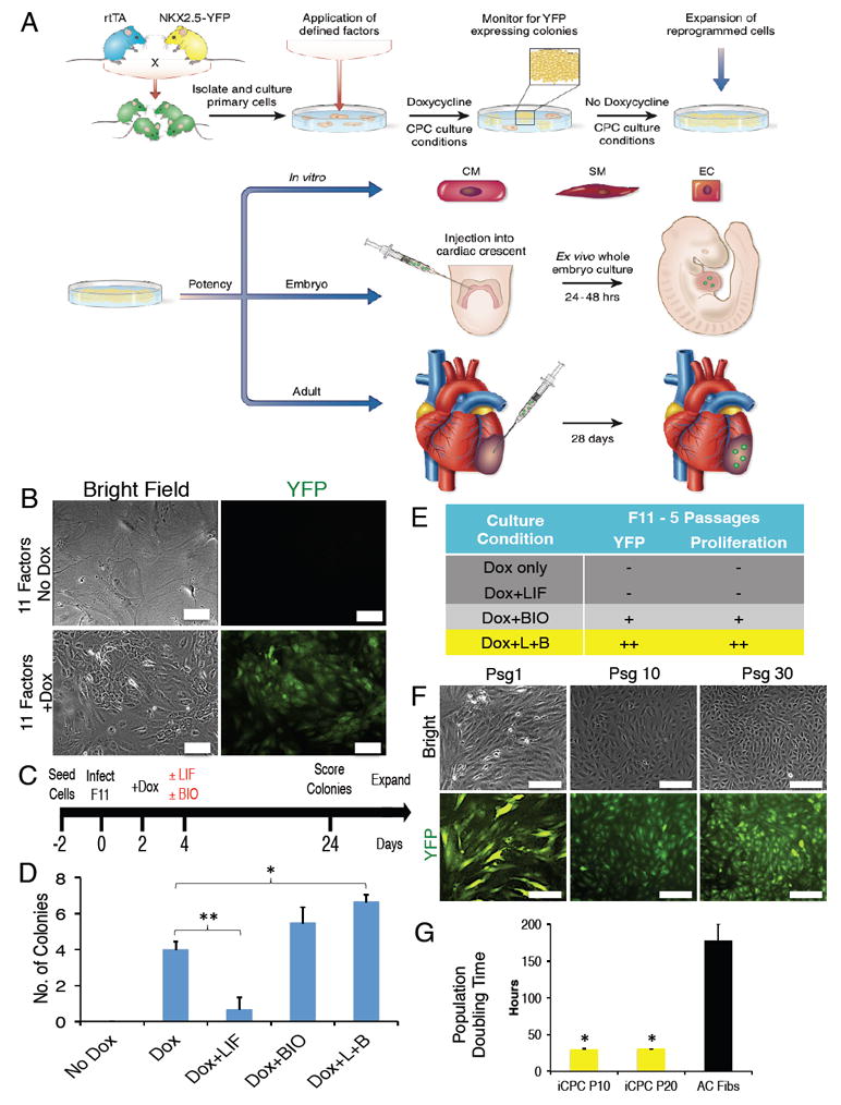
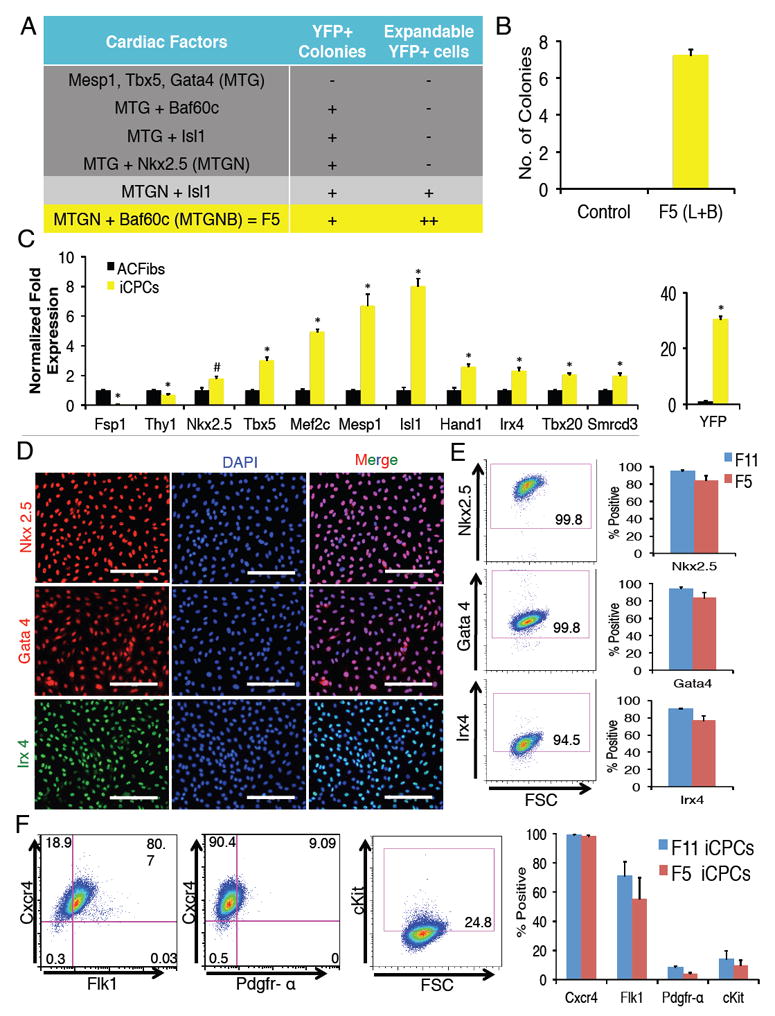
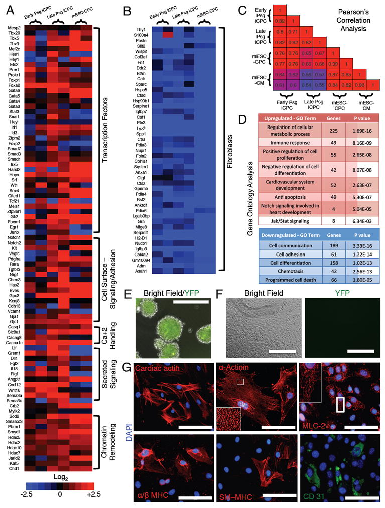
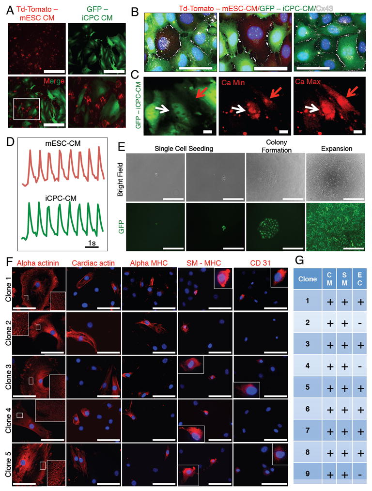
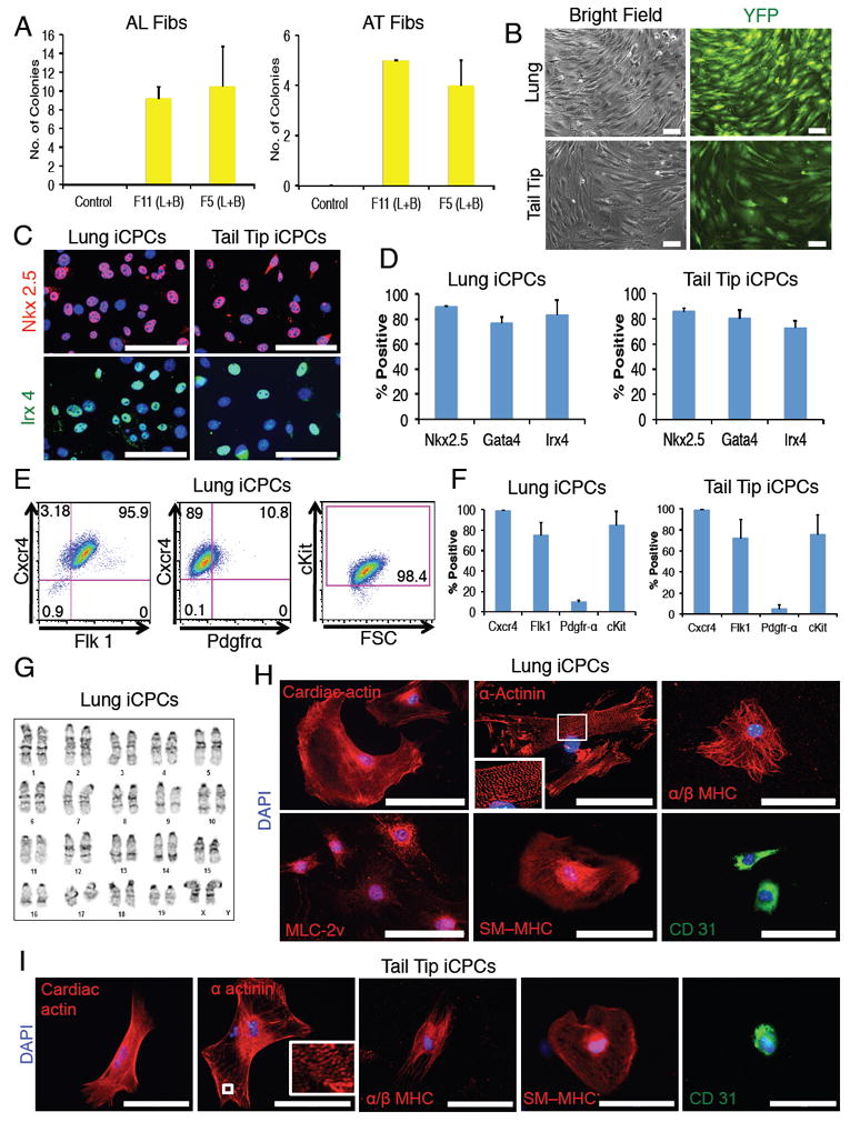
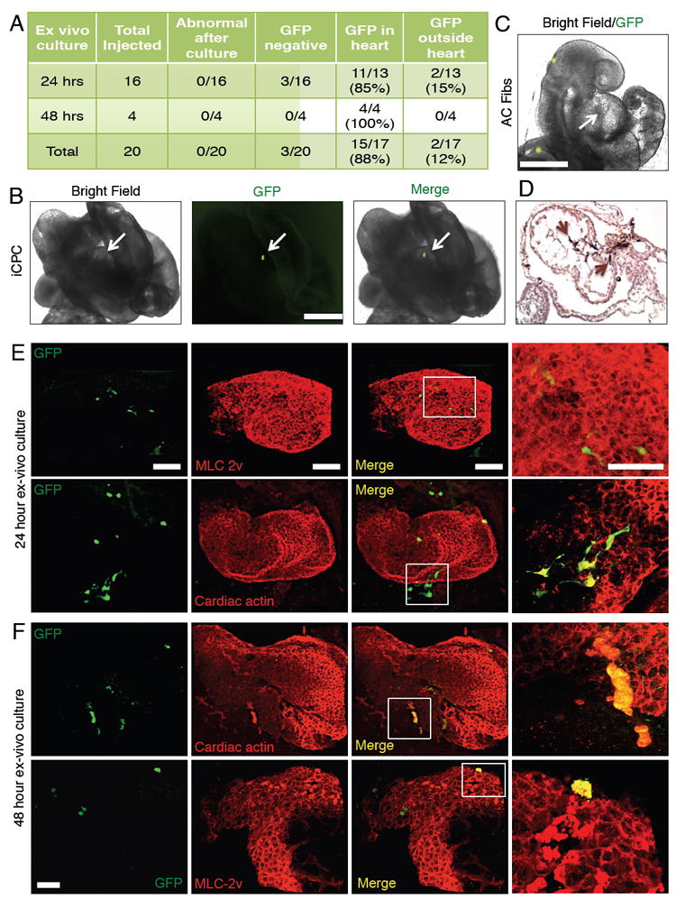
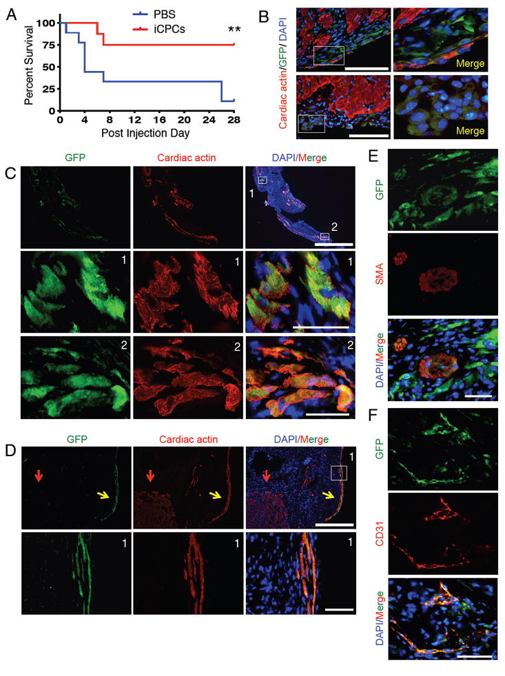
Several studies have reported reprogramming of fibroblasts into induced cardiomyocytes; however, reprogramming into proliferative induced cardiac progenitor cells (iCPCs) remains to be accomplished. Here we report that a combination of 11 or 5 cardiac factors along with canonical Wnt and JAK/STAT signaling reprogrammed adult mouse cardiac, lung, and tail tip fibroblasts into iCPCs. The iCPCs were cardiac mesoderm-restricted progenitors that could be expanded extensively while maintaining multipotency to differentiate into cardiomyocytes, smooth muscle cells, and endothelial cells in vitro. Moreover, iCPCs injected into the cardiac crescent of mouse embryos differentiated into cardiomyocytes. iCPCs transplanted into the post-myocardial infarction mouse heart improved survival and differentiated into cardiomyocytes, smooth muscle cells, and endothelial cells. Lineage reprogramming of adult somatic cells into iCPCs provides a scalable cell source for drug discovery, disease modeling, and cardiac regenerative therapy.


Figures

Figure 1
Figure 1. Screening for iCPC Inducing Factors and Optimal Culture Conditions
(A) Schematic representation of experimental design depicting direct reprogramming of adult fibroblasts to iCPCs by defined factors and culture conditions, expansion of iCPCs, and in vitro as well as in vivo differentiation of iCPCs into cardiac-lineage cells. (B) Infection with a combination of 11 cardiac factors induced Nkx2.5-EYFP expression in AC Fibs only after dox induction. (C) Strategy to test the impact of culture conditions on F11 reprogramming efficiency as well as the ability of EYFP+ cells to maintain a proliferative state. (D) Number of EYFP+ colonies formed (per 50,000 starting cells) in the respective culture conditions (**p<0.01, *p<0.05). (E) Impact of culture conditions on EYFP+ colonies expanded up to 5 psgs scoring for EYFP+ expression and proliferative ability (Dox only: n=8, Dox+LIF: n=3, Dox+BIO: n=4, Dox+LIF+BIO: n=9). (L=LIF, B=BIO). (F) F11 iCPCs maintained EYFP expression and proliferative ability for at least 30 psgs after dox withdrawal. (G) Population doubling time for psg 10 (P10) and psg 20 (P20) F11 iCPCs as compared to uninfected AC Fibs (n=3). Data presented as mean. Error bars = SEM. Scale bar = 100 μm in B, 200 μm in F. See also Figure S1, S2 and Movie S1.
Figure 2
Figure 2. Cardiac Factors Stably Reprogram Adult Cardiac Fibroblasts into Proliferative iCPCs
(A) Factor combinations tested both for ability to produce Nkx2.5-EYFP+ colonies and to expand them for at least 5 psgs without dox. (n=3-5) (B) Number of EYFP+ colonies produced after infection with 5 factors (MTGNB) and culture in iCPC induction medium for 3 weeks (per 50,000 seeded cells, n=8). (L=LIF, B=BIO). (C) qPCR analysis of F11 iCPCs showed upregulation of CPC markers and downregulation of fibroblast markers. Data represent normalized fold expression relative to uninfected AC Fibs (*p<0.01, #p<0.05). (D) Immunofluorescence labeling of F11 iCPCs showed nuclear localization of cardiac TFs Nkx2.5, Gata4, and Irx4. (E) Flow cytometry analysis revealed that majority of F11 iCPCs expressed cardiac TFs. F11 & F5 iCPCs showed comparable expression of cardiac TFs. (F) Flow cytometry analyses showed that F11 iCPCs expressed cell surface makers such as Cxcr4, Flk1, Pdgfr-α, cKit that are associated with CPCs (n=3). F11 & F5 iCPCs showed comparable expression of CPC associated cell surface markers. Error bars = SEM. Scale bars = 200 μm. See also Figure S1, S2 and S3 (for F5 iCPCs).
Figure 3
Figure 3. iCPCs Exhibit Cardiac-Mesoderm Restricted Gene Expression, are Multipotent and Differentiate into Cardiomyocytes, Smooth Muscle Cells and Endothelial Cells in vitro
(A-B) Heat maps of RNA Seq data illustrating differentially expressed genes in early psg (1-3), late psg (8-10) iCPCs and mESC-CPCs (Wamstad et al., 2012) as compared to AC Fibs. (n=2, biological replicates in each group). (A) Genes involved in embryonic cardiovascular development were increasingly upregulated in iCPC with psgs. (B) Fibroblasts genes were strongly downregulated. (C) Pearson’s correlation analysis of all expressed genes among low psg iCPCs, high psg iCPCs, mESC-CPCs and mESC-CMs (Wamstad et al., 2012). Numbers indicate Pearson’s R values. (D) Gene ontology analysis performed for upregulated and downregulated genes in late psg iCPCs as compared to AC Fibs. (E) F11 iCPCs aggregated in cardiac differentiation medium were EYFP+ at day 2. (F) F11 iCPC aggregates were plated and cultured in low serum conditions and lost Nkx2.5-EYFP expression by day 20. (G) Immunocytochemistry on plated cells revealed expression of CM markers such as cardiac actin, α-actinin (note highly organized sarcomere staining), MLC-2v, α/β MHC, a SM marker SM-MHC and EC marker CD31. Scale bars = 400 μm in E & F, 100 μm in G. See also Figure S3 and S5
Figure 4
Figure 4. iCPC-CMs Show Contraction, Calcium Transients upon Co-culture with mESC-CMs and Single Cell Derived iCPC Clones Exhibit Cardiovascular Potency
(A) iCPC-CMs infected with a GFP expressing lentivirus co-cultured with mESC-CMs that expressed td-tomato. No cell fusion was detected. (B) Cx43 immunolabeling showed that iCPC-CMs developed gap junctions with mESC-CMs and other iCPC-CMs. (C) iCPC-CMs showed synchronous calcium transients with mESC-CMs 3 weeks after co-culture. White arrow = iCPC-CM, red arrow = mESC-CM. (D) Time course of calcium transients. (E) Images show clonal expansion of single cell F5 iCPCs. F5 iCPCs derived from AC Fibs were seeded in low-density cultures to obtain isolated single cells. Single cell derived colonies were then picked and expanded. (F) Differentiation of iCPC clones followed by immunostaining for cardiac lineage markers revealed that that iCPC clones were either tripotent (differentiated into CMs, SMs and ECs) or bipotent (differentiated into CMs, SMs). (G) Table showing the cardiac lineage potency of various iCPC clones. CM=cardiomyocytes, SM=smooth muscle cell, EC=endothelial cell. Scale bars = 200 μm in A, 50 μm in B, 10 μm in C, 1 second in D, 400 μm in E (single cell seeding), 1000 μm in E (colony formation, expansion), 100 μm in F. See also Figure S3, S5, Movie S2 and S3.
Figure 5
Figure 5. Cardiac Factors Stably Reprogram Adult Lung and Adult Tail-tip Fibroblasts into Proliferative and Multipotent iCPCs
(A) Number of Nkx2.5-EYFP+ colonies produced (per 50,000 seeded cells) after infection of adult lung & adult tail-tip fibroblasts with 11 or 5 factors and culture in iCPC induction medium for 3 weeks (n=4). (L=LIF, B=BIO). (B) EYFP+ cells reprogrammed using 5 factors could be stably expanded without doxycycline for at least 10 psgs (Lung) and 5 psgs (Tail). (C) Immunolabeling revealed F5 lung-iCPCs & tail-tip-iCPCs had nuclear localization of CPC TFs (merged images are depicted, Red = Nkx2.5, Green = Irx4, Blue = DAPI), quantified in (D). (E&F) Flow cytometry analysis revealed that F5 lung-iCPCs & tail-tip-iCPCs expressed cell surface markers associated with CPCs (n=3). (G) F5 lung-iCPCs showed a normal diploid karyotype. (H) Lung-iCPCs were multipotent and differentiated into CMs (cardiac actin, α-actinin, MLC-2v, α/β MHC), SMs (SM-MHC) and ECs (CD31). Note highly organized sarcomere staining for α-actinin. (I) Tail-tip-iCPCs were multipotent and differentiated into cardiomyocytes (cardiac actin, α-actinin, α/β MHC), smooth muscle cells (SM-MHC) and endothelial cells (CD31). Note organized sarcomere staining for α-actinin. Data presented as mean, error bars = SEM. Scale bars = 100μm. See also Figure S2, S3.
Figure 6
Figure 6. iCPCs Localize to Developing Heart Tube and Differentiate into Cardiomyocytes Upon Injection into the Cardiac Crescent of Mouse Embryos
(A) Shows the number of embryos injected with F11 iCPCs and the location of iCPC-derived cells 24 or 48 hrs after whole embryo culture. (B) F11 iCPCs (labeled with GFP expressing lentivirus) injected into the cardiac crescent of mouse embryos colonized the developing heart tube as assessed after 24 hrs of whole embryo culture. See also Movie S4 for 24 hr cultured embryos and Movie S5 for 48 hr cultured embryos. (C) AC Fibs (labeled with GFP expressing lentivirus) injected into the cardiac crescent were excluded from the developing heart tube (arrow) as assessed after 24 hrs of whole embryo culture. (D) Histological sections of iCPC-injected embryos were stained for GFP antibody (dark brown color). iCPC-derived cells (brown arrows) integrated with host cell in the developing heart tube. (E) 24 hrs cultured iCPC injected embryos were immunostained in whole mount preparations for CM markers and GFP. 3D reconstruction images show iCPCs differentiated into CMs, as indicated by co-expression of CM markers and GFP. (F) iCPC-CMs attained shape/size similar to native CMs after 48 hrs culture. Scale bar = 500 μm in B-C, and100 μm in E-F. See also Figure S6, Movie S4-S7.
Figure 7
Figure 7. iCPCs Differentiate into Cardiac Lineage Cells In vivo and Improve Survival in Mice Post Myocardial Infarction
(A) Kaplan-Meier survival analysis revealed that iCPC injection significantly improved survival in treated animals as compared to control. (n=8 for iCPCs, n=9 for PBS) **p<0.01, Mantel-Cox test. (B) AC Fibs derived F5 iCPCs (labeled with GFP expressing lentivirus) were injected into the MI border zone and hearts were analyzed 4 days post injection. Immunolabeling of tissue sections revealed that a majority of surviving cells were localized on the edge of the scar tissue and showed faint expression of cardiac actin. Merged images are depicted. Scale bars = 100μm. (C) Injected iCPCs differentiated into cardiomyocytes based on organized cardiac actin immunolabeling (28 days after injection). Lower two rows of images are from insets in the top row. Scale bars = 1000μm, 50μm in insets. Tissue section represents the apex region of the left ventricular wall. (D) Immunolabeling for GFP and cardiac actin revealed that some iCPC-derived cardiomyocytes aligned and attained rod-shaped morphology 28 days after injection. Lower row of images are insets from top row. Red arrow = host CM, yellow arrow = iCPC-derived CM. Scale bars = 400μm, 50μm in insets. (E) iCPCs differentiated into smooth muscle cells within scar tissue based on co-expression of GFP and smooth muscle actin (SMA), as assessed 28 days after injection. Scale bar = 50μm. (F) iCPCs differentiated into endothelial cells within scar tissue based on co-expression of GFP and CD31, as assessed 28 days after injection. All images are from Mouse 1. Scale bar = 50μm. See also Figure S7 (Mouse 2 and 3).

Comment in

Similar articles

Cited by

References

    1. Abu-Issa R, Kirby ML. Heart field: from mesoderm to heart tube. Annual review of cell and developmental biology. 2007;23:45–68. - PubMed
    1. Birket MJ, Ribeiro MC, Verkerk AO, Ward D, Leitoguinho AR, den Hartogh SC, Orlova VV, Devalla HD, Schwach V, Bellin M, et al. Expansion and patterning of cardiovascular progenitors derived from human pluripotent stem cells. Nature biotechnology. 2015;33:970–979. - PubMed
    1. Bondue A, Tannler S, Chiapparo G, Chabab S, Ramialison M, Paulissen C, Beck B, Harvey R, Blanpain C. Defining the earliest step of cardiovascular progenitor specification during embryonic stem cell differentiation. J Cell Biol. 2011;192:751–765. - PMC - PubMed
    1. Brambrink T, Foreman R, Welstead GG, Lengner CJ, Wernig M, Suh H, Jaenisch R. Sequential expression of pluripotency markers during direct reprogramming of mouse somatic cells. Cell stem cell. 2008;2:151–159. - PMC - PubMed
    1. Cao N, Liang H, Huang J, Wang J, Chen Y, Chen Z, Yang HT. Highly efficient induction and long-term maintenance of multipotent cardiovascular progenitors from human pluripotent stem cells under defined conditions. Cell research. 2013;23:1119–1132. - PMC - PubMed

Publication types

Substances