

Dual Receptor Recognizing Cell Penetrating Peptide for Selective Targeting, Efficient Intratumoral Diffusion and Synthesized Anti-Glioma Therapy
- PMID: 26877777
- PMCID: PMC4729767
- DOI: 10.7150/thno.13532
Dual Receptor Recognizing Cell Penetrating Peptide for Selective Targeting, Efficient Intratumoral Diffusion and Synthesized Anti-Glioma Therapy
Abstract
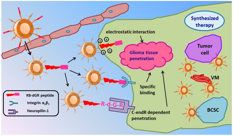
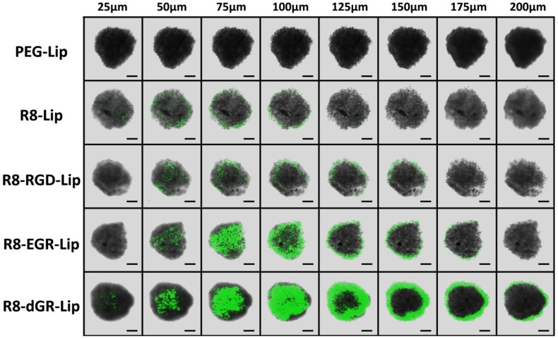
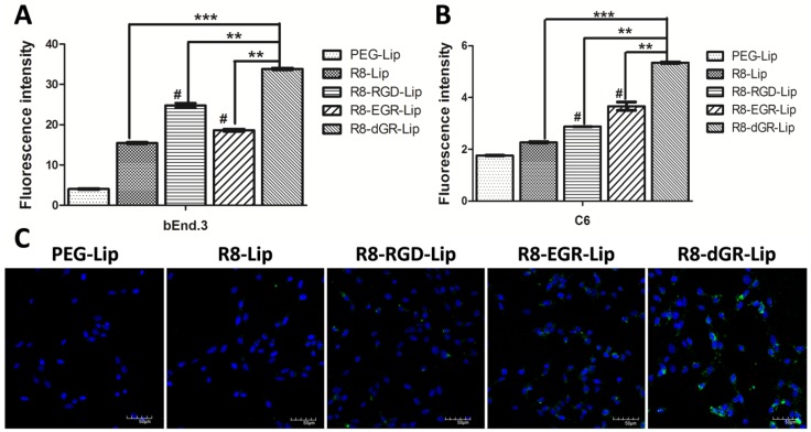
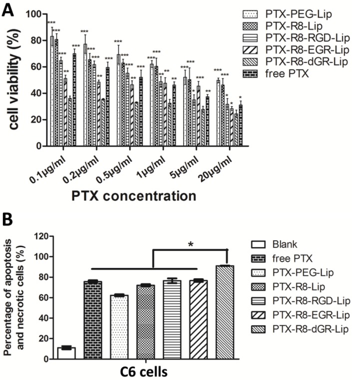
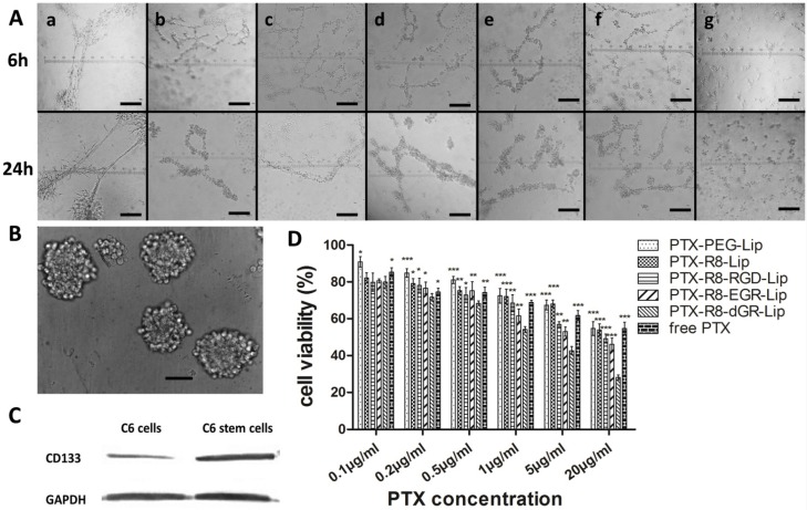
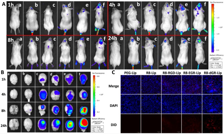
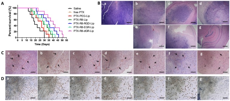
Cell penetrating peptides (CPPs) were widely used for drug delivery to tumor. However, the nonselective in vivo penetration greatly limited the application of CPPs-mediated drug delivery systems. And the treatment of malignant tumors is usually followed by poor prognosis and relapse due to the existence of extravascular core regions of tumor. Thus it is important to endue selective targeting and stronger intratumoral diffusion abilities to CPPs. In this study, an RGD reverse sequence dGR was conjugated to a CPP octa-arginine to form a CendR (R/KXXR/K) motif contained tandem peptide R8-dGR (RRRRRRRRdGR) which could bind to both integrin αvβ3 and neuropilin-1 receptors. The dual receptor recognizing peptide R8-dGR displayed increased cellular uptake and efficient penetration ability into glioma spheroids in vitro. The following in vivo studies indicated the active targeting and intratumoral diffusion capabilities of R8-dGR modified liposomes. When paclitaxel was loaded in the liposomes, PTX-R8-dGR-Lip induced the strongest anti-proliferation effect on both tumor cells and cancer stem cells, and inhibited the formation of vasculogenic mimicry channels in vitro. Finally, the R8-dGR liposomal drug delivery system prolonged the medium survival time of intracranial C6 bearing mice by 2.1-fold compared to the untreated group, and achieved an exhaustive anti-glioma therapy including anti-tumor cells, anti-vasculogenic mimicry and anti-brain cancer stem cells. To sum up, all the results demonstrated that R8-dGR was an ideal dual receptor recognizing CPP with selective glioma targeting and efficient intratumoral diffusion, which could be further used to equip drug delivery system for effective glioma therapy.
Keywords: Anti-glioma; C-end Rule; Cell penetrating peptides; Glioma targeting; Tumor penetration.
Conflict of interest statement
Competing Interests: The authors have declared that no competing interest exists.
Figures
Similar articles
-
Paclitaxel loaded liposomes decorated with a multifunctional tandem peptide for glioma targeting.Biomaterials. 2014 Jun;35(17):4835-47. doi: 10.1016/j.biomaterials.2014.02.031. Epub 2014 Mar 17. Biomaterials. 2014. PMID: 24651033
-
Multifunctional Tandem Peptide Modified Paclitaxel-Loaded Liposomes for the Treatment of Vasculogenic Mimicry and Cancer Stem Cells in Malignant Glioma.ACS Appl Mater Interfaces. 2015 Aug 5;7(30):16792-801. doi: 10.1021/acsami.5b04596. Epub 2015 Jul 22. ACS Appl Mater Interfaces. 2015. PMID: 26173814
-
A pH-responsive cell-penetrating peptide-modified liposomes with active recognizing of integrin αvβ3 for the treatment of melanoma.J Control Release. 2015 Nov 10;217:138-50. doi: 10.1016/j.jconrel.2015.09.009. Epub 2015 Sep 12. J Control Release. 2015. PMID: 26368312
-
Tumor penetrating peptides for improved drug delivery.Adv Drug Deliv Rev. 2017 Feb;110-111:3-12. doi: 10.1016/j.addr.2016.03.008. Epub 2016 Apr 1. Adv Drug Deliv Rev. 2017. PMID: 27040947 Free PMC article. Review.
-
Analytical and drug delivery strategies for short peptides: From manufacturing to market.Anal Biochem. 2025 Jan;696:115699. doi: 10.1016/j.ab.2024.115699. Epub 2024 Oct 24. Anal Biochem. 2025. PMID: 39461693 Review.
Cited by
-
Intelligent Photosensitive Mesenchymal Stem Cells and Cell-Derived Microvesicles for Photothermal Therapy of Prostate Cancer.Nanotheranostics. 2018 Nov 11;3(1):41-53. doi: 10.7150/ntno.28450. eCollection 2019. Nanotheranostics. 2018. PMID: 30662822 Free PMC article.
-
Peptide-Hitchhiking for the Development of Nanosystems in Glioblastoma.ACS Nano. 2024 Jul 2;18(26):16359-16394. doi: 10.1021/acsnano.4c01790. Epub 2024 Jun 11. ACS Nano. 2024. PMID: 38861272 Free PMC article. Review.
-
Dual drug-loaded nano-platform for targeted cancer therapy: toward clinical therapeutic efficacy of multifunctionality.J Nanobiotechnology. 2020 Sep 4;18(1):123. doi: 10.1186/s12951-020-00681-8. J Nanobiotechnology. 2020. PMID: 32887626 Free PMC article.
-
Liposomes for the Treatment of Brain Cancer-A Review.Pharmaceuticals (Basel). 2023 Jul 25;16(8):1056. doi: 10.3390/ph16081056. Pharmaceuticals (Basel). 2023. PMID: 37630971 Free PMC article. Review.
-
Preparation and Application of Cell Membrane-Camouflaged Nanoparticles for Cancer Therapy.Theranostics. 2017 Jun 25;7(10):2575-2592. doi: 10.7150/thno.20118. eCollection 2017. Theranostics. 2017. PMID: 28819448 Free PMC article. Review.
References
-
- Milletti F. Cell-penetrating peptides: classes, origin, and current landscape. Drug discovery today. 2012;17:850–60. - PubMed
-
- Kuai R, Yuan W, Li W, Qin Y, Tang J, Yuan M. et al. Targeted delivery of cargoes into a murine solid tumor by a cell-penetrating peptide and cleavable poly (ethylene glycol) comodified liposomal delivery system via systemic administration. Molecular pharmaceutics. 2011;8:2151–61. - PubMed
-
- Wang H, Zhao Y, Wang H, Gong J, He H, Shin MC. et al. Low-molecular-weight protamine-modified PLGA nanoparticles for overcoming drug-resistant breast cancer. Journal of Controlled Release. 2014;192:47–56. - PubMed
Publication types
MeSH terms
Substances
LinkOut - more resources
Full Text Sources
Other Literature Sources
Medical
