. 2016 Jan 1;7(1):11-27.
doi: 10.1039/C5MD00344J. Epub 2015 Sep 21.

Mechanisms of Resistance to Aminoglycoside Antibiotics: Overview and Perspectives

Affiliations

Mechanisms of Resistance to Aminoglycoside Antibiotics: Overview and Perspectives

Sylvie Garneau-Tsodikova et al. Medchemcomm. .

Abstract

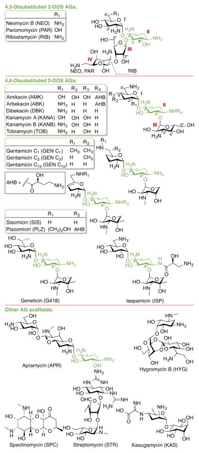
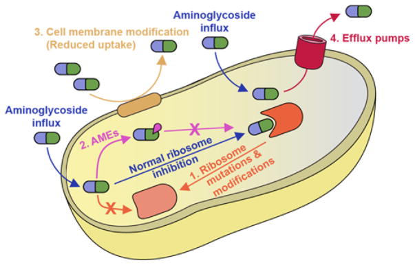
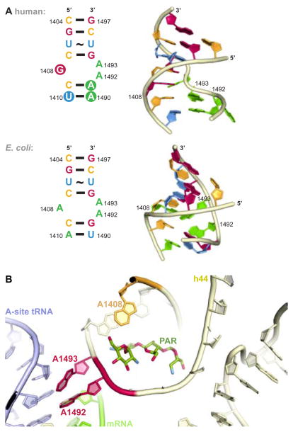
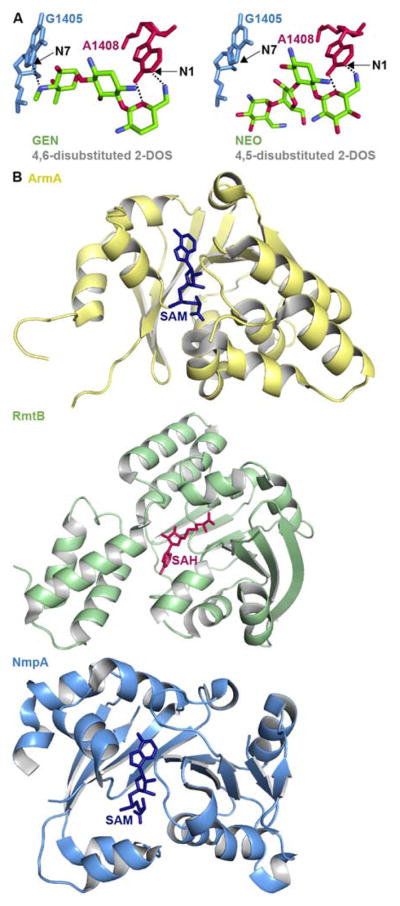
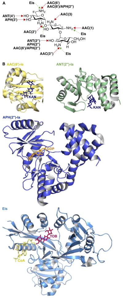
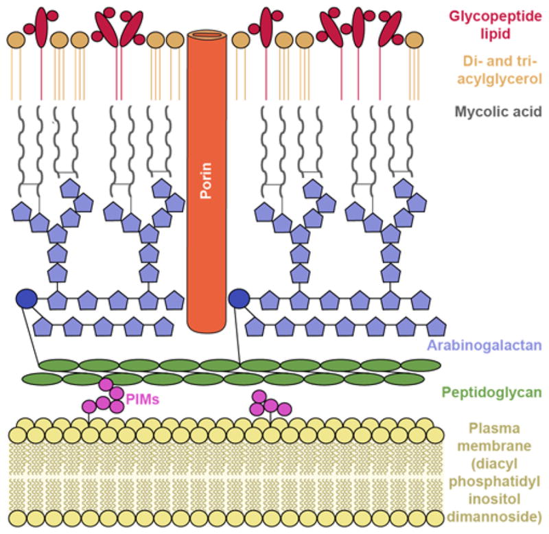
Aminoglycoside (AG) antibiotics are used to treat many Gram-negative and some Gram-positive infections and, importantly, multidrug-resistant tuberculosis. Among various bacterial species, resistance to AGs arises through a variety of intrinsic and acquired mechanisms. The bacterial cell wall serves as a natural barrier for small molecules such as AGs and may be further fortified via acquired mutations. Efflux pumps work to expel AGs from bacterial cells, and modifications here too may cause further resistance to AGs. Mutations in the ribosomal target of AGs, while rare, also contribute to resistance. Of growing clinical prominence is resistance caused by ribosome methyltransferases. By far the most widespread mechanism of resistance to AGs is the inactivation of these antibiotics by AG-modifying enzymes. We provide here an overview of these mechanisms by which bacteria become resistant to AGs and discuss their prevalence and potential for clinical relevance.


Figures

Fig. 1
Fig. 1
Structures of AGs presented in this review.
Fig. 2
Fig. 2
Schematic overview of mechanisms of resistance to AGs discussed in this review.
Fig. 3
Fig. 3
A. Comparison of internal loop of h44 of human and bacterial (E. coli) A-site. B. Structure of PAR bound to h44.
Fig. 4
Fig. 4
A. Structure of the 4,6-disustituted 2-DOS AG GEN and 4,5-disubstituted 2-DOS AG NEO in close proximity to ribosome (PDB code 4V53 and 4V52, respectively) with positions methylated by RMTases. B. Representative example of crystal structures of the RMTases ArmA as yellow cartoon with SAM as navy stick (PBD code 3FZG), RmtB as green cartoon with SAH as red stick (PBD code 3FRH), and NmpA as pale blue cartoon with SAM as navy stick (PBD code 3P2K).
Fig. 5
Fig. 5
A. Chemical modifications and positions affected by various AMEs on KANB. B. Representative example of recent crystal structures for AACs (AAC(6′)-Ie as yellow cartoon with KANA as navy stick; PDB code 4QC6; from the bifunctional enzyme AAC(6′)-Ie/APH(2″)-Ia), APHs (APH(2″)-Ia as dark blue cartoon with GDP as orange stick; PDB code 4ORK; from the bifunctional enzyme AAC(6′)-Ie/APH(2″)-Ia), ANTs (ANT(2″)-Ia as green cartoon with KANA as navy stick; PDB code 4WQL; normally a monomer), and Eis as pale blue cartoon with CoA as yellow stick and TOB as red stick (PDB code 4JD6; normally an hexamer) resistance enzymes. Note: Only the monomer of each of these enzymes is shown.
Fig. 6
Fig. 6
AG transport in Gram-negative bacteria. Influx of AGs occurs through hydrophilic porin protein channels. Efflux of AGs may occur through active transport pumps such as the RND-type efflux pump (e.g., AcrAD-TolC, as shown.)
Fig. 7
Fig. 7
Mycobacterium cell wall.

References

    1. Becker B, Cooper MA. ACS Chem BIol. 2013;8:105–115. - PubMed
    1. Fosso MY, Li Y, Garneau-Tsodikova S. Med Chem Comm. 2014;5:1075–1091. - PMC - PubMed
    1. Green KD, Garneau-Tsodikova S. Front Microbiol. 2013;4:208. - PMC - PubMed
    1. Blair JM, Webber MA, Baylay AJ, Ogbolu DO, Piddock LJ. Nat Rev Microbiol. 2015;13:42–51. - PubMed
    1. Fernandez L, Breidenstein EB, Hancock RE. Drug Resist Updates. 2011;14:1–21. - PubMed