Nephrotic syndrome complicated with portal, splenic, and superior mesenteric vein thrombosis
- PMID: 26877968
- PMCID: PMC4714169
- DOI: 10.1016/j.krcp.2014.07.001
Nephrotic syndrome complicated with portal, splenic, and superior mesenteric vein thrombosis
Abstract
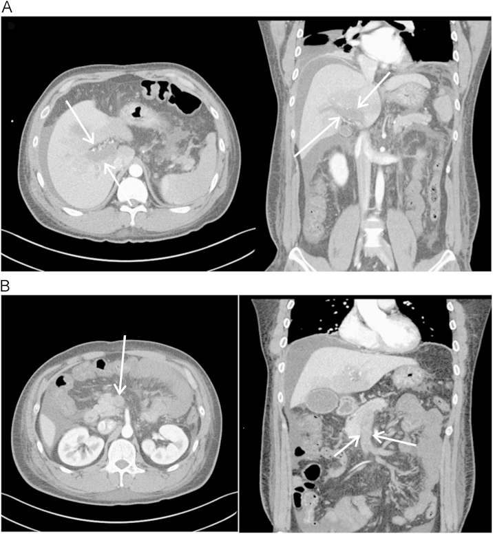
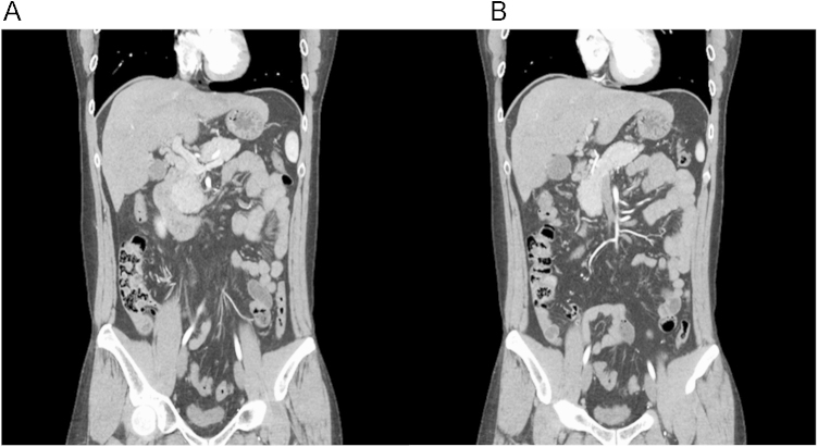
Thromboembolism is a major complication of nephrotic syndrome. Renal vein thrombosis and deep vein thrombosis are relatively common, especially in membranous nephropathy. However, the incidence of portal vein and superior mesenteric vein (SMV) thrombosis in patients with nephrotic syndrome is very rare. To date, several cases of portal vein thrombosis treated by anticoagulation therapy, not by thrombolytic therapy, have been reported as a complication of nephrotic syndrome. Here, we report a case of portal, splenic, and SMV thrombosis in a patient with a relapsed steroid dependent minimal change disease who was treated successfully with anticoagulation and thrombolytic therapy using urokinase. Radiologic findings and his clinical conditions gradually improved. Six months later, a complete remission of the nephrotic syndrome was observed and the follow-up computed tomography scan showed the disappearance of all portal vein, splenic vein, and SMV thrombi.
Keywords: Complications; Nephrotic syndrome; Thrombolytic therapy; Venous thrombosis.
Figures
Similar articles
-
A case report of minimal change nephrotic syndrome complicated with portal, splenic and superior mesenteric vein thrombosis.Clin Nephrol. 2012 Jun;77(6):505-9. doi: 10.5414/cn107372. Clin Nephrol. 2012. PMID: 22595395
-
Nephrotic syndrome with portal, splenic and renal vein thrombosis. A case report.Nephron. 2002;92(3):680-4. doi: 10.1159/000064107. Nephron. 2002. PMID: 12372955 Review.
-
[Transradial approach for transcatheter selective superior mesenteric artery urokinase infusion therapy in patients with acute extensive portal and superior mesenteric vein thrombosis].Zhonghua Yi Xue Za Zhi. 2012 Jun 5;92(21):1448-52. Zhonghua Yi Xue Za Zhi. 2012. PMID: 22944028 Chinese.
-
Low-molecular-weight heparin successfully used to treat a nephrotic patient complicated by superior mesenteric vein thrombosis and portal vein thrombosis.Med Princ Pract. 2011;20(2):196-9. doi: 10.1159/000319925. Epub 2011 Jan 20. Med Princ Pract. 2011. PMID: 21252580
-
Applications of percutaneous mechanical thrombectomy in transjugular intrahepatic portosystemic shunt and portal vein thrombosis.Tech Vasc Interv Radiol. 2003 Mar;6(1):59-69. doi: 10.1053/tvir.2003.36433. Tech Vasc Interv Radiol. 2003. PMID: 12772131 Review.
Cited by
-
Portal Vein Thrombosis: A Rare Complication of Nephrotic Syndrome.Indian J Nephrol. 2018 May-Jun;28(3):236-239. doi: 10.4103/ijn.IJN_25_17. Indian J Nephrol. 2018. PMID: 29962677 Free PMC article.
-
Tricky acute mesenteric ischemia: what can we do?Gastroenterol Rep (Oxf). 2025 Jul 7;13:goaf067. doi: 10.1093/gastro/goaf067. eCollection 2025. Gastroenterol Rep (Oxf). 2025. PMID: 40625657 Free PMC article. Review.
-
Isolated superior mesenteric vein thrombosis in an adult with nephrotic syndrome due to minimal change disease: a case report.J Med Case Rep. 2025 Apr 1;19(1):149. doi: 10.1186/s13256-025-05130-4. J Med Case Rep. 2025. PMID: 40170178 Free PMC article.
-
Nephrotic syndrome presented as a portal vein thrombosis: a case report.Ann Med Surg (Lond). 2023 Apr 6;85(5):2112-2114. doi: 10.1097/MS9.0000000000000482. eCollection 2023 May. Ann Med Surg (Lond). 2023. PMID: 37229005 Free PMC article.
-
[General recommendations for the management of glomerular diseases-2023].Wien Klin Wochenschr. 2023 Aug;135(Suppl 5):696-704. doi: 10.1007/s00508-023-02265-6. Epub 2023 Sep 20. Wien Klin Wochenschr. 2023. PMID: 37728654 Free PMC article. German.
References
-
- Harris RC, Ismail N. Extrarenal complications of the nephrotic syndrome. Am J Kidney Dis. 1994;23:477–497. - PubMed
-
- Llach F. Hypercoagulability, renal vein thrombosis, and other thrombotic complications of nephrotic syndrome. Kidney Int. 1985;28:429–439. - PubMed
-
- Sagripanti A, Barsotti G. Hypercoagulability, intraglomerular coagulation, and thromboembolism in nephrotic syndrome. Nephron. 1995;70:271–281. - PubMed
-
- Kumar S, Sarr MG, Kamath PS. Mesenteric venous thrombosis. N Engl J Med. 2001;345:1683–1688. - PubMed
-
- Brunaud L, Antunes L, Collinet-Adler S, Marchal F, Ayav A, Bresler L, Boissel P. Acute mesenteric venous thrombosis: case for nonoperative management. J Vasc Surg. 2001;34:673–679. - PubMed
LinkOut - more resources
Full Text Sources
Other Literature Sources