. 2016 Apr;186(4):777-93.
doi: 10.1016/j.ajpath.2015.11.024. Epub 2016 Feb 13.

Initial Suppression of Transforming Growth Factor-β Signaling and Loss of TGFBI Causes Early Alveolar Structural Defects Resulting in Bronchopulmonary Dysplasia

Affiliations

Initial Suppression of Transforming Growth Factor-β Signaling and Loss of TGFBI Causes Early Alveolar Structural Defects Resulting in Bronchopulmonary Dysplasia

Shawn K Ahlfeld et al. Am J Pathol. 2016 Apr.

Abstract

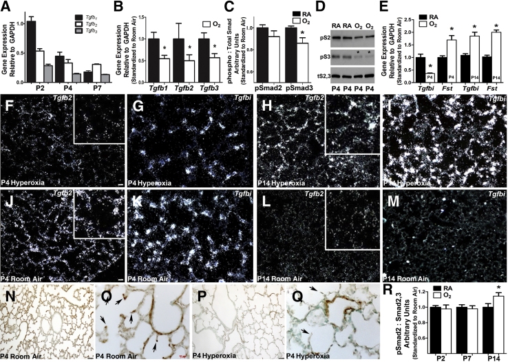
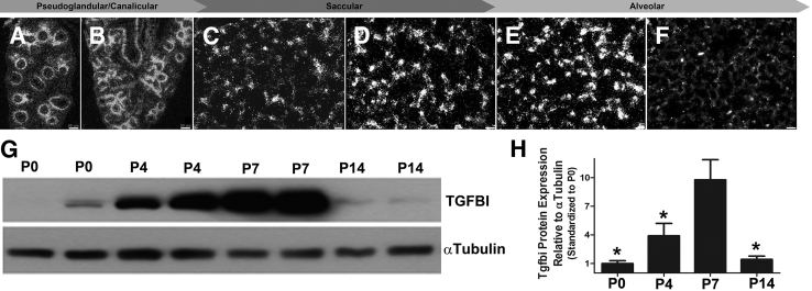
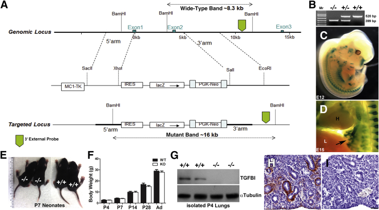
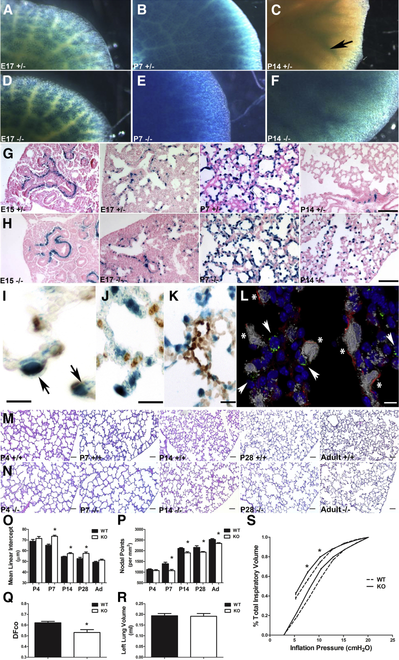
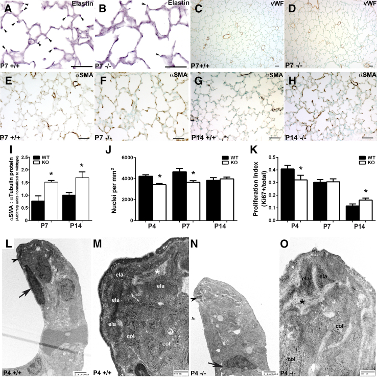
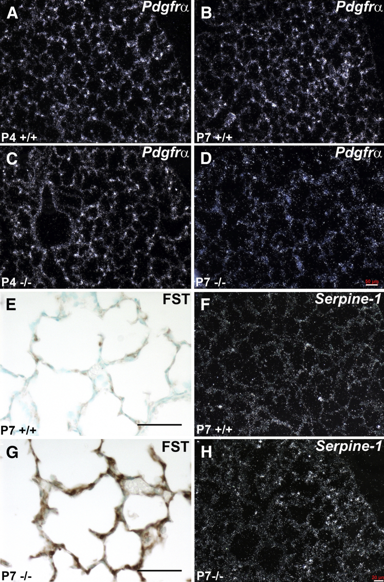
Septation of the gas-exchange saccules of the morphologically immature mouse lung requires regulated timing, spatial direction, and dosage of transforming growth factor (TGF)-β signaling. We found that neonatal hyperoxia acutely initially diminished saccular TGF-β signaling coincident with alveolar simplification. However, sustained hyperoxia resulted in a biphasic response and subsequent up-regulation of TGF-β signaling, ultimately resulting in bronchopulmonary dysplasia. Significantly, we found that the TGF-β-induced matricellular protein (TGFBI) was similarly biphasically altered in response to hyperoxia. Moreover, genetic ablation revealed that TGFBI was required for normal alveolar structure and function. Although the phenotype was not neonatal lethal, Tgfbi-deficient lungs were morphologically abnormal. Mutant septal tips were stunted, lacked elastin-positive tips, exhibited reduced proliferation, and contained abnormally persistent alveolar α-smooth muscle actin myofibroblasts. In addition, Tgfbi-deficient lungs misexpressed TGF-β-responsive follistatin and serpine 1, and transiently suppressed myofibroblast platelet-derived growth factor α differentiation marker. Finally, despite normal lung volume, Tgfbi-null lungs displayed diminished elastic recoil and gas exchange efficiency. Combined, these data demonstrate that initial suppression of the TGF-β signaling apparatus, as well as loss of key TGF-β effectors (like TGFBI), underlies early alveolar structural defects, as well as long-lasting functional deficits routinely observed in chronic lung disease of infancy patients. These studies underline the complex (and often contradictory) role of TGF-β and indicate a need to design studies to associate alterations with initial appearance of phenotypical changes suggestive of bronchopulmonary dysplasia.


Figures

Figure 1
Figure 1
Hyperoxia acutely diminishes saccular lung transforming growth factor (TGF)-β superfamily signaling and misexpression of down-stream targets. A: Quantitative PCR (qPCR) analysis of relative whole lung transforming growth factor (Tgf) β1, Tgfβ2, and Tgfβ3 ligand mRNA expression levels during normal postnatal saccular (P2, P4) and early alveolar (P7) lung development in room air (RA) control mice. B: qPCR assessment of Tgfβ1, Tgfβ2, and Tgfβ3 mRNA expression reveals all three ligands are suppressed in P4 lungs exposed continuously to 85% hyperoxia (oxygen) when compared with normal RA. C and D: Morphometric quantitation (C) of Western measurement of phosphorylated Smad2 and Smad3 protein levels (D) confirms that intracellular TGF-β canonical Smad-mediated signaling is suppressed in P4 lungs (asterisk) exposed to 85% O2, when compared with room air (RA) P4 littermate lungs and total Smad2 and Smad3 levels. E: qPCR mRNA profiling of 84 TGF-β–responsive genes reveals that, in addition to Tgfβ ligands, two downstream targets are statistically misexpressed (asterisk) at P4 and P14 after 85% O2 exposure. Specifically, Tgfbi mRNA levels are suppressed, whereas follistatin (Fst) mRNA levels increase via 85% O2 when compared with RA controls at P4. However, both Tgfbi and Fst are elevated via 85% O2 when compared with RA controls at P14. F–I:In situ hybridization of altered Tgfβ2 (F and H) and Tgfbi (G and I) at P4 and P14 mRNA expression within alveolar tissue sections in animals continuously exposed to 85% O2. Note the more punctate Tgfbi saccular wall expression when compared with Tgfβ2. J–M:In situ hybridization verification of normal Tgfβ2 (J and L) and Tgfbi (K and M) at P4 and P14 in RA control animals. Enlarged insets (F, H, J, and L) show expression at developing septal tips. N–Q: Immunohistochemical analysis confirms that the reduction in mRNA expression translates to a reduction in TGFBI protein expression in P4 lungs from animals continuously exposed to 85% O2 when compared with RA. Arrows highlight expression at developing septal tips (O and Q). R: Western analysis of phosphorylated Smad2 levels in P2 to P14 lungs reveals TGF-β canonical Smad signaling is subsequently elevated in P14 85% O2 exposed lungs (asterisk). Data are represented as means ± SEM (A–C, E, and R). N = 4 lungs from separate animals per group per time point (AR). P < 0.05 by t-test (RA versus hyperoxia at each time point; A–C, E, and R). Scale bars: 50 μm (F, G, N, and P); 10 μm (O). Original magnifications: ×10 (N and P); ×40 (O and Q). GAPDH, glyceraldehyde-3-phosphate dehydrogenase.
Figure 2
Figure 2
Expression of transforming growth factor-β–induced matricellular protein (TGFBI) during lung development. A–F:In situ hybridization analysis of Tgfbi spatiotemporal mRNA expression in distal lung sections throughout the pseudoglandular (E15; A), canalicular (E17; B), saccular [postnatal day (P) 0 (C) and P4 (D)], and alveolar [P7 (E) and P14 (F)] stages of lung development. Note widespread Tgfbi expression by discrete cells throughout the saccular and alveolar walls that peaks P4 to P7 but is dramatically reduced by P14. G: Representative immunoblot of duplicate lung TGFBI protein expression (top panel) from P0, P4, P7, and P14 whole lung homogenates with α-tubulin loading control (bottom panel). H: Densitometry quantification confirms peak lung TGFBI expression coincides with alveolar-septal development at P4 to P7, followed by significant down-regulation by P14. Densitometry normalized with α-tubulin. P < 0.05 versus P7, by one-way analysis of variance with Tukey's post test. N = 4 lungs per group (AH). Scale bars: 50 μm (A and B); 20 μm (C); 10 μm (DF).
Figure 3
Figure 3
Systemic loss of Tgfbi does not affect postnatal viability. A: Strategy for generation of TgfbilacZ mice. B: PCR genotyping detection of wild-type (WT; top panels) and null TgfbilacZ (bottom panels) alleles in Tgfbi null (−/−), heterozygous (+/−), and WT (+/+) animals. C and D: Whole-mount X-Gal staining demonstrating TgfbilacZ reporter expression in heterozygote E12 (C) and E15 embryos (arrow indicates lacZ within the developing lung buds; D). E and F: Null pups (left panel) are grossly indistinguishable from wild-type pups (right panel) at P7 and exhibit similar growth characteristics from P4 through to adulthood (Ad). G: Western blot analysis verifies the absence of transforming growth factor-β–induced matricellular protein (TGFBI) from within duplicate null P4 lungs when compared with age-matched WT lungs and α-tubulin loading control. H and I: Immunohistochemical staining for TGFBI in E15 lung sections from WT (H) and null (I) littermate embryos confirms in utero TGFBI absence in nulls. Data are represented as means ± SEM (F). N = 5 to 9 animals per group per time point (F). Scale bar = 50 μm (H and I).
Figure 4
Figure 4
Phenotypic analysis of Tgfbi knockout (KO) lungs. A–F: Whole-mount X-Gal staining indicating TgfbilacZ reporter expression in heterozygous (+/−) lungs at E17 (A), postnatal day (P) 7 (B), and P14 (arrow indicates lacZ restricted around large airways; C) compared with TgfbilacZ nulls (−/−) at E17 (D) and P7 (E) and P14 (F). F: Although there is similar lacZ spatiotemporal expression at E17 and P7, there is widespread ectopic lacZ reporter expression within the P14-null distal airspaces. G and H: Histological analysis confirms above whole-mount reporter patterns and ectopic TgfbilacZ expression in developing septal tips of P14 nulls (H) compared with heterozygous controls (G). I–K: Immunohistochemical analysis of X-Gal–stained sections demonstrates colocalization of α-smooth muscle actin (α-SMA) protein (I, marker of alveolar myofibroblasts) with TgfbilacZ-expressing cells (arrows) but not with Pro-SPC (marker of alveolar type II cells; J) or platelet endothelial cell adhesion molecule (PECAM) marker (K). L: Confocal coimmunolocalization of TGFBI-driven lacZ (gray) with α-SMA (red), Pro-SPC (green), and nuclear DAPI (blue) markers. There is colocalization of lacZ with α-SMA (asterisk), but not with Pro-SPC (arrowheads). M and N: Hematoxylin and eosin–stained inflation-fixed lung sections throughout alveolar lung development in wild-type (M) and Tgfbi nulls (N) from P4 through 12 months/adulthood (Ad). O and P: Quantification of distal airspace development of sections in M and N via mean linear intercept (O) and nodal point density (P) in wild-type (WT) and Tgfbi nulls reveals that during the period of rapid alveolar septation, loss of Tgfbi retards alveolar septation, resulting in larger and/or simpler distal airspaces from P7 onwards. Q: Functional analysis by way of measurement of the diffusing factor for carbon monoxide (DFco) in adult WT and Tgfbi nulls reveals a persistent functional insufficiency. R: However, left lung volume, as assessed by water displacement in adult WT and Tgfbi nulls, is equivalent. S: Assessment of lung elastance by pressure-volume loops in adult WT (broken line) and Tgfbi nulls (solid line) confirms nulls exhibit decreased elastic recoil. Data are represented as means ± SEM (O–R). N = 4 to 9 separate animals per group per time point (AS). P < 0.05 by t-test (WT versus Tgfbi null at each time point). Scale bars: 50 μm (G, H, M, and N); 5 μm (I and L); 10 μm (J); 20 μm (K).
Figure 5
Figure 5
Structural and ultrastructural examination of neonatal lung alveolar septation defects in Tgfbi-null mice. A–F: Immunohistochemical analysis of elastin (A and B), von Willebrand factor (vWF; C and D), and α-smooth muscle actin (α-SMA; E and F) localization in postnatal day (P) 7 wild-type (WT; +/+) and Tgfbi null (−/−) lungs. There is a significant lack of elastin-positive P7 null tips (septal tips indicated via arrows in A +/+ and B −/−), but vWF and α-SMA are similarly distributed in WT (C and E) and null (D and F) P7 tips. G and H: Compared with WT animals (G), there is ectopic expression of α-SMA protein in alveolar septal tips of P14 Tgfbi nulls (H). I: Western verification of elevated α-SMA levels in P7 and P14 Tgfbi null lungs. J and K: Assessment of alveolar tissue cellularity (J) and proliferation index (K) in WT and Tgfbi nulls at P4, P7, and P14. L–O: Transmission electron micrographs of septal tips at P4 in WT (L, enlarged in M) and Tgfbi null (N, enlarged in O) lungs. Normal collections of collagen microfilaments (col) and parallel collagen bundles (asterisk) are observed within both WT and blunted Tgfbi null septal tips; however, amorphous elastin (ela) deposition is diminished in stunted Tgfbi null septal tips (N, arrowhead) when compared with WT (L, arrowhead). Data are represented as means ± SEM (I–K). N = 5 to 6 separate animals per group per time point (AO). P < 0.05 by t-test (WT versus Tgfbi null at each time point). Scale bars: 50 μm (A–H ); 2 μm (K and M); 500 nm (L and N). KO, knockout.
Figure 6
Figure 6
Dynamic expression deviations in myofibroblast progenitor and downstream transforming growth factor (TGF)-β family signaling markers in Tgfbi null lungs during alveolar septal development. A–D:In situ hybridization detection of platelet-derived growth factor receptor α (Pdgfrα) mRNA expression in developing alveoli in wild-type (A and B) and Tgfbi nulls (C and D), at postnatal day (P) 4 and P7, reveals suppression of Pdgfrα in P7 null lungs. E and G: Immunohistochemical localization of follistatin (FST) protein within P7 wild-type (E) and Tgfbi-null (G) lungs indicates excessive FST deposition in null alveoli. F and H:In situ hybridization of serpine mRNA expression in developing alveoli in P7 wild-type (F) and Tgfbi-null (H) detects up-regulation within the null lung periphery. Scale bars: 50 μm (AF); 20 μm (G and H).

Similar articles

Cited by

References

    1. Kimura J., Deutsch G.H. Key mechanisms of early lung development. Pediatr Dev Pathol. 2007;10:335–347. - PubMed
    1. Burri P.H. Fetal and postnatal development of the lung. Annu Rev Physiol. 1984;46:617–628. - PubMed
    1. Ahlfeld S.K., Conway S.J. Aberrant signaling pathways of the lung mesenchyme and their contributions to the pathogenesis of bronchopulmonary dysplasia. Birth Defects Res A Clin Mol Teratol. 2012;94:3–15. - PMC - PubMed
    1. McGowan S.E. Paracrine cellular and extracellular matrix interactions with mesenchymal progenitors during pulmonary alveolar septation. Birth Defects Res A Clin Mol Teratol. 2014;100:227–239. - PubMed
    1. Ahlfeld S.K., Conway S.J. Assessment of inhibited alveolar-capillary membrane structural development and function in bronchopulmonary dysplasia. Birth Defects Res A Clin Mol Teratol. 2014;100:168–179. - PMC - PubMed

Publication types

MeSH terms

Substances

LinkOut - more resources