. 2016 Feb 16:6:21769.
doi: 10.1038/srep21769.

LGI1 acts presynaptically to regulate excitatory synaptic transmission during early postnatal development

Affiliations

LGI1 acts presynaptically to regulate excitatory synaptic transmission during early postnatal development

Morgane Boillot et al. Sci Rep. .

Abstract

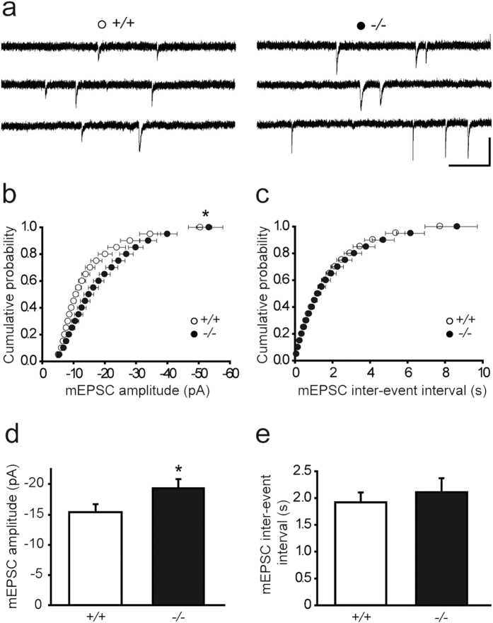
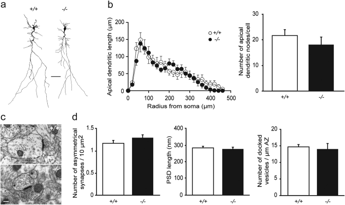
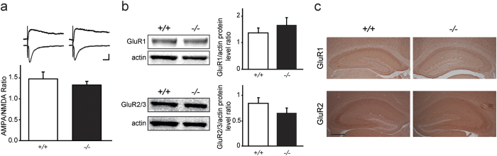
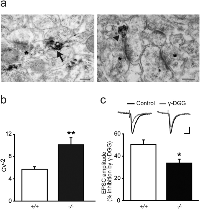
The secreted leucine-rich glioma inactivated 1 (LGI1) protein is an important actor for human seizures of both genetic and autoimmune etiology: mutations in LGI1 cause inherited temporal lobe epilepsy, while LGI1 is involved in antibody-mediated encephalitis. Remarkably, Lgi1-deficient (Lgi1(-/-)) mice recapitulate the epileptic disorder and display early-onset spontaneous seizures. To understand how Lgi1-deficiency leads to seizures during postnatal development, we here investigated the early functional and structural defects occurring before seizure onset in Lgi1(-/-) mice. We found an increased excitatory synaptic transmission in hippocampal slices from Lgi1(-/-) mice. No structural alteration in the morphology of pyramidal cell dendrites and synapses was observed at this stage, indicating that Lgi1-deficiency is unlikely to trigger early developmental abnormalities. Consistent with the presynaptic subcellular localization of the protein, Lgi1-deficiency caused presynaptic defects, with no alteration in postsynaptic AMPA receptor activity in Lgi1-/- pyramidal cells before seizure onset. Presynaptic dysfunction led to increased synaptic glutamate levels, which were associated with hyperexcitable neuronal networks. Altogether, these data show that Lgi1 acts presynaptically as a negative modulator of excitatory synaptic transmission during early postnatal development. We therefore here reveal that increased presynaptic glutamate release is a key early event resulting from Lgi1-deficiency, which likely contributes to epileptogenesis.


Figures

Figure 1
Figure 1. Hippocampal glutamatergic synaptic transmission is increased in Lgi1−/− mice.
Cumulative distributions and means of mEPSC amplitude (b,d) and inter-event interval (c,e), with sample traces above (a), in CA1 pyramidal cells from Lgi1+/+ (n = 18) and Lgi1−/− mice (n = 20) at P8. Scale bar: 20 pA, 500 ms. mEPSC amplitude cumulative distribution and mean, but not inter-event interval, differ significantly between Lgi1−/− and Lgi1+/+ mice (p < 0.05, Kolmogorov-Smirnov and t-test, respectively).
Figure 2
Figure 2. Dendritic arborization and excitatory synapses are unaltered in hippocampal pyramidal cells from Lgi1−/− mice.
(a) Representative dendritic morphology of neurobiotin-filled CA1 pyramidal cells from Lgi1+/+ and Lgi1−/− mice at P9. Scale bar: 50 μm. (b) Sholl analysis reveals similar apical dendritic length (p > 0.05, two-way ANOVA) and number of apical dendritic nodes (p > 0.05, Mann-Whitney) between Lgi1+/+ (n = 9) and Lgi1−/− mice (n = 14). (c) Electron micrographs of asymmetrical synapses in CA1 stratum radiatum from Lgi1+/+ and Lgi1−/− mice at P8. Scale bar: 200 nm. (d) No change was found in asymmetrical synapse density (n = 2900 μm2 from 9 Lgi1+/+ and 6 Lgi1−/− mice), PSD length (n = 40 synapses from 9 Lgi1+/+ and 6 Lgi1−/− mice) and number of docked vesicles (n = 20 synapses from 9 Lgi1+/+ and 6 Lgi1−/− mice) (p > 0.05, Mann-Whitney). PSD, postsynaptic density; AZ, active zone.
Figure 3
Figure 3. Postsynaptic AMPAR density is unchanged in Lgi1−/− mice.
(a) Similar ratios of AMPA to NMDA eEPSCs in CA1 pyramidal cells from P8 Lgi1+/+ (n = 6) and Lgi1−/− mice (n = 7; p > 0.05, t-test), with sample traces above. Scale bars: 20 ms, 50 pA. (b) Top: Representative Western blot and quantification of GluR1 and actin bands showing similar GluR1 protein levels in the hippocampus of P8 Lgi1+/+ (n = 4) and Lgi1−/− mice (n = 5; p > 0.05, t-test). Bottom: Representative Western blot and quantification of GluR2/3 and actin bands showing similar GluR2/3 protein levels in the hippocampus of P8 Lgi1+/+ (n = 3) and Lgi1−/− mice (n = 3; p > 0.05, t-test). (c) Representative immunohistochemical staining for GluR1 (top) and extracellular GluR2 (bottom) subunits in hippocampal sections of Lgi1+/+ (n = 3) and Lgi1−/− (n = 3) mice showing similar expression of the AMPAR subunits at P8. Scale bar: 400 μm.
Figure 4
Figure 4. Presynaptic alterations lead to increased synaptic glutamate levels in Lgi1−/− mice.
(a) Hippocampal electron micrographs from an Lgi1+/+ mouse showing the subcellular localization of Lgi1, immunostained with patient CSF, in presynaptic elements (arrow) and thin neurites (arrowhead). Non-reactive synaptic contacts are labeled with asterisks. Scale bars: 200 nm. (b) Coefficient of variation (CV) analysis of AMPAR eEPSC amplitude reveals an increase in CV−2 in CA1 pyramidal cells from P8 Lgi1−/− (n = 23) compared to Lgi1+/+ mice (n = 22; p < 0.05, t-test). (c) Synaptic glutamate levels, revealed by γ-DGG (500 μM) inhibition of eEPSC amplitude, are enhanced in P8 Lgi1−/− (n = 6) compared to Lgi1+/+ mice (n = 7; p < 0.05, t-test).
Figure 5
Figure 5. Lgi1−/− mice display glutamatergically-driven spontaneous interictal-like discharges.
(a) Scheme of a hippocampal slice illustrating the arrangement of electrodes for field potential (FP) and whole-cell patch clamp dual recordings. (b) Representative trace of CA1 FP (top) and simultaneous whole-cell patch-clamp recording (bottom) of an Lgi1−/− CA1 pyramidal cell (resting potential, −70 mV), showing spontaneous interictal-like activities at P9 in normal ACSF. Scale bars: FP: 200 μV, 5 s; whole-cell recording: 20 mV, 5 s. Right: Expanded traces showing an interictal-like network activity (top) that coincides with an intracellular depolarizing plateau potential with burst firing (bottom). Scale bars: FP: 100 μV, 1 s; whole-cell recording: 20 mV, 1 s. Quantification of (c) interictal activity and (d) depolarizing plateau potential in Lgi1−/− CA1 pyramidal cells (n = 14). (e) Representative traces of CA1 FP (top) and simultaneous pyramidal cell intracellular recordings (bottom) (resting potential, −70 mV) in an Lgi1−/− slice before (left) and after application (right) of glutamate receptor antagonists NBQX (10 μM) and APV (40 μM), which entirely block interictal-like discharges. Scale bars: FP: 100 μV, 10 s; whole-cell recording: 50 mV, 10 s. AP, action potential.

References

    1. Kalachikov S. et al. Mutations in LGI1 cause autosomal-dominant partial epilepsy with auditory features. Nat. Genet. 30, 335–341 (2002). - PMC - PubMed
    1. Morante-Redolat J. M. et al. Mutations in the LGI1/Epitempin gene on 10q24 cause autosomal dominant lateral temporal epilepsy. Hum. Mol. Genet. 11, 1119–1128 (2002). - PubMed
    1. Michelucci R., Pasini E. & Nobile C. Lateral temporal lobe epilepsies: clinical and genetic features. Epilepsia 50 Suppl 5, 52–54 (2009). - PubMed
    1. Irani S. R. et al. Antibodies to Kv1 potassium channel-complex proteins leucine-rich, glioma inactivated 1 protein and contactin-associated protein-2 in limbic encephalitis, Morvan’s syndrome and acquired neuromyotonia. Brain 133, 2734–2748 (2010). - PMC - PubMed
    1. Lai M. et al. Investigation of LGI1 as the antigen in limbic encephalitis previously attributed to potassium channels: a case series. Lancet. Neurol. 9, 776–785 (2010). - PMC - PubMed

Publication types

Substances