Review
. 2016 Apr;21(4):450-63.
doi: 10.1038/mp.2016.1. Epub 2016 Feb 16.

Overshadowed by the amygdala: the bed nucleus of the stria terminalis emerges as key to psychiatric disorders

Affiliations
Review

Overshadowed by the amygdala: the bed nucleus of the stria terminalis emerges as key to psychiatric disorders

M A Lebow et al. Mol Psychiatry. 2016 Apr.

Erratum in

Abstract

The bed nucleus of the stria terminalis (BNST) is a center of integration for limbic information and valence monitoring. The BNST, sometimes referred to as the extended amygdala, is located in the basal forebrain and is a sexually dimorphic structure made up of between 12 and 18 sub-nuclei. These sub-nuclei are rich with distinct neuronal subpopulations of receptors, neurotransmitters, transporters and proteins. The BNST is important in a range of behaviors such as: the stress response, extended duration fear states and social behavior, all crucial determinants of dysfunction in human psychiatric diseases. Most research on stress and psychiatric diseases has focused on the amygdala, which regulates immediate responses to fear. However, the BNST, and not the amygdala, is the center of the psychogenic circuit from the hippocampus to the paraventricular nucleus. This circuit is important in the stimulation of the hypothalamic-pituitary-adrenal axis. Thus, the BNST has been largely overlooked with respect to its possible dysregulation in mood and anxiety disorders, social dysfunction and psychological trauma, all of which have clear gender disparities. In this review, we will look in-depth at the anatomy and projections of the BNST, and provide an overview of the current literature on the relevance of BNST dysregulation in psychiatric diseases.


Conflict of interest statement

The authors declare no conflict of interest.

Figures

Figure 1
Figure 1
Afferent projections of the rodent BNST. Diverse brain areas project to the different sub-nuclei of the BNST and often to several sub-nuclei (adapted from refs 10, 11, 12, 13, 23). (I) Purple arrows represent incoming projections to the anterolateral (al), anteromedial (am) and ventral (fu) areas of the BNST at the level of the anterior commissure (ac). (II) Orange arrows represent projections to the anterolateral (al) and anteromedial (am) BNST areas posterior to the anterior commissure (ac). (III) Green arrows represent projections to the posterior BNST comprising of three nuclei: the principle (pr), the interfascicular (if) and the transverse (tr). 3v, third ventricle; ac, anterior commissure; al, anterolateral BNST; am, anteromedial BNST; Amy, amygdala; BNST, bed nucleus stria terminalis; d, dorsal nucleus; dm, dorsomedial nucleus; DR, dorsal raphe; FC, frontal cortex; fu, fusiform nucleus; GP, globus pallidus; hypo, hypothalamus; if, interfascicular nucleus; ju, juxtacapsular nucleus; LC, locus coeruleus; LS, lateral septum; mg, magnocellular nucleus; MPO, medial preoptic area; NTS, nucleus solitary tract; OB, olfactory bulb; ov, oval nucleus; pr, principle nucleus; PVN, paraventricular nucleus; rh, rhomboid nucleus; st, striatum; tr, transverse nucleus; VS, ventral subiculum; VTA, ventral tegmental area. PowerPoint slide
Figure 2
Figure 2
Efferent projections of the rodent BNST. The BNST sends diverse and unique projections from different subregions (adapted from refs 10, 11, 12, 13, 23). (I) Purple arrows represent outgoing projections from the anterolateral (al), anteromedial (al) and ventral (fu) areas of the BNST at the level of the anterior commissure (ac). (II) Orange arrows represent projections from anterolateral and medial BNST areas posterior to the anterior commissure (ac). (III) Green arrows represent projections from the posterior BNST comprising of three nuclei: the principle (pr), the interfascicular (if) and the transverse (tr). 3v, third ventricle; ac, anterior commissure; al, anterolateral BNST; am, anteromedial BNST; Amy, amygdala; BNST, bed nucleus stria terminalis; d, dorsal nucleus; dm, dorsomedial nucleus; DR, dorsal raphe; FC, frontal cortex; fu, fusiform nucleus; GP, globus pallidus; hypo, hypothalamus; if, interfascicular nucleus; ju, juxtacapsular nucleus; LC, locus coeruleus; LS, lateral septum; mg, magnocellular nucleus; MPO, medial preoptic area; NTS, nucleus solitary tract; OB, olfactory bulb; ov, oval nucleus; pr, principle nucleus; PVN, paraventricular nucleus; rh, rhomboid nucleus; st, striatum; tr, transverse nucleus; VS, ventral subiculum; VTA, ventral tegmental area. PowerPoint slide
Figure 3
Figure 3
Heatmap representing subpopulations of receptors, neurotransmitters and transporters in all subregions of the BNST. Adapted from the study by Bota et al. with the following genes added: DARPP32, CGRP, VIP, PAC1, PACAP,, CRFR1, CRFR2,, PR,, A1-Adr,, A2-Adr,, B1-Adr,, , B2-Adr., The original data were extracted from the study by Bota et al. which categorized the expression levels from 0 to 9, with 12 having no data. In our version, we binned all the genes previously published and newly added from 0 to 3, 4 to 6, 6 to 9 and no data into 4 ranks of strong, moderate, weak expression and no data to combine all the data into the qualitative expression heatmap. A1-Adr, adrenergic receptor alpha-1; A2-1 GABA A, GABRA2, GABA a2 receptor; A2-Adr, adrenergic receptor alpha-2; A3-1 GABA A, GABA3, GABA a3 receptor, B1-1 GABA A, GABA beta 1 receptor; AII, angiotensin I; AR, androgen receptor; AVP, vasopression; B1-Adr, adrenergic receptor beta 1; B2-Adr, adrenergic receptor beta 2; B3-1 GABA A, GABA beta 3 receptor; CaB, calbindin; CB-1R, cannabinoid receptor 1; CCK, cholecystokinin; CGRP, calcitonin gene-related peptide; ChAT, choline acetyltransferase; CR, calretinin; CRFR1, cortiotrophin releasing factor receptor type 1; CRFR2, corticotrophin-releasing factor, receptor type 2; D1, Drd1A, dopamine receptor type 1A; D2, Drd2A, dopamine receptor type 2A; DARPP32, dopamine- and cAMP-regulated phosphoprotein; DOR, delta opioid receptor; DYN, dynorphin; ENK, enkelphalin; ER, estrogen receptors; G1-1 GABA A, GABA, gamma 1 receptor; GAD65, glutamate decarboxylase 65; GAD67, glutamate decarboxylase 67; GAL, galanin; GluR7, GRIK3, kainate receptor; GlurR5, GRIK1 kainate receptor; Htr1A, 5Ht1A, serotonin receptor type 1; KA2, GRIK 5, kainate receptor; KOR, kappa-opioid receptor; mGluR2, metabotropic glutamate receptor 2; mGluR7A, metabotropic glutamate receptor 7A; mGluR7B, metabotropic glutamate receptor 7B; MOR, mu-opioid receptor; nChR, nicotinic acetylcholine receptor; NK3, tachykinin receptor 3; NKB, neurokinin B; NPY, neuropeptide Y; NPY1, neuropeptide Y receptor Y1; NPY2, neuropeptide Y receptor Y2; NPY4, neuropeptide Y receptor Y4; NPY5, neuropeptide Y receptor Y5; NT, neurotensin; Oxt-1A, oxytocin receptor 1A; Oxt-1B, oxytocin receptor 1B; PAC1, Adcyap1r1, pituitary adenylate cyclase-activating polypeptide type 1; PACAP, pituitary adenylate cyclase-activating polypeptide; PR, progesterone receptor; SOM, somatostatin; SP, substance P; VGLUT1, vesicular glutamate transporter 1; VGLUT2, vesicular glutamate transporter 2; VGLUT3, vesicular glutamate transporter 3; VIP, vasoactive intestinal peptide. PowerPoint slide
Figure 4
Figure 4
Anatomy and subdivisions (red text) of the human anterior BNST (left panel) and posterior BNST (right panel). The orange dots depict the network of brain areas with either anatomical connection or which have been implicated in its functional connectivity. Image adapted from Atlas of the Human Brain (http://www.thehumanbrain.info). ac, anterior commissure; Acc, nucleus accumbens; BLA, basal lateral nucleus of the amygdala; BM, basal medial nucleus of the amygdala; BNST, bed nucleus of the stria terminalis; BNSTc, bed nucleus of the stria terminalis central subdivision; BNSTL, bed nucleus of the stria terminalis lateral subdivision; BNSTm, bed nucleus of the stria terminalis medial subdivision; BNSTv, bed nucleus of the stria terminalis ventral subdivision; Cd, caudate; DB, diagonal band; Ent, entorhinal cortex; FPu, putaminal fundus region; GPe, globus pallidus external; Ic, internal capsule; LA, lateral nucleus of the anygdala; LH, lateral hypothalamus; LS, lateral septum; mPFC, medial prefrontal cortex; MPO, medial preoptic nucleus; oc, optic chiasm; PirF, piriform cortex; Pu, putamen; PVN, paraverntricular nucleus; Sch, suprachiasmatic nucleus; VP, ventral pallidum; vPFC, ventral prefrontal cortex. PowerPoint slide
Figure 5
Figure 5
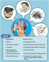
Summary representing the behaviors that the BNST regulates and implicated pathology in event of dysfunction of connectivity or neurotransmitter populations. BNST, bed nucleus of the stria terminalis. PowerPoint slide

Comment in

Similar articles

Cited by

References

    1. Dumont EC. What is the bed nucleus of the stria terminalis? Prog Neuropsychopharmacol Biol Psychiatry. 2009;33:1289–1290. doi: 10.1016/j.pnpbp.2009.07.006. - DOI - PMC - PubMed
    1. Davis M, Walker DL, Miles L, Grillon C. Phasic vs sustained fear in rats and humans: role of the extended amygdala in fear vs anxiety. Neuropsychopharmacology. 2010;35:105–135. doi: 10.1038/npp.2009.109. - DOI - PMC - PubMed
    1. Coria-Avila GA, Manzo J, Garcia LI, Carrillo P, Miquel M, Pfaus JG. Neurobiology of social attachments. Neurosci Biobehav Rev. 2014;43:173–182. doi: 10.1016/j.neubiorev.2014.04.004. - DOI - PubMed
    1. Nelson RJ, Trainor BC. Neural mechanisms of aggression. Nat Rev Neurosci. 2007;8:536–546. doi: 10.1038/nrn2174. - DOI - PubMed
    1. Marsh AA. What can we learn about emotion by studying psychopathy? Front Hum Neurosci. 2013;7:181. doi: 10.3389/fnhum.2013.00181. - DOI - PMC - PubMed

Publication types