

A novel fusion imaging system for endoscopic ultrasound
- PMID: 26879165
- PMCID: PMC4770620
- DOI: 10.4103/2303-9027.175882
A novel fusion imaging system for endoscopic ultrasound
Abstract
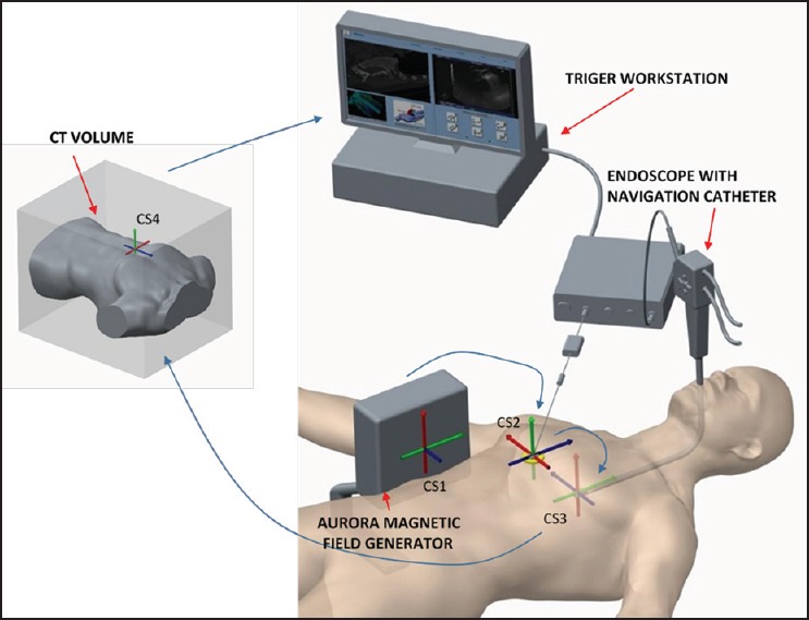
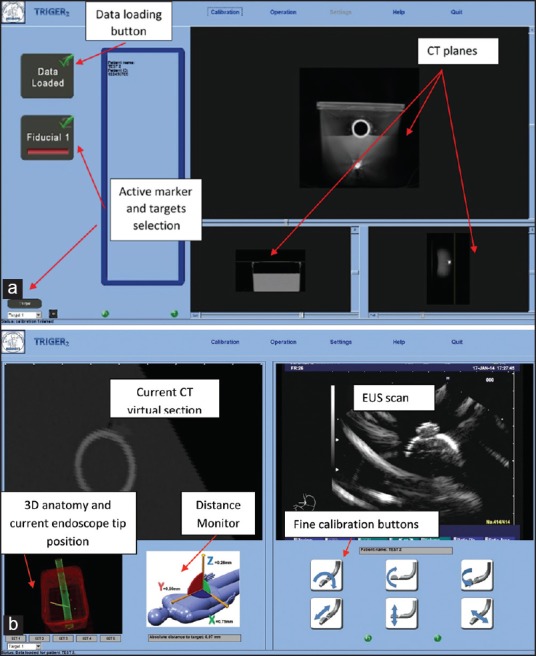
Background and objective: Navigation of a flexible endoscopic ultrasound (EUS) probe inside the gastrointestinal (GI) tract is problematic due to the small window size and complex anatomy. The goal of the present study was to test the feasibility of a novel fusion imaging (FI) system which uses electromagnetic (EM) sensors to co-register the live EUS images with the pre-procedure computed tomography (CT) data with a novel navigation algorithm and catheter.
Methods: An experienced gastroenterologist and a novice EUS operator tested the FI system on a GI tract bench top model. Also, the experienced gastroenterologist performed a case series of 20 patients during routine EUS examinations.
Results: On the bench top model, the experienced and novice doctors reached the targets in 67 ± 18 s and 150 ± 24 s with a registration error of 6 ± 3 mm and 11 ± 4 mm, respectively. In the case series, the total procedure time was 24.6 ± 6.6 min, while the time to reach the clinical target was 8.7 ± 4.2 min.
Conclusions: The FI system is feasible for clinical use, and can reduce the learning curve for EUS procedures and improve navigation and targeting in difficult anatomic locations.
Conflict of interest statement
Figures
References
-
- Vilmann P, Seicean A, Sãftoiu A. Tips to overcome technical challenges in EUS-guided tissue acquisition. Gastrointest Endosc Clin N Am. 2014;24:109–24. - PubMed
-
- Wani S, Coté GA, Keswani R, et al. Learning curves for EUS by using cumulative sum analysis: Implications for American Society for Gastrointestinal Endoscopy recommendations for training. Gastrointest Endosc. 2013;77:558–65. - PubMed
-
- Barthet M. Endoscopic ultrasound teaching and learning. Minerva Med. 2007;98:247–51. - PubMed
-
- Bhutani MS, Wong RF, Hoffman BJ. Training facilities in gastrointestinal endoscopy: An animal model as an aid to learning endoscopic ultrasound. Endoscopy. 2006;38:932–4. - PubMed
LinkOut - more resources
Full Text Sources
Other Literature Sources
