Review
. 2016 Mar;18(3):28.
doi: 10.1007/s11886-016-0705-1.

Intracoronary Imaging in the Detection of Vulnerable Plaques

Affiliations
Review

Intracoronary Imaging in the Detection of Vulnerable Plaques

Jonathan A Batty et al. Curr Cardiol Rep. 2016 Mar.

Abstract

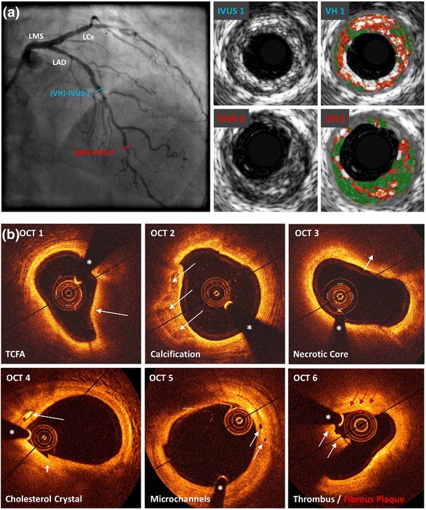
Coronary artery disease is the result of atherosclerotic changes to the coronary arterial wall, comprising endothelial dysfunction, vascular inflammation and deposition of lipid-rich macrophage foam cells. Certain high-risk atherosclerotic plaques are vulnerable to disruption, leading to rupture, thrombosis and the clinical sequelae of acute coronary syndrome. Though recognised as the gold standard for evaluating the presence, distribution and severity of atherosclerotic lesions, invasive coronary angiography is incapable of identifying non-stenotic, vulnerable plaques that are responsible for adverse cardiovascular events. The recognition of such limitations has impelled the development of intracoronary imaging technologies, including intravascular ultrasound, optical coherence tomography and near-infrared spectroscopy, which enable the detailed evaluation of the coronary wall and atherosclerotic plaques in clinical practice. This review discusses the present status of invasive imaging technologies; summarises up-to-date, evidence-based clinical guidelines; and addresses questions that remain unanswered with regard to the future of intracoronary plaque imaging.

Keywords: Coronary artery disease; Imaging; Interventional cardiology; Intravascular ultrasonography; Near-infrared spectroscopy; Optical coherence tomography.


Figures

Fig. 1
Fig. 1
Examples of intracoronary imaging modalities. a (VH-)IVUS. The coronary angiogram shows the left anterior descending artery (RAO cranial), demonstrating minimal stenosis, but multiple vulnerable plaques are visualised on IVUS. Cross-sectional images demonstrate calcified TCFA (blue line, IVUS1 and VH1) and non-calcified TCFA (red line, IVUS2 and VH2). Keys to VH-IVUS: dark green fibrous tissue, light green fibro-fatty tissue, red necrotic core, white dense calcium. b OCT. Several examples of features associated with plaque vulnerability are presented, including: OCT1 TCFA (arrow indicates thin fibrous cap), OCT2 coronary arterial calcification (arrows indicate well-demarcated calcification), OCT3 necrotic core (arrows indicate lipid pool/necrotic core), OCT4 presence of cholesterol microcrystal (arrow indicates well-demarcated crystal structure), OCT5 microchannels (arrows indicate two separate channels) associated with a non-obstructive lesion and OCT6 (white arrows indicate low-attenuation white thrombus; red arrows indicate highly fibrous plaque). Diag diagonal artery, IVUS intravascular ultrasound, LAD left anterior descending artery, LCx left circumflex artery, LMS left main stem, OCT optical coherence tomography, OM obtuse marginal artery, prob probability, RAO right anterior oblique view, TCFA thin-cap fibroatheroma, VH virtual histology. Asterisk indicates guidewire artefact; dagger indicates seam line artefact

References

    1. Mozaffarian D, Benjamin EJ, Go AS, et al. Heart disease and stroke statistics—2015 update: a report from the American Heart Association. Circulation. 2015;131(4):e29–322. doi: 10.1161/CIR.0000000000000152. - DOI - PubMed
    1. Nichols M, Townsend N, Scarborough P, et al. Cardiovascular disease in Europe 2014: epidemiological update. Eur Heart J. 2014;35(42):2950–9. doi: 10.1093/eurheartj/ehu299. - DOI - PubMed
    1. Ryan TJ. The coronary angiogram and its seminal contributions to cardiovascular medicine over five decades. Circulation. 2002;106(6):752–6. doi: 10.1161/01.CIR.0000024109.12658.D4. - DOI - PubMed
    1. Topol EJ, Nissen SE. Our preoccupation with coronary luminology. The dissociation between clinical and angiographic findings in ischemic heart disease. Circulation. 1995;92(8):2333–42. doi: 10.1161/01.CIR.92.8.2333. - DOI - PubMed
    1. Little WC, Constantinescu M, Applegate RJ, et al. Can coronary angiography predict the site of a subsequent myocardial infarction in patients with mild-to-moderate coronary artery disease? Circulation. 1988;78(5 Pt 1):1157–66. doi: 10.1161/01.CIR.78.5.1157. - DOI - PubMed

MeSH terms

LinkOut - more resources