

Endothelial Mineralocorticoid Receptor Mediates Diet-Induced Aortic Stiffness in Females
- PMID: 26879229
- PMCID: PMC4798906
- DOI: 10.1161/CIRCRESAHA.115.308269
Endothelial Mineralocorticoid Receptor Mediates Diet-Induced Aortic Stiffness in Females
Abstract
Rationale: Enhanced activation of the mineralocorticoid receptors (MRs) in cardiovascular tissues increases oxidative stress, maladaptive immune responses, and inflammation with associated functional vascular abnormalities. We previously demonstrated that consumption of a Western diet (WD) for 16 weeks results in aortic stiffening, and that these abnormalities were prevented by systemic MR blockade in female mice. However, the cell-specific role of endothelial cell MR (ECMR) in these maladaptive vascular effects has not been explored.
Objective: We hypothesized that specific deletion of the ECMR would prevent WD-induced increases in endothelial sodium channel activation, reductions in bioavailable nitric oxide, increased vascular remodeling, and associated increases in vascular stiffness in females.
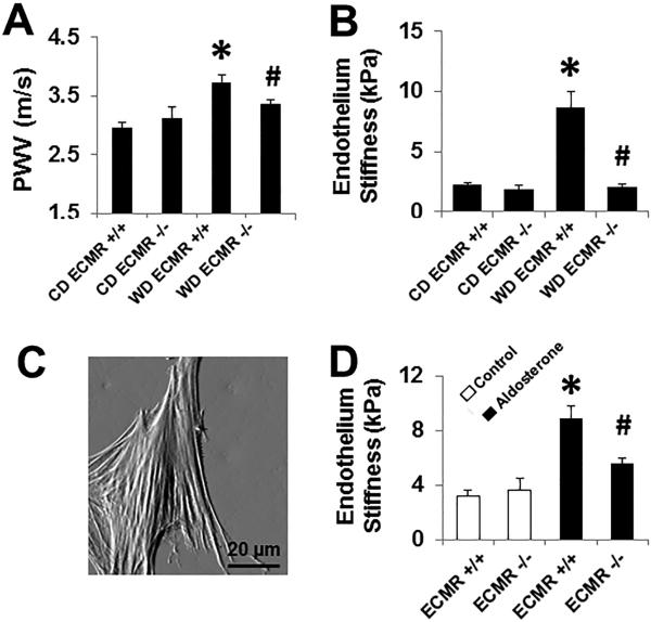
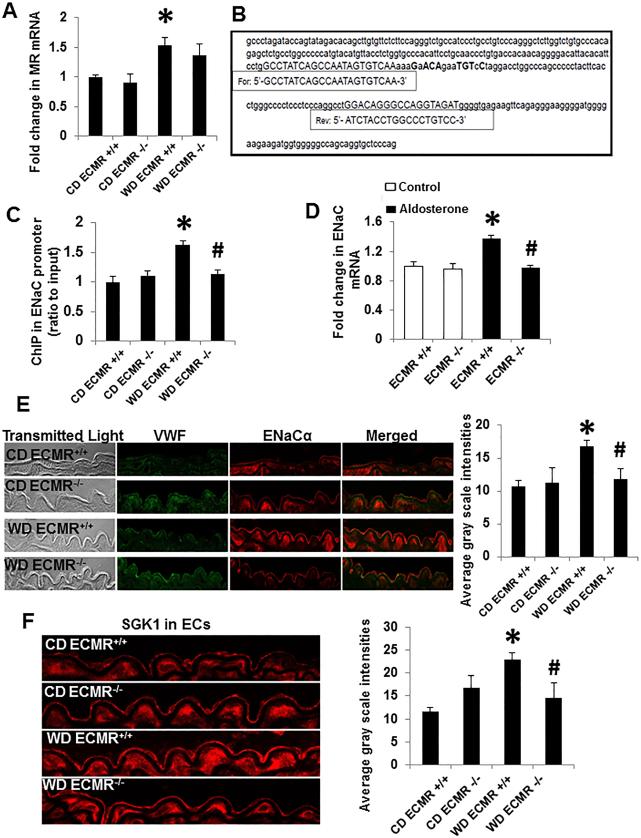
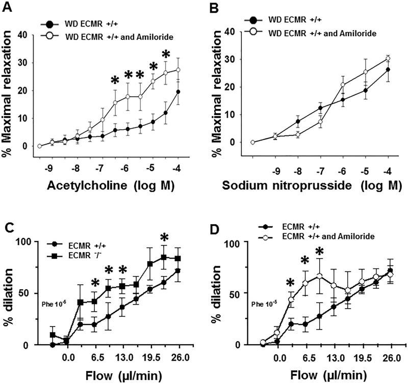
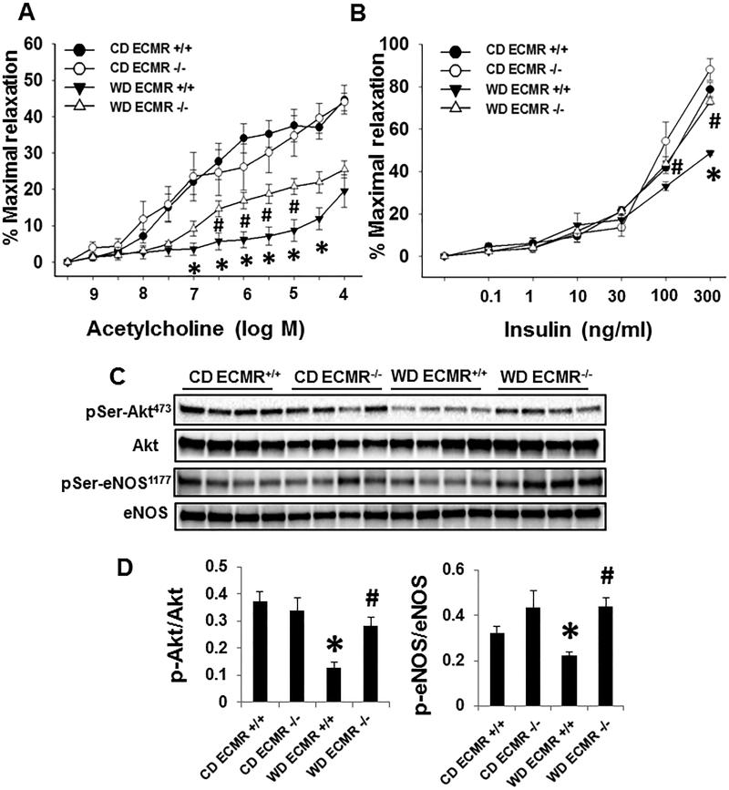
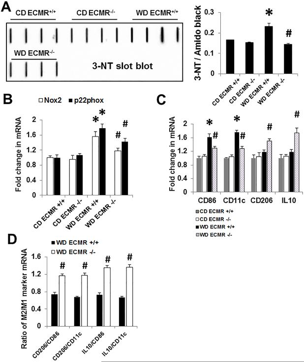
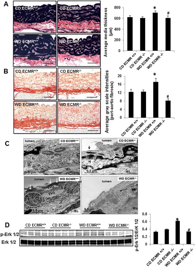
Methods and results: Four-week-old female ECMR knockout and wild-type mice were fed either mouse chow or WD for 16 weeks. WD feeding resulted in aortic stiffness and endothelial dysfunction as determined in vivo by pulse wave velocity and ex vivo by atomic force microscopy, and wire and pressure myography. The WD-induced aortic stiffness was associated with enhanced endothelial sodium channel activation, attenuated endothelial nitric oxide synthase activation, increased oxidative stress, a proinflammatory immune response and fibrosis. Conversely, cell-specific ECMR deficiency prevented WD-induced aortic fibrosis and stiffness in conjunction with reductions in endothelial sodium channel activation, oxidative stress and macrophage proinflammatory polarization, restoration of endothelial nitric oxide synthase activation.
Conclusions: Increased ECMR signaling associated with consumption of a WD plays a key role in endothelial sodium channel activation, reduced nitric oxide production, oxidative stress, and inflammation that lead to aortic remodeling and stiffness in female mice.
Keywords: Western diet; inflammation; macrophages; nitric oxide; vascular stiffness.
© 2016 American Heart Association, Inc.
Figures
Similar articles
-
Diet-Induced Obesity Promotes Kidney Endothelial Stiffening and Fibrosis Dependent on the Endothelial Mineralocorticoid Receptor.Hypertension. 2019 Apr;73(4):849-858. doi: 10.1161/HYPERTENSIONAHA.118.12198. Hypertension. 2019. PMID: 30827147 Free PMC article.
-
Low-Dose Mineralocorticoid Receptor Blockade Prevents Western Diet-Induced Arterial Stiffening in Female Mice.Hypertension. 2015 Jul;66(1):99-107. doi: 10.1161/HYPERTENSIONAHA.115.05674. Epub 2015 May 26. Hypertension. 2015. PMID: 26015449 Free PMC article.
-
Western diet induces renal artery endothelial stiffening that is dependent on the epithelial Na+ channel.Am J Physiol Renal Physiol. 2020 May 1;318(5):F1220-F1228. doi: 10.1152/ajprenal.00517.2019. Epub 2020 Apr 13. Am J Physiol Renal Physiol. 2020. PMID: 32281419 Free PMC article.
-
Role of the vascular endothelial sodium channel activation in the genesis of pathologically increased cardiovascular stiffness.Cardiovasc Res. 2022 Jan 7;118(1):130-140. doi: 10.1093/cvr/cvaa326. Cardiovasc Res. 2022. PMID: 33188592 Free PMC article. Review.
-
The endothelial mineralocorticoid receptor: mediator of the switch from vascular health to disease.Curr Opin Nephrol Hypertens. 2017 Mar;26(2):97-104. doi: 10.1097/MNH.0000000000000306. Curr Opin Nephrol Hypertens. 2017. PMID: 27930384 Free PMC article. Review.
Cited by
-
Smooth Muscle Cell-Mineralocorticoid Receptor as a Mediator of Cardiovascular Stiffness With Aging.Hypertension. 2018 Apr;71(4):609-621. doi: 10.1161/HYPERTENSIONAHA.117.10437. Epub 2018 Feb 20. Hypertension. 2018. PMID: 29463624 Free PMC article.
-
Diet-Induced Obesity Promotes Kidney Endothelial Stiffening and Fibrosis Dependent on the Endothelial Mineralocorticoid Receptor.Hypertension. 2019 Apr;73(4):849-858. doi: 10.1161/HYPERTENSIONAHA.118.12198. Hypertension. 2019. PMID: 30827147 Free PMC article.
-
The Renin Angiotensin Aldosterone System in Obesity and Hypertension: Roles in the Cardiorenal Metabolic Syndrome.Med Clin North Am. 2017 Jan;101(1):129-137. doi: 10.1016/j.mcna.2016.08.009. Med Clin North Am. 2017. PMID: 27884224 Free PMC article. Review.
-
The immunology of hypertension.J Exp Med. 2018 Jan 2;215(1):21-33. doi: 10.1084/jem.20171773. Epub 2017 Dec 15. J Exp Med. 2018. PMID: 29247045 Free PMC article. Review.
-
Mineralocorticoid Receptors Mediate Diet-Induced Lipid Infiltration of Skeletal Muscle and Insulin Resistance.Endocrinology. 2022 Oct 11;163(11):bqac145. doi: 10.1210/endocr/bqac145. Endocrinology. 2022. PMID: 36039677 Free PMC article.
References
-
- Petersen KS, Blanch N, Keogh JB, Clifton PM. Effect of weight loss on pulse wave velocity: systematic review and meta-analysis. Arterioscler Thromb Vasc Biol. 2015;35:243–52. - PubMed
-
- DeMarco VG, Habibi J, Jia G, Aroor AR, Ramirez-Perez FI, Martinez-Lemus LA, Bender SB, Garro M, Hayden MR, Sun Z, Meininger GA, Manrique C, Whaley-Connell A, Sowers JR. Low-Dose Mineralocorticoid Receptor Blockade Prevents Western Diet-Induced Arterial Stiffening in Female Mice. Hypertension. 2015;66:99–107. - PMC - PubMed
-
- Rutter MK, Parise H, Benjamin EJ, Levy D, Larson MG, Meigs JB, Nesto RW, Wilson PW, Vasan RS. Impact of glucose intolerance and insulin resistance on cardiac structure and function: sex-related differences in the Framingham Heart Study. Circulation. 2003;107:448–54. - PubMed
Publication types
MeSH terms
Substances
Grants and funding
LinkOut - more resources
Full Text Sources
Other Literature Sources
Molecular Biology Databases
