. 2017 Jan;222(1):41-59.
doi: 10.1007/s00429-016-1199-8. Epub 2016 Feb 15.

Expression of Kir4.1 and Kir5.1 inwardly rectifying potassium channels in oligodendrocytes, the myelinating cells of the CNS

Affiliations

Expression of Kir4.1 and Kir5.1 inwardly rectifying potassium channels in oligodendrocytes, the myelinating cells of the CNS

C Brasko et al. Brain Struct Funct. 2017 Jan.

Abstract

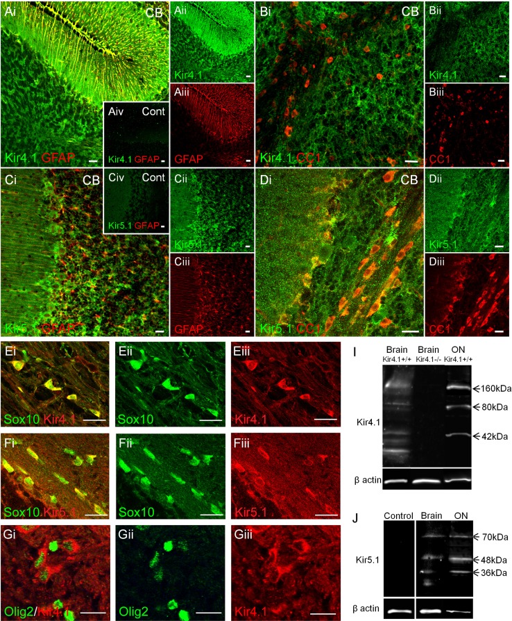
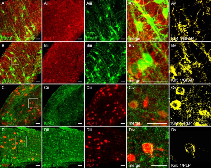
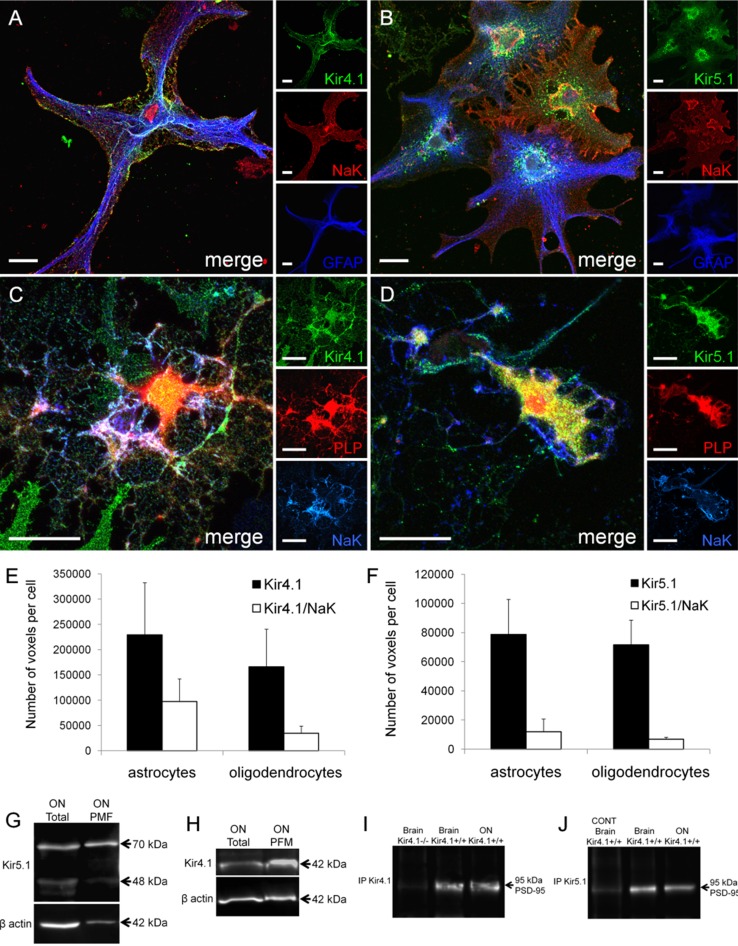
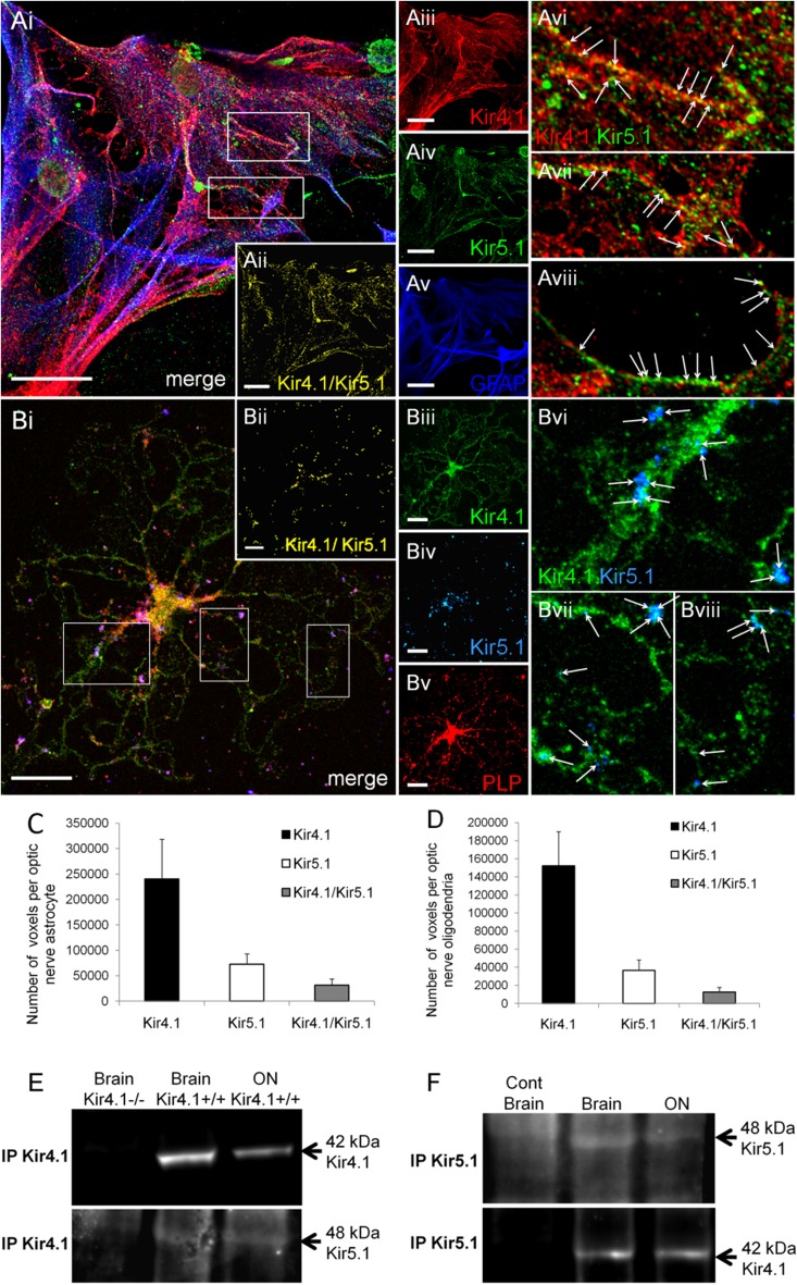
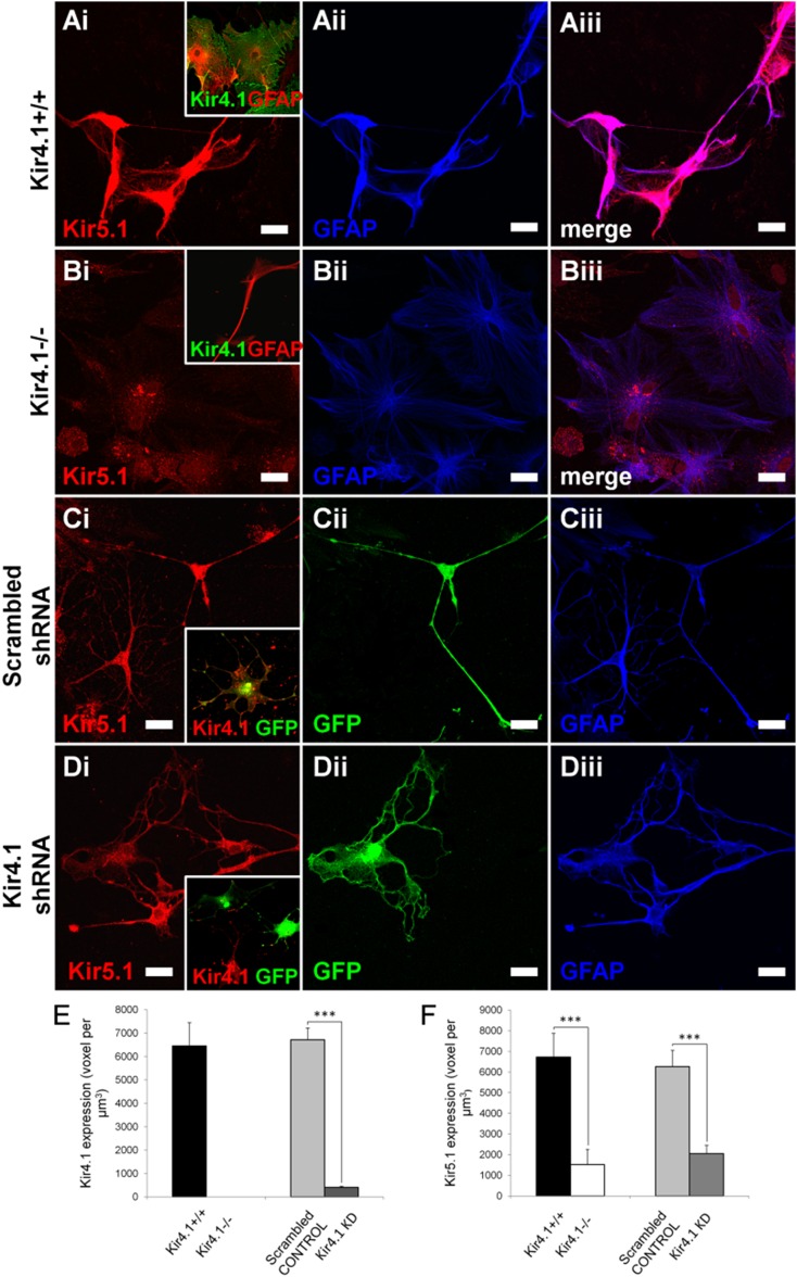
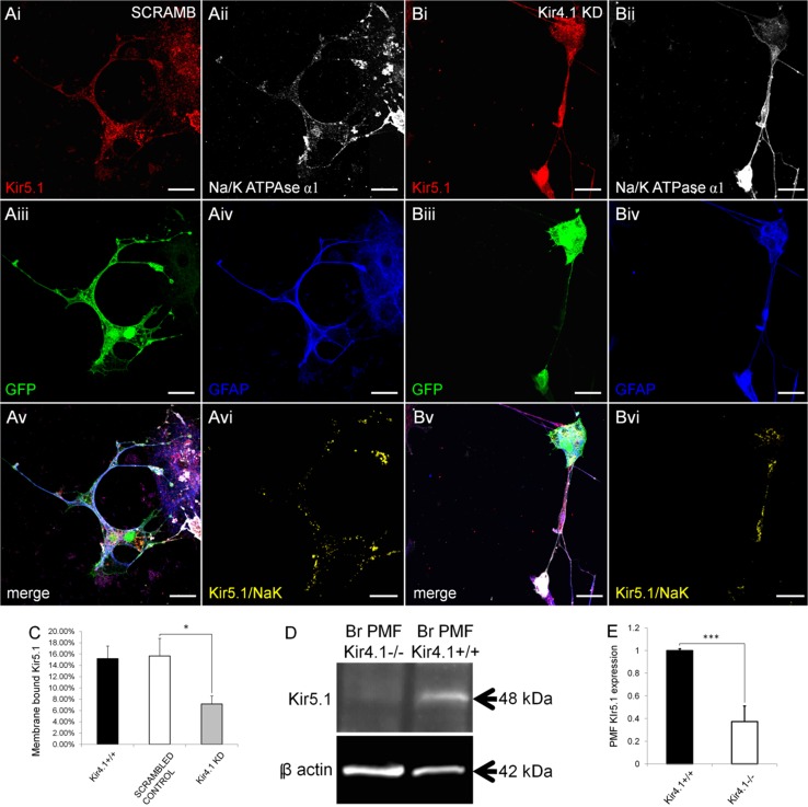
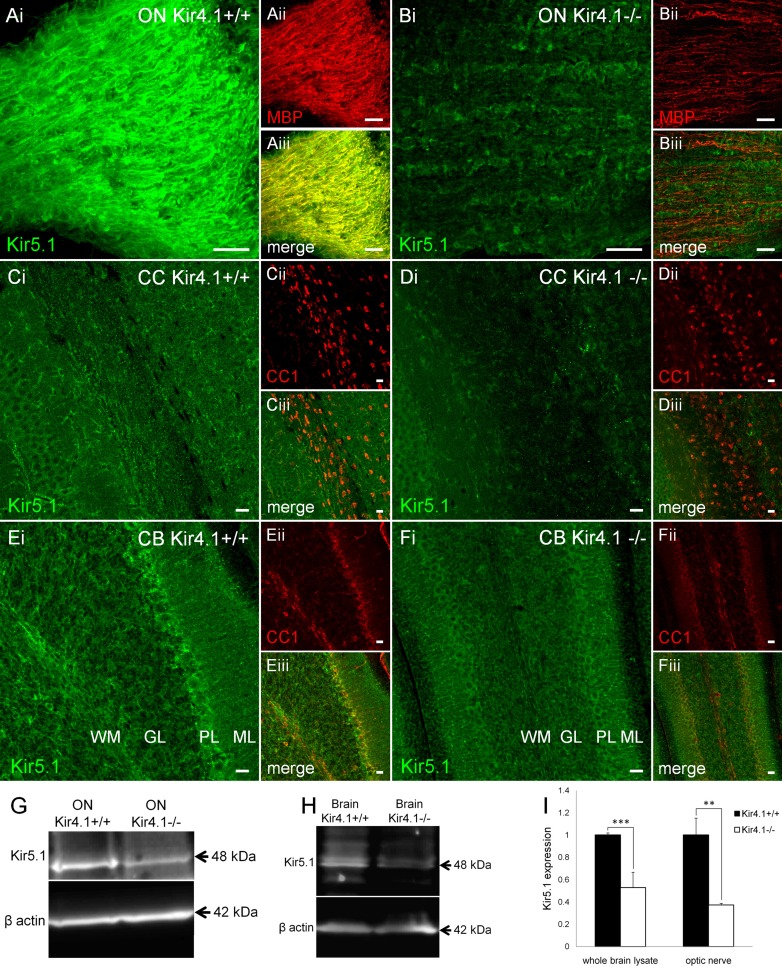
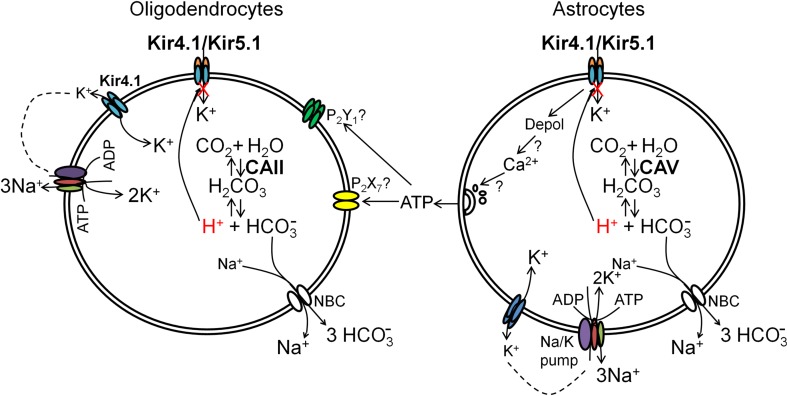
The inwardly rectifying K+ channel subtype Kir5.1 is only functional as a heteromeric channel with Kir4.1. In the CNS, Kir4.1 is localised to astrocytes and is the molecular basis of their strongly negative membrane potential. Oligodendrocytes are the specialised myelinating glia of the CNS and their resting membrane potential provides the driving force for ion and water transport that is essential for myelination. However, little is known about the ion channel profile of mature myelinating oligodendrocytes. Here, we identify for the first time colocalization of Kir5.1 with Kir4.1 in oligodendrocytes in white matter. Immunolocalization with membrane-bound Na+/K+-ATPase and western blot of the plasma membrane fraction of the optic nerve, a typical CNS white matter tract containing axons and the oligodendrocytes that myelinate them, demonstrates that Kir4.1 and Kir5.1 are colocalized on oligodendrocyte cell membranes. Co-immunoprecipitation provides evidence that oligodendrocytes and astrocytes express a combination of homomeric Kir4.1 and heteromeric Kir4.1/Kir5.1 channels. Genetic knock-out and shRNA to ablate Kir4.1 indicates plasmalemmal expression of Kir5.1 in glia is largely dependent on Kir4.1 and the plasmalemmal anchoring protein PSD-95. The results demonstrate that, in addition to astrocytes, oligodendrocytes express both homomeric Kir4.1 and heteromeric Kir4.1/Kir5.1 channels. In astrocytes, these channels are essential to their key functions of K+ uptake and CO2/H+ chemosensation. We propose Kir4.1/Kir5.1 channels have equivalent functions in oligodendrocytes, maintaining myelin integrity in the face of large ionic shifts associated with action potential propagation along myelinated axons.

Keywords: Astrocyte; Glia; Inward rectifying potassium channel; Oligodendrocyte; Potassium regulation; White matter.


Conflict of interest statement

The authors declare that they have no conflicts of interest. Informed consent All procedures performed in studies involving animals were in accordance with the ethical standards of the institution at which the studies were conducted. Animal rights All applicable international, national, and/or institutional guidelines for the care and use of animals were followed.

Figures

Fig. 1
Fig. 1
Expression of Kir4.1 and Kir5.1 in oligodendrocytes and astrocytes in the cerebellum. Immunolabelling for Kir4.1 and Kir5.1, in combination with GFAP for astrocytes (A, C), and APC/CC1 for oligodendrocytes (B, D). Immunolabelling for Kir4.1 (E) and Kir5.1 (F) in mice in which EGFP is under the control of the oligodendrocyte-specific Sox10 promoter. G Double immunolabelling for Kir4.1 (red) and the oligodenrocyte-specific marker Olig2 (green). Insets in Aiv and Civ illustrate negative controls, in the Kir4.1 KO mouse (Aiv) and following preincubation with the Kir5.1 blocking peptide (Civ). Scale bars 20 μm. Western blot analysis of the brain and optic and nerve for Kir4.1 (I) and Kir5.1 (J); bands were absent in the negative controls, in the Kir4.1 knock-out mouse (I) following preincubation in the Kir5.1 blocking peptide (J)
Fig. 2
Fig. 2
Expression of Kir4.1 and Kir5.1 in optic nerve oligodendrocytes and astrocytes. Immunolabelling for Kir4.1 (A, C) and Kir5.1 (B, D), in GFAP-GFP mice to identify astrocytes (A, B) and PLP-DsRED mice to identify oligodendrocytes (C, D). Cellular expression of Kir4.1 and Kir5.1 is demonstrated by the generation of colocalisation channels (Av, Bv, Cv, Dv) from confocal z-stacks (Aiv, Biv, Civ, Div), and green and red channels of equal intensity appear yellow. Scale bars 20 μm
Fig. 3
Fig. 3
Plasmalemmal expression of Kir4.1 and Kir5.1 subunit in optic nerve glia. Immunolocalization of Kir4.1 and Kir5.1 with the membrane bound Na–K-ATPase α1 subunit in optic nerve explants of astrocytes identified by GFAP (A, B) and oligodendrocytes identified by PLP-DsRed (C, D). Scale bars 20 μm. Quantification in astrocytes and oligodendrocytes of total number of voxels immunopositive for Kir4.1 and Kir5.1, compared to voxels that were identified as colocalized for Kir4.1/Na–K-ATPase (E) and Kir5.1/Na–K-ATPase (F); data are mean ± SEM, n = 13 cells for each analysis. Western blot analysis of Kir5.1 (G) and Kir4.1 (H) in total optic nerve lysate and plasma membrane fraction. Co-immunoprecipitation of Kir4.1 (I) and Kir5.1 (J) with PSD95, in total brain and optic nerve (ON) lysate; negative controls were Kir4.1 knock-out mice (−/−) for Kir4.1 and preincubation with the blocking peptide for Kir5.1
Fig. 4
Fig. 4
Co-expression of Kir4.1 and Kir5.1 in optic nerve oligodendrocytes and astrocytes. Co-immunolocalization of Kir4.1 and Kir5.1 in optic nerve explant cultures, in astrocytes identified by GFAP immunolabelling (A) and oligodendrocytes identified by PLP-DsRED (B). The overlay and individual channels are illustrated, together with the co-localisation channel for Kir4.1/Kir5.1 (Aii, Bii). Boxed areas on overlay images (Ai, Bi) are enlarged in AviAviii and BviBviii, to illustrate punctate colocalization of Kir4.1 and Kir5.1 along processes (some indicated by arrows). Scale bars 20 μm. Quantification of the number of voxels that were positive for Kir4.1 and Kir5.1 alone and of Kir4.1/Kir5.1 together, in astrocytes (C, n = 15) and oligodendrocytes (D, n = 13); data are mean ± SEM. Co-immunoprecipitation of Kir4.1 with Kir5.1 (E) and of Kir5.1 with Kir4.1 (F) from total brain and optic nerve (ON) lysates; negative controls were Kir4.1 knock-out mice (−/−) for Kir4.1, and using the blocking peptide for Kir5.1
Fig. 5
Fig. 5
Glial Kir5.1 expression is reduced in the absence of Kir4.1 subunit. Immunolabelling for Kir5.1 was determined in optic nerve explants cultures, comparing wild-type mice (A, Kir4.1+/+) with Kir4.1 knock-out mice (B, Kir4.1−/−), and following transfection with scrambled shRNA (C) or Kir4.1 shRNA (D); transfected cells were identified by the expression of GFP (appears green) and insets demonstrate Kir4.1 expression in controls (Ai, Ci) and complete ablation in Kir4.1−/− mice (Bi) and Kir4.1 shRNA (Di). Scale bars 10 μm. Quantification of expression of Kir4.1 (E) and Kir5.1 (F) in Kir4.1+/+, Kir4.1−/−, scrambled control and Kir4.1shRNA glia; analysis was performed on 10–12 cells in each group, and data are expressed as mean ± SEM number of voxels per µm3, ***p < 0.001, one-tailed t test
Fig. 6
Fig. 6
Specific reduction in plasmalemmal Kir5.1 in the absence of Kir4.1. Immunolocalization of Kir5.1 with the membrane bound Na–K-ATPase α1 subunit in optic nerve explant astrocytes identified by expression of GFAP, following transfection with scrambled shRNA (A) or Kir4.1 shRNA (B); transfected cells were identified by co-transfection with GFP (appears green) and the co-localization channel indicates voxels in which Kir5.1 and Na–K-ATPase immunolabelling was at the same intensity (Avi, Bvi). Scale bars 20 μm. C Quantification of plasmalemmal Kir5.1 expressed as percentage of total Kir5.1 + voxels (data are mean ± SEM, n = 11–13 per group; *p < 0.05, one-tailed t test). D, E Western blot of Kir5.1 expression in the brain plasma membrane fraction from Kir4.1+/+ wild-type and Kir4.1−/− knock-out mice (D) and mean (±SEM) integrated density normalised against β-actin (E, n = 3, ***p < 0.001, one-tailed t test)
Fig. 7
Fig. 7
Reduction of Kir5.1 in oligodendrocytes and myelin in the absence of Kir4.1. Immunolocalization of Kir5.1 with myelin basic protein, MBP (A, B) and the oligodenrocyte marker APC/CC1 (CF), in brain tissue from wild-type Kir4.1+/+ mice (A, C, E) compared to Kir4.1−/− knock-out mice (B, D, F). Scale bars 20 μm. Western blot analysis of Kir5.1 from total lysates of optic nerve (G) and brain (H) from wild-type Kir4.1+/+ and Kir4.1−/− knock-out mice, and mean (±SEM) integrated density normalised against β-actin (I, n = 3, **p < 0.01; ***p < 0.001, one-tailed t test)
Fig. 8
Fig. 8
Functional implications of homomeric Kir4.1 and heteromeric Kir4.1/Kir5.1 channels in oligodendrocytes. Oligodendroglial expression of Kir4.1 channels indicates they may be important in uptake of excess K+ released during axonal action potential propagation, a function largely attribiuted to astrocytes. Due to their wrapping of axons, oligodendrocytes are exposed to large ionic and pH shifts during axonal electrical activity, and it is likely weakly rectifying homomeric Kir4.1 and strongly rectifying Kir4.1/Kir5.1 heteromeric channels are important in maintaining the negative resting membrane potential, which is essential for oligodendroglial and myelin integrity. Weakly rectifying homomeric Kir4.1 channels may preferentially extrude K+ and supply extracellular K+ for the Na+–K+-pumps, as described in transporting epithelia. In contrast, the pH sensitivity of heteromeric Kir4.1/Kir5.1 channels is likely to have a role in the CO2/pH chemosensation in glia, involving carbonic anhydrase that is enriched in astrocytes and oligodendrocytes. Furthermore, intracellular acidification and inhibition of Kir4.1/Kir5.1 channels has been shown to trigger release of ATP from astrocytes, which would act on oligodendroglial P2X and P2Y receptors to provide a mechanism of astrocyte–oligodendrocyte signaling in response to metabolic challenges, which has important implications for white matter physiology and pathology

Similar articles

Cited by

References

    1. Azim K, Rivera A, Raineteau O, Butt AM. GSK3beta regulates oligodendrogenesis in the dorsal microdomain of the subventricular zone via Wnt-beta-catenin signaling. Glia. 2014;62:778–779. doi: 10.1002/glia.22641. - DOI - PubMed
    1. Barlow AL, Macleod A, Noppen S, Sanderson J, Guerin CJ. Colocalization analysis in fluorescence micrographs: verification of a more accurate calculation of Pearson’s correlation coefficient. Microsc Microanal. 2010;16:710–724. doi: 10.1017/S143192761009389X. - DOI - PubMed
    1. Bay V, Butt AM. Relationship between glial potassium regulation and axon excitability: a role for glial Kir4.1 channels. Glia. 2013;60:651–660. doi: 10.1002/glia.22299. - DOI - PubMed
    1. Bhat RV, Axt KJ, Fosnaugh JS, Smith KJ, Johnson KA, Hill DE, Kinzler KW, Baraban JM. Expression of the APC tumor suppressor protein in oligodendroglia. Glia. 1996;17:169–174. doi: 10.1002/(SICI)1098-1136(199606)17:2<169::AID-GLIA8>3.0.CO;2-Y. - DOI - PubMed
    1. Bolton S, Butt AM. Cyclic AMP-mediated regulation of the resting membrane potential in myelin-forming oligodendrocytes in the isolated intact rat optic nerve. Exp Neurol. 2006;202:36–43. doi: 10.1016/j.expneurol.2006.05.009. - DOI - PubMed

Publication types

Substances