

Expanded Stem Cells, Stromal-Vascular Fraction, and Platelet-Rich Plasma Enriched Fat: Comparing Results of Different Facial Rejuvenation Approaches in a Clinical Trial
- PMID: 26879294
- PMCID: PMC5127465
- DOI: 10.1093/asj/sjv231
Expanded Stem Cells, Stromal-Vascular Fraction, and Platelet-Rich Plasma Enriched Fat: Comparing Results of Different Facial Rejuvenation Approaches in a Clinical Trial
Abstract
Background: In a previous study, the authors demonstrated that treatment with expanded adipose-derived stem cells or stromal vascular fraction (SVF)-enriched fat modify the pattern of the dermis in human beings, representing a skin rejuvenation effect. Considering that expanded stem cells require a cell factor, the authors wanted to assess similar results by replacing them with platelet-rich plasma (PRP), which is easier to obtain and for which an empirical regenerative effect has been already described.
Objectives: To determine if PRP injection could replace the cutaneous regenerative effect of adipose-derived stem cells.
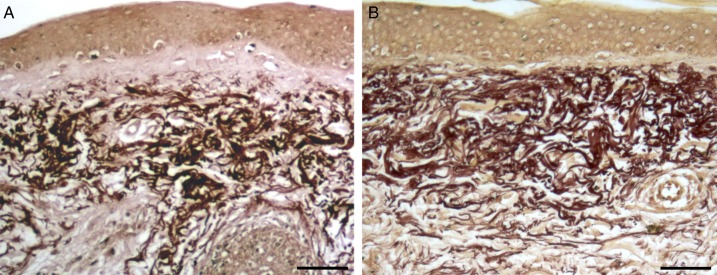
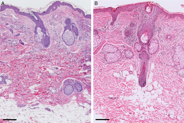
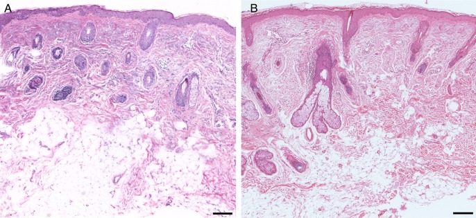
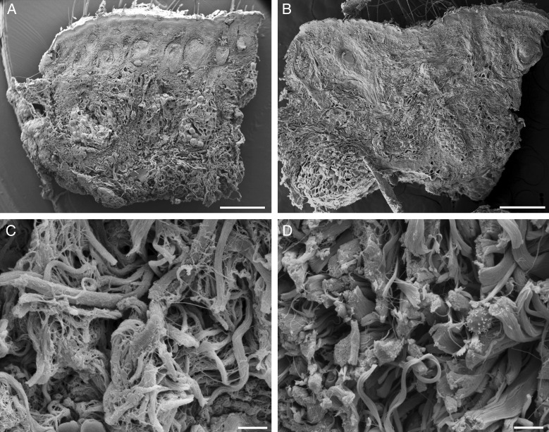
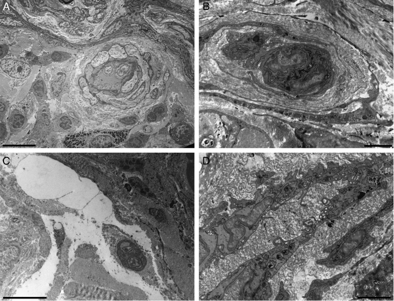
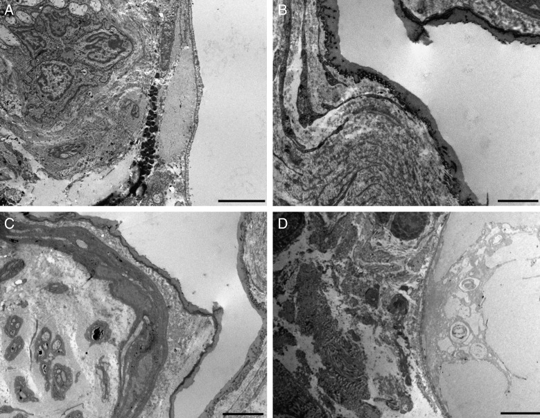
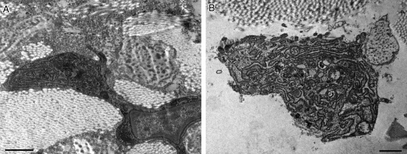
Methods: This study was performed in 13 patients who were candidates for facelift. The patients underwent sampling of fat by liposuction from the abdomen and submitted to one of three protocols: injection of SVF-enriched fat or expanded adipose-derived stem cells or fat plus PRP in the preauricular areas. Fragments of skin were removed before and 3 months after treatment and analyzed by optical and electron microscopy.
Results: The use of fat plus PRP led to the presence of more pronounced inflammatory infiltrates and a greater vascular reactivity, increasing in vascular permeability and a certain reactivity of the nervous component. The addition of PRP did not improve the regenerative effect.
Conclusion: The use of PRP did not have significant advantages in skin rejuvenation over the use of expanded adipose-derived stem cells or SVF-enriched fat. The effect of increased vascular reactivity may be useful in pathological situations in which an intense angiogenesis is desirable, such as tissular ischemia.
© 2016 The American Society for Aesthetic Plastic Surgery, Inc. Reprints and permission: journals.permissions@oup.com.
Figures
Comment in
-
Commentary on: Expanded Stem Cells, Stromal-Vascular Fraction, and Platelet-Rich Plasma Enriched Fat: Comparing Results of Different Facial Rejuvenation Approaches in a Clinical Trial.Aesthet Surg J. 2016 Mar;36(3):271-4. doi: 10.1093/asj/sjv239. Aesthet Surg J. 2016. PMID: 26879295 Free PMC article. No abstract available.
Similar articles
-
Antiaging treatment of the facial skin by fat graft and adipose-derived stem cells.Plast Reconstr Surg. 2015 Apr;135(4):999-1009. doi: 10.1097/PRS.0000000000001123. Plast Reconstr Surg. 2015. PMID: 25811565
-
Fat, Stem Cells, and Platelet-Rich Plasma.Clin Plast Surg. 2016 Jul;43(3):473-88. doi: 10.1016/j.cps.2016.03.017. Clin Plast Surg. 2016. PMID: 27363761 Review.
-
Effect of Use of Platelet-Rich Plasma (PRP) in Skin with Intrinsic Aging Process.Aesthet Surg J. 2018 Feb 15;38(3):321-328. doi: 10.1093/asj/sjx137. Aesthet Surg J. 2018. PMID: 29040421
-
Skin Rejuvenation and Volume Enhancement with the Micro Superficial Enhanced Fluid Fat Injection (M-SEFFI) for Skin Aging of the Periocular and Perioral Regions.Aesthet Surg J. 2017 Jan;37(1):14-23. doi: 10.1093/asj/sjw084. Epub 2016 May 30. Aesthet Surg J. 2017. PMID: 27241362
-
Platelet-rich plasma, the ultimate secret for youthful skin elixir and hair growth triggering.J Cosmet Dermatol. 2018 Jun;17(3):423-430. doi: 10.1111/jocd.12404. Epub 2017 Sep 8. J Cosmet Dermatol. 2018. PMID: 28887865 Review.
Cited by
-
The Crosstalk of Adipose-Derived Stem Cells (ADSC), Oxidative Stress, and Inflammation in Protective and Adaptive Responses.Int J Mol Sci. 2020 Dec 4;21(23):9262. doi: 10.3390/ijms21239262. Int J Mol Sci. 2020. PMID: 33291664 Free PMC article. Review.
-
Innovations in Skin and Soft Tissue Aging-A Systematic Literature Review and Market Analysis of Therapeutics and Associated Outcomes.Aesthetic Plast Surg. 2023 Aug;47(4):1609-1622. doi: 10.1007/s00266-023-03322-1. Epub 2023 May 8. Aesthetic Plast Surg. 2023. PMID: 37154849 Free PMC article.
-
Mechanism of action and therapeutic effects of oxidative stress and stem cell-based materials in skin aging: Current evidence and future perspectives.Front Bioeng Biotechnol. 2023 Jan 9;10:1082403. doi: 10.3389/fbioe.2022.1082403. eCollection 2022. Front Bioeng Biotechnol. 2023. PMID: 36698629 Free PMC article. Review.
-
Injectable Tissue Replacement and Regeneration: Anatomic Fat Grafting to Restore Decayed Facial Tissues.Plast Reconstr Surg Glob Open. 2019 Aug 12;7(8):e2293. doi: 10.1097/GOX.0000000000002293. eCollection 2019 Aug. Plast Reconstr Surg Glob Open. 2019. PMID: 31592023 Free PMC article.
-
Commentary on: Expanded Stem Cells, Stromal-Vascular Fraction, and Platelet-Rich Plasma Enriched Fat: Comparing Results of Different Facial Rejuvenation Approaches in a Clinical Trial.Aesthet Surg J. 2016 Mar;36(3):271-4. doi: 10.1093/asj/sjv239. Aesthet Surg J. 2016. PMID: 26879295 Free PMC article. No abstract available.
References
-
- D'Amico RA, Rubin JP, Neumeister MW et al. . American Society of Plastic Surgeons/Plastic Surgery Foundation Regenerative Medicine Task Force. A Report of the ASPS Task Force on regenerative medicine: opportunities for plastic surgery. Plast Reconstr Surg. 2013;1312:393-399. - PubMed
-
- Rigotti G, Marchi A, Sbarbati A. Adipose-derived mesenchymal stem cells: past, present, and future. Aesthet Plast Surg. 2009;333:271-273. - PubMed
-
- Del Vecchio D, Rohrich RJ. A classification of clinical fat graft: different problems, different solutions. Plast Reconstr Surg. 2012;1303:511-522. - PubMed
-
- Bosset S, Barré P, Chalon A et al. . Skin ageing: clinical and histopathologic study of permanent and reducible wrinkles. Eur J Dermatol. 2002;123:247-252. - PubMed
-
- Kim JH, Jung M, Kim HS et al. . Adipose-derived stem cells as a new therapeutic modality for ageing skin. Exp Dermatol. 2011;205:383-387. - PubMed
Publication types
MeSH terms
LinkOut - more resources
Full Text Sources
Other Literature Sources
Medical
Research Materials
