Tracheal Involvement in Crohn Disease: the First Case in Korea
- PMID: 26879553
- PMCID: PMC4821520
- DOI: 10.5946/ce.2015.059
Tracheal Involvement in Crohn Disease: the First Case in Korea
Erratum in
-
Erratum: Tracheal Involvement in Crohn Disease: the First Case in Korea.Clin Endosc. 2016 May;49(3):310. doi: 10.5946/ce.2015.059.e1. Clin Endosc. 2016. PMID: 27230104 Free PMC article.
Abstract
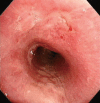
Respiratory involvement in Crohn disease (CD) is rare condition with only about a dozen reported cases. We report the first case of CD with tracheal involvement in Korea. An 18-year-old woman with CD was hospitalized because of coughing, dyspnea, and fever sustained for 3 weeks. Because she had stridor in her neck, we performed computed tomography of the neck, which showed circumferential wall thickening of the larynx and hypopharynx. Bronchoscopy revealed mucosal irregularity, ulceration, and exudates debris in the proximal trachea, and bronchial biopsy revealed chronic inflammation with granulation tissue. Based on these findings, we suspected CD with tracheal involvement and began administering intravenous methylprednisolone at 1 mg/kg per day, after which her symptoms and bronchoscopic findings improved.
Keywords: Crohn disease; Inflammatory bowel diseases; Tracheobronchial involvement.
Conflict of interest statement
Figures


References
-
- Spira A, Grossman R, Balter M. Large airway disease associated with inflammatory bowel disease. Chest. 1998;113:1723–1726. - PubMed
-
- Henry MT, Davidson LA, Cooke NJ. Tracheobronchial involvement with Crohn’s disease. Eur J Gastroenterol Hepatol. 2001;13:1495–1497. - PubMed
-
- Asami T, Koyama S, Watanabe Y, et al. Tracheobronchitis in a patient with Crohn’s disease. Intern Med. 2009;48:1475–1478. - PubMed
-
- Camus P, Colby TV. The lung in inflammatory bowel disease. Eur Respir J. 2000;15:5–10. - PubMed
-
- Kraft SC, Earle RH, Roesler M, Esterly JR. Unexplained bronchopulmonary disease with inflammatory bowel disease. Arch Intern Med. 1976;136:454–459. - PubMed
LinkOut - more resources
Full Text Sources
Other Literature Sources

