. 2016 Feb 16:6:21595.
doi: 10.1038/srep21595.

(-)-Epigallocatechin-3-Gallate Enhances Hepatitis C Virus Double-Stranded RNA Intermediates-Triggered Innate Immune Responses in Hepatocytes

Affiliations

(-)-Epigallocatechin-3-Gallate Enhances Hepatitis C Virus Double-Stranded RNA Intermediates-Triggered Innate Immune Responses in Hepatocytes

Yizhong Wang et al. Sci Rep. .

Abstract

(-)-Epigallocatechin-3-gallate (EGCG), a major polyphenol component of green tea, has recently been identified as an inhibitor of hepatitis C virus (HCV) entry. Here, we examined whether EGCG can enhance hepatocyte-mediated intracellular innate immunity against HCV. HCV dsRNAs (Core, E1-P7, NS-3'NTR and NS5A) induced interferon-λ1 (IFN-λ1) expression in human hepatocytes. These HCV dsRNAs also induced the expression of Toll-like receptor 3 (TLR3), retinoic acid-inducible gene I (RIG-I) and several antiviral IFN-stimulated genes (ISGs) expression. Although EGCG treatment of hepatocytes alone had little effect on TLR3 and RIG-I signaling pathways, EGCG significantly enhanced HCV dsRNAs-induced the expression of IFN-λ1, TLR3, RIG-I and antiviral ISGs in hepatocytes. Furthermore, treatment of HCV-infected hepatocytes with EGCG and HCV dsRNAs inhibited viral replication. Given that EGCG has the ability to enhance HCV dsRNAs-induced intracellular antiviral innate immunity against HCV, suggesting the potential application of EGCG as a new anti-HCV agent for HCV therapy.


Figures

Figure 1
Figure 1. HCV dsRNAs induce IFN-λ1 expression.
Huh7 cells were stimulated with the HCV dsRNAs (Core, E1-P7, NS-3′NTR, NS5A, 1 μg/mL), same dose of poly I:C (1 μg/mL) was used as a positive control. Total RNA extracted from cells after 24 h exposure was subjected to the real time RT-PCR for the mRNA levels of IFN-λ1 and GAPDH. The data are expressed IFN-λ1 mRNA (A) levels relative (fold) to the control (vehicle only, which defined as 1). Supernatant (SN) was collected from the cell cultures 48 h post exposure for ELISA to measure the protein levels of IFN-λ1 (B). The results shown are mean ± SD of triplicate, representative of three independent experiments.
Figure 2
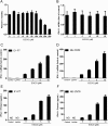
Figure 2. EGCG enhances HCV dsRNAs-induced IFN-λ1.
(A) Cyotoxicity test of EGCG on Huh7 cells. Huh7 cells were treated with concentrations of EGCG as indicated for 72 h. The cells viability was determined using an MTS-based viability assay by determining OD at 490 nm. (B) Effect of EGCG on IFN-λ1 expression in Huh7 cells. Huh7 cells were treated with EGCG at the indicated concentrations for 24 h. Total RNA from cells was subjected to IFN-λ1 gene expression by real time RT-PCR. (C–F) Effect of EGCG on dsRNAs-induced IFN-λ1 expression in Huh7 cells. Huh7 cells were treated with EGCG as indicated for 1 h prior to HCV dsRNAs (E1-P7, NS-3′NTR) stimulation. Total RNA extracted from cells after 24 h stimulation was subjected to the real time RT-PCR for the mRNA levels of IFN-λ1 and GAPDH. The data are expressed IFN-λ1 mRNA (C,D) levels relative (fold) to the control (vehicle only, which defined as 1). After 48 h stimulation, supernatant (SN) was collected from the cell cultures for ELISA to measure the protein level of IFN-λ1 (E,F). The results shown are mean ± SD of triplicate, representative of three independent experiments.
Figure 3
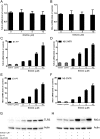
Figure 3. EGCG increases HCV dsRNAs-induced TLR3 and RIG-I expression.
(A,B) Effect of EGCG on TLR3 and RIG-I expression in Huh7 cells. Huh7 cells were treated with EGCG at the indicated concentrations for 24 h. Total RNA extracted from cells was subjected to TLR3 (A) and RIG-I (B) gene expression by real time RT-PCR. (C–G) Effect of EGCG on dsRNAs-induced TLR3 and RIG-I expression in Huh7 cells. Huh7 cells were treated with EGCG as indicated for 1 h prior to HCV dsRNAs (E1-P7, NS-3′NTR) stimulation. Total RNA extracted from cells after 24 h stimulation was subjected to the real time RT-PCR for the mRNA levels of TLR3, RIG-I and GAPDH. The data are expressed as TLR3 (C,D) and RIG-I mRNA (E,F) levels relative (fold) to the control (vehicle only, which defined as 1). The results shown are mean ± SD of triplicate, representative of three independent experiments. After 48 h stimulation, cell lysates were collected from the cell cultures 48 h after stimulation for Western blot to measure the protein level of TLR3 (G) and RIG-I (H). Three independent experiments were performed and one representative experiment is shown.
Figure 4
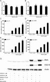
Figure 4. EGCG enhances HCV dsRNAs-induced ISGs expression in Huh7 cells.
(A,B) Effect of EGCG on ISG15 and MxA expression in Huh7 cells. Huh7 cells were treated with EGCG at the indicated concentrations for 24 h. Total RNA from cells was subjected to ISG15 (A) and MxA (B) gene expression by real time RT-PCR. (C–H) Effect of EGCG on dsRNAs-induced ISG15 and MxA expression in Huh7 cells. Huh7 cells were treated with EGCG as indicated for 1 h prior to HCV dsRNAs (E1-P7, NS-3′NTR) stimulation. Total RNA extracted from cells 24 h after stimulation was subjected to the real time RT-PCR for the mRNA levels of ISG15 and MxA and GAPDH. The data are expressed ISG15 (C,D) and MxA mRNA (E,F) levels relative (fold) to the control (vehicle only, which defined as 1). The results shown are mean ± SD of triplicate, representative of three experiments. Cell lysates were collected from the cell cultures 48 h after stimulation for Western blot to measure the protein level of ISG15 and MxA (G,H). Three independent experiments were performed and one representative work is shown.
Figure 5
Figure 5. The combination of EGCG treatment and HCV dsRNAs stimulation inhibits HCV replication.
(A,B) Effect of EGCG on HCV JFH-1 replication. JFH-1-infected Huh7 cells (72 h postinfection) were treated with EGCG at the indicated concentrations for 48 h. Intracellular (A) and extracellular (B) RNA extracted from JFH-1-infected Huh7 cells or culture supernatant (SN) was subjected to the real time RT-PCR for HCV and GAPDH RNA quantification. (C–G) Effect of the combination of EGCG treatment and HCV dsRNAs stimulation on HCV replication. JFH-1-infected Huh7 cells (72 h postinfection) were treated with EGCG at the indicated concentrations for 1 h prior to HCV dsRNAs (E1-P7, NS-3′NTR) stimulation. Intracellular (C,D) and extracellular (E,F) RNA was extracted from JFH-1-infected Huh7 cells or culture SN after 48 h stimulation was subjected to the real time RT-PCR for HCV and GAPDH RNA quantification. Intracellular HCV RNA level is expressed as HCV RNA levels relative (%) to the control (vehicle only, which are defined as 100%). Extracellular is expressed as copies/mL. The results shown are mean ± SD of triplicate cultures, representative of three experiments.
Figure 6
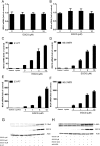
Figure 6. Effect of EGCG on HCV dsRNAs-induced IFN-λ1 expression in JFH-1-infected Huh7 cells.
JFH-1-infected Huh7 cells (72 h postinfection) were treated with EGCG as indicated for 1 h prior to HCV dsRNAs (E1-P7, NS-3′NTR) stimulation. Total RNA extracted from cells after 24 h stimulation was subjected to the real time RT-PCR for the mRNA levels of IFN-λ1 and GAPDH. The data are expressed IFN-λ1 mRNA (A,B) levels relative (fold) to the control (vehicle only, which defined as 1). After 48 h stimulation, supernatant (SN) was collected from the cell cultures for ELISA to measure the protein level of IFN-λ1 (C,D). The results shown are mean ± SD of triplicate, representative of three experiments.
Figure 7
Figure 7. Effect of EGCG on HCV dsRNAs-induced ISGs expression in JFH-1-infected Huh7 cells.
(A,B) Effect of EGCG on ISG15 and MxA expression in JFH-1- infected Huh7 cells. JFH-1-infected Huh7 cells (72 h postinfection) were treated with EGCG at the indicated concentrations for 24 h. Total RNA from cells was subjected to ISG15 (A) and MxA (B) gene expression by real time RT-PCR. (C–H) EGCG enhances dsRNAs-induced ISG15 and MxA expression in JFH-1-infected Huh7 cells. JFH-1-infected Huh7 cells (72 h postinfection) were treated with EGCG as indicated for 1 h prior to HCV dsRNAs (E1-P7, NS-3′NTR) stimulation. Total RNA extracted from cells after 24 h stimulation was subjected to the real time RT-PCR for the mRNA levels of ISG15 and MxA and GAPDH. The data are expressed ISG15 (C,D) and MxA mRNA (E,F) levels relative (fold) to the control (vehicle only, which defined as 1). The results shown are mean ± SD of triplicate, representative of three experiments. After 48 h stimulation, cell lysates were collected from the cell cultures for Western blot to measure the protein level of ISG15 and MxA (G). Three independent experiments were performed and one representative experiment is shown.
Figure 8
Figure 8. Roles of TLR3 and RIG-I signaling in HCV dsRNA-induced antiviral factors expression.
(A–C) Huh7 cells were pretreated with Bafilomycin A1 (25–100 nM) for 1 h prior to E1-P7 stimulation (1 μg/mL). After 24 h E1-P7 stimulation, total RNA extracted from cells was subjected to the real-time RT-PCR for mRNA levels of IFN-λ1 (A), MxA (B) and ISG15 (C). (D–G) Huh7 cells were transfected with siRNA against RIG-I (100 nM) or siRNA control (100  nM) for 24 h prior to E1-P7 stimulation (1 μg/mL). After 24 h E1-P7 stimulation, total RNA extracted from cells was subjected to the real-time RT-PCR for mRNA levels of RIG-I (D), IFN-λ1 (E), ISG15 (F) and MxA (G). The data are expressed as mRNA levels stimulated for relative (fold) to the control (without stimulation, which is defined as 1). The results are mean ± SD of three different experiments.

Similar articles

Cited by

References

    1. Alter M. J. Prevention of spread of hepatitis C. Hepatology 36, S93–98 (2002). - PubMed
    1. Chen S. L. & Morgan T. R. The natural history of hepatitis C virus (HCV) infection. Int J Med Sci 3, 47–52 (2006). - PMC - PubMed
    1. Battaglia A. M. & Hagmeyer K. O. Combination therapy with interferon and ribavirin in the treatment of chronic hepatitis C infection. Ann Pharmacother 34, 487–494 (2000). - PubMed
    1. Gale M. Jr. & Foy E. M. Evasion of intracellular host defence by hepatitis C virus. Nature 436, 939–945 (2005). - PubMed
    1. Li K. et al.. Immune evasion by hepatitis C virus NS3/4A protease-mediated cleavage of the Toll-like receptor 3 adaptor protein TRIF. Proc Natl Acad Sci USA 102, 2992–2997 (2005). - PMC - PubMed

Publication types

MeSH terms