. 2016 Feb;44(2):173-88.
doi: 10.1177/0192623315625859. Epub 2016 Feb 14.

Recommendations from the INHAND Apoptosis/Necrosis Working Group

Affiliations

Recommendations from the INHAND Apoptosis/Necrosis Working Group

Susan A Elmore et al. Toxicol Pathol. 2016 Feb.

Abstract

Historically, there has been confusion relating to the diagnostic nomenclature for individual cell death. Toxicologic pathologists have generally used the terms "single cell necrosis" and "apoptosis" interchangeably. Increased research on the mechanisms of cell death in recent years has led to the understanding that apoptosis and necrosis involve different cellular pathways and that these differences can have important implications when considering overall mechanisms of toxicity, and, for these reasons, the separate terms of apoptosis and necrosis should be used whenever differentiation is possible. However, it is also recognized that differentiation of the precise pathway of cell death may not be important, necessary, or possible in routine toxicity studies and so a more general term to indicate cell death is warranted in these situations. Morphological distinction between these two forms of cell death can sometimes be straightforward but can also be challenging. This article provides a brief discussion of the cellular mechanisms and morphological features of apoptosis and necrosis as well as guidance on when the pathologist should use these terms. It provides recommended nomenclature along with diagnostic criteria (in hematoxylin and eosin [H&E]-stained sections) for the most common forms of cell death (apoptosis and necrosis). This document is intended to serve as current guidance for the nomenclature of cell death for the International Harmonization of Nomenclature and Diagnostic Criteria Organ Working Groups and the toxicologic pathology community at large. The specific recommendations are:Use necrosis and apoptosis as separate diagnostic terms.Use modifiers to denote the distribution of necrosis (e.g., necrosis, single cell; necrosis, focal; necrosis, diffuse; etc.).Use the combined term apoptosis/single cell necrosis whenThere is no requirement or need to split the processes, orWhen the nature of cell death cannot be determined with certainty, orWhen both processes are present together. The diagnosis should be based primarily on the morphological features in H&E-stained sections. When needed, additional, special techniques to identify and characterize apoptosis can also be used.

Keywords: INHAND; apoptosis; cell death; guidance; necrosis; single cell necrosis.


Figures

Figure 1
Figure 1
Summary diagram of cell death nomenclature recommendations.
Figure 2
Figure 2
Apoptosis of exocrine pancreatic cells with cytoplasmic and nuclear condensation and nuclear fragmentation (arrows). Image courtesy of NTP Archives.
Figure 3
Figure 3
Apoptosis in the outflow tract cushions of the embryonic day 13.5 mouse heart. A Transverse section (A) through the developing heart demonstrates the newly formed conal septum (arrow) separating the two ventricular outlets, pulmonary trunk and aorta. Foci of apoptotic cell debris (B, arrows) are found in the cushion tissue surrounding the newly formed conal septum. A mitotic cell (arrowhead) is also present. Previously published in Toxicologic Pathology; Savolainen et al. 2009 Jun; 37(4): 395-414.
Figure 4
Figure 4
Apoptosis between digits in the developing mouse. Limb buds in the mouse embryo (A) have a webbed appearance with interdigital tissue that has not yet regressed (arrow). Higher magnification (B) shows scattered apoptotic cellular debris (outlined by arrows) in the interdigital tissue. Images courtesy of Julie Foley, NIEHS.
Figure 5
Figure 5
Thymocytes undergo a process of positive and negative selection in the thymus to produce T cells that recognize self-major histocompatability complex (MHC) molecules but do not recognize self-peptides. Positive selection occurs in the cortex whereas negative selection occurs in the medulla. This process is illustrated by these apoptotic lymphocytes (arrows) engulfed by tingible body macrophages in the cortex (A; positive selection) and medulla (B; negative selection) of an adult mouse thymus.
Figure 6
Figure 6
Germinal centers (GC) are unique sites in peripheral lymphoid tissue where clonal selection of B cells takes place in response to stimulation by various antigens. To select a proper B-cell clone for antibody-mediated immunity, multiple apoptotic signals synchronize in the GC, both in negative and positive selection pathways. This process is illustrated here by the apoptosis of lymphocytes in the developing germinal center of a rat mesenteric lymph node (arrows).
Figure 7
Figure 7
Apoptosis of surface epithelial cells is a normal process of cell turnover in the villi of the small intestine. The apoptotic cells are stained a dark golden brown (arrows) using in situ end-labeling (ISEL) of fragmented DNA. Image courtesy of NTP Archives.
Figure 8
Figure 8
Epithelial apoptosis (arrows) in the initial segment of the rat epididymis with luminal cell debris. This is commonly seen in response to reduced testosterone levels because the epididymis is an androgen dependent tissue. Similar changes can be seen in the seminal vesicles and prostate.
Figure 9
Figure 9
Example of apoptosis in the liver characterized by scattered single apoptotic hepatocytes (A & B, arrows) that are smaller than adjacent unaffected hepatocytes with hypereosinophilic cytoplasm (cytoplasmic condensation), and pyknotic and fragmented nuclei (B, arrows). Some cells are in a more advanced stage of apoptosis and have “rounded up” (B, arrowhead). Tingible body macrophages are not a feature in the liver, presumably due to the fast and efficient removal of cell debris by Kupffer cells. Previously published in Toxicologic Pathology; Elmore et al. (2014) Jan;42(1):12-44.
Figure 10
Figure 10
Experimental induction of apoptosis in the thymus cortex of a male 3-month-old Sprague Dawley rat dosed with 1mg/kg dexamethasone and necropsied 12 hours later (B) and compared to the thymus cortex from a concurrent control rat (A). The majority of cortical thymocytes in the treated rat (B) have undergone apoptosis. Most are in late stages of apoptosis with small, rounded hyperchromatic apoptotic bodies (arrows). Some have been engulfed by tingible body macrophages (arrowhead).
Figure 11
Figure 11
Tingible body macrophages (arrows) with engulfed apoptotic bodies in the thymus cortex of a male 3-month-old Sprague Dawley rat dosed with 1mg/kg dexamethasone and necropsied 24 hours later. There are also scattered free apoptotic bodies (small, dark, hyperchromatic) within the cortical parenchyma that have not yet been engulfed.
Figure 12
Figure 12
Focus of inflammation in the liver with an apoptotic cell (arrow), considered a “bystander effect”. The apoptotic cell did not rupture and incite the inflammatory response. Rather, the inflammatory cells created an adverse environment for this adjacent hepatocyte.
Figure 13
Figure 13
Focus of focal necrosis and inflammation in the liver (arrows) characterized by a focal group of contiguous cells with cell swelling, loss of cellular detail, neutrophils, and cell debris. Previously published in Toxicologic Pathology; Elmore et al. (2014) Jan;42(1):12-44.
Figure 14
Figure 14
Example of single cell necrosis in the liver. There is marked cell swelling and karyorrhexis in a necrotic hepatocyte (arrow) and a nearby small focus of inflammation (arrowhead), most likely secondary to cell rupture. Previously published in Toxicologic Pathology; Elmore et al. (2014) Jan;42(1):12-44.
Figure 15
Figure 15
Example of a toxic insult that resulted in apoptosis and necrosis in the heart. Focus of apoptotic and necrotic cardiomyocytes and macrophages in a 14-week-old rat treated with ephedrine and caffeine (Howden, Hanlon et al. 2005, Nyska, Murphy et al. 2005). This lesion could be diagnosed as apoptosis/single cell necrosis.
Figure 16
Figure 16
Thymus lymphocyte apoptosis with classic necrosis in a male 3-month-old Sprague Dawley rat dosed with 1mg/kg dexamethasone and necropsied 24-48 hours later. Over time, this lesion progressed from a strictly apoptotic phenotype to a mostly necrotic phenotype. In figure A the necrotic cell debris is identified as scattered eosinophilic material (cytoplasmic remnants) (long arrows) admixed with very small basophilic debris (nuclear remnants). The apoptotic bodies are identified as small, round, densely basophilic structures (A, short arrows). More severe lymphocyte apoptosis and classic necrosis is illustrated in Figure B. The abundant pale eosinophilic material is evidence of necrosis (B, long arrows) while the small, round, darkly basophilic structures are apoptotic bodies (B. short arrows). Necrotic nuclear debris is also present. Figure C is another example of marked lymphocyte apoptosis (short arrows) and classic necrosis (long arrows). Lymphocyte apoptosis and necrosis with inflammation is illustrated in Figure D. Because classic necrosis is the predominant lesion, this could be diagnosed as necrosis with discussion of apoptotic (D, short arrows) and inflammatory cells (D, long arrows) in the pathology narrative.
Figure 17
Figure 17
Example of hepatocellular apoptosis (A-D, arrows) and single cell necrosis (A-D, arrowheads) occurring together in the liver. The degenerating/necrotic cells are large and swollen with pale eosinophilic cytoplasm and karyolysis whereas the apoptotic cells are small and shrunken with hypereosinophilic cytoplasm and pyknotic/fragmented nuclei. Note the lack of tingible body macrophages.
Figure 18
Figure 18
Examples of necrosis and apoptosis of kidney tubule epithelial cells. Scattered renal tubules show necrosis of the epithelium; each cluster of tubules most likely represents a single convoluted tubule (A, arrows). This would have a diagnosis of renal tubular necrosis. In figure B there are tubules with occasional desquamated epithelial cells that have an apoptotic morphology (arrows) but that could also be mixed with necrotic cells. In this case one could use either apoptosis or necrosis if there were confidence in the type of cell death or the combination term apoptosis/single cell necrosis could be used. Images courtesy of Dr. John Seely.
Figure 19
Figure 19
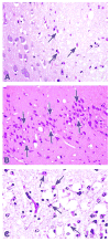
Examples of classic red dead neurons and apoptotic neurons. Classic red dead neurons (arrows) in the cortex of a rat exposed to carbonyl sulfide via inhalation (A) (Morgan, et al, 2005). ‘Red dead’ pyramidal neurons (outlined by arrows) in the hippocampus from a mouse exposed to an excitotoxic amino acid (kainic acid) (B). According to recent reports in the literature, these cells are likely undergoing a mixture of apoptosis and necrosis (Wang et al., 2005). Cells morphologically similar to apoptotic neurons (arrows, C). Images A and B courtesy of Dr. Jim Morrison. Image C courtesy of Dr. Roland Auer.

Similar articles

Cited by

References

    1. Bergsbaken T, Fink SL, Cookson BT. Pyroptosis: host cell death and inflammation. Nat Rev Microbiol. 2009;7:99–109. - PMC - PubMed
    1. Chimal-Monroy J, Abarca-Buis RF, Cuervo R, Diaz-Hernandez M, Bustamante M, Rios-Flores A, Romero-Suarez S, Farrera-Hernandez A. Molecular control of cell differentiation and programmed cell death during digit development. IUBMB Life. 2011;63:922–9. - PubMed
    1. Cho YS, Challa S, Moquin D, Genga R, Ray TD, Guildford M, Chan FK. Phosphorylation-driven assembly of the RIP1-RIP3 complex regulates programmed necrosis and virus-induced inflammation. Cell. 2009;137:1112–23. - PMC - PubMed
    1. Dekkers MP, Barde YA. Developmental biology. Programmed cell death in neuronal development. Science. 2013;340:39–41. - PubMed
    1. Denecker G, Vercammen D, Declercq W, Vandenabeele P. Apoptotic and necrotic cell death induced by death domain receptors. Cell Mol Life Sci. 2001;58:356–70. - PMC - PubMed

Publication types