Review
. 2016 May;13(2):173-88.
doi: 10.1177/1479972316631139. Epub 2016 Feb 15.

Clinical review: Endobronchial valve treatment for emphysema

Affiliations
Review

Clinical review: Endobronchial valve treatment for emphysema

Nabil Jarad. Chron Respir Dis. 2016 May.

Abstract

Breathlessness and impaired quality of life are prominent features in patients with severe emphysema even when conventional methods of treatment are optimal. Lung volume reduction using endobronchial management for emphysema has emerged as a new method to relieve symptoms and improve lung function tests in this group. The endobronchial valves (EBVs) are the most widely used treatment. This article outlines current criteria of patients' selection with literature review and evidence of efficacy. Complications of EBV insertion as well as current shortfalls of this method of treatment are also discussed.

Keywords: COPD; Emphysema; FEV1; valves; volume reduction.


Conflict of interest statement

Declaration of Conflicting Interests: The author(s) declared no potential conflicts of interest with respect to the research, authorship, and/or publication of this article.

Figures

Figure 1.
Figure 1.
A schematic adaptation of FEV1/age graph in susceptible smokers with COPD. The progressive decline in FEV1 is associated with increased tendency for lung exacerbations. The ECLIPSE study found that a group of patients can have exacerbations at higher level of FEV1. The dashed blue lines are the level of FEV1 at which each set of symptoms/complication of COPD are broadly occur. FEV1: forced expiratory volume in 1 second; COPD: chronic obstructive pulmonary disease.
Figure 2.
Figure 2.
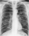
A chest radiograph of a patient with emphysema showing hyperinflated lung fields and flat diaphragm. The white lines represent the position of diaphragm in normal lungs.
Figure 3.
Figure 3.
Radiological emphysema as a proportion of all lung fields (y axis) on HRCT scan imaging according to progressive severity of impairment of lung function tests (x axis). HRCT: high-resolution computed tomography.
Figure 4.
Figure 4.
Types of emphysema on section of the lungs at post-mortem from coal miners. (a) Homogenous emphysema; (b) predominantly upper lobe emphysema; and (c) predominantly lower lobe emphysema.
Figure 5.
Figure 5.
Upper lobes emphysema on coronal images of HRCT (left) and lower lobe emphysema (right). HRCT: high-resolution computed tomography.
Figure 6.
Figure 6.
The 5-year probability of death in a subset of patients in the NETT trial – patients included in this graph were those with poor exercise capacity and upper lobe predominant disease. NETT: National Emphysema Treatment Trial.
Figure 7.
Figure 7.
The three sizes of the Zephyr EBVs. From left to right: 5.5 mm wide, 4.0 mm wide and 4.0 mm wide with short length (low profile). EBVs: endobronchial valves.
Figure 8.
Figure 8.
Zephyr EBV during inspiration (a) and expiration (b). The left panel is a schematic presentation (Pulmonx with permission). The right panel are valves in situ. Please note that the ‘duckbill’ is closed during inspiration and open during expiration to allow air and secretions out. EBV: endobronchial valve.
Figure 9.
Figure 9.
A 64-year-old patient with upper lobe emphysema prior to valve insertion (a) and 4 weeks after valve insertion (b). Please note the collapse in the left upper lobe and the elevation of the left hemi-diaphragm (b).
Figure 10.
Figure 10.
Complete (intact) interlobar fissures on the right lung (left) and the left lung (right). The patient is likely to respond to insertion of EBV. EBV: endobronchial valve.
Figure 11.
Figure 11.
Incomplete fissure between the left upper and the left lower lobe (right). Incomplete horizontal fissure between the right upper lobe and the right middle lobe (left). The second patient is not likely to respond to insertion of an EBV in the right upper lobe but may respond to treatment of combined right upper lobe and right middle lobe. EBV: endobronchial valve.
Figure 12.
Figure 12.
Change in FEV1 and SGRQ at 6 months in a subgroup of patients enrolled in the European VENT study. Please note the marked improvement in both outcome measures in patients with intact fissure and when there is a volume reduction (correct placement) in the target lobe after the procedure. FEV1: forced expiratory volume in 1 second; SGRQ: St George’s Respiratory Questionnaire.,
Figure 13.
Figure 13.
Long-term (5 years) change in FEV1 in those with complete interlobar fissures (fissures) compared to those with incomplete fissures (no fissure). FEV1: forced expiratory volume in 1 second.
Figure 14.
Figure 14.
The Chartis catheter before (a) and after (b) balloon inflation. The inflated balloon is occluding the left upper lobe (c). The Chartis console showing positive collateral ventilation trace (d).
Figure 15.
Figure 15.
Three patterns of recordings of flow using the Chartis catheter. Decline in flow with time (orange recording) seen on the upper panel representing CV negative. The flow is unchanged with time on the middle panel representing CV positive pattern. The absence of flow typically seen in highly destructed lobes is demonstrated in the lower panel. The blue recording represents the pressure in the target lobe and helps to identify an adequate balloon seal. CV: collateral ventilation.
Figure 16.
Figure 16.
Proportion of patients in whom target lobe volume reduction of over 350 ml (responders) when CV was not detected compared to those in whom CV was detected using the Chartis catheter. Adapted from Herth et al.. CV: collateral ventilation.
Figure 17.
Figure 17.
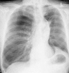
Left upper lobe pneumonia in a patient with EBV in the left upper lobe bronchus. Note the reduction in the left lung volume and the pulling up of the left hemi-diaphragm. The patient was treated successfully with intravenous then oral antibiotics. Following the treatment, the patient experienced a good clinical response to EBV insertion. EBV: endobronchial valve.
Figure 18.
Figure 18.
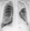
Left-sided stable pneumothorax. Please note a successful valve insertion with collapse in the left upper lobe and elevation of the left hemi-diaphragm.
Figure 19.
Figure 19.
Severe pneumothorax and surgical emphysema 2 days after insertion of EBVs in the left upper lobe of a 42-year-old lady. The valves are visible in the left hilum successfully occluding the left upper lobe. The lower aspect of a collapsed left upper lobe is also seen. Pneumothorax in this patient did not respond to the intercostal chest drain due to the formation of bronchopleural fistula. Valves needed to be removed. EBV: endobronchial valve.
Figure 20.
Figure 20.
Dislodging of valves. (a) A valve dislodged shortly after insertion and situated near the main carina; (b) a coughing up valve in a 42-year-old patient; (c) two well-placed valves in the right lower lobe bronchi in a 72-year-old patient; and (d) a displaced valve with mucous impact on the valve inserted in the apical segment of the left lower lobe bronchus.
Figure 21.
Figure 21.
Granulation tissue causing leak around the valve. Valve was removed, and another valve was successfully inserted 2 months later after healing of tissue granulation.
Figure 22.
Figure 22.
Percentage of patients attaining the MCID 6 months after EBV insertion compared with the control group. MCID: minimal clinically important difference; EBV: endobronchial valve; RV: residual volume; 6MWD: 6-minute walk distance; SGRQ: St George’s Respiratory Questionnaire.

References

    1. Global initiative for Chronic Obstructive Pulmonary Disease (GOLD initiative), http://www.goldcopd.org/
    1. Hurst JR, Vestbo J, Anzueto A, et al. Susceptibility to exacerbation in chronic obstructive pulmonary disease. N Engl J Med 2010; 363: 1128–1138. - PubMed
    1. Donaldson GC, Wedzicha JA. COPD exaerbations – 1. Epidemiology. Thorax 2006; 61: 164–168. - PMC - PubMed
    1. Celli BR, Barnes PJ. Exacerbations of chronic obstructive pulmonary disease. Eur Resp J 2007; 29: 1224–1238. - PubMed
    1. Kelly JL, Bamsey O, Smith C, et al. Health status assessment in routine clinical practice: the chronic obstructive pulmonary disease assessment test score in outpatients. Respiration 2012; 84: 193–199. - PubMed

LinkOut - more resources