Review
. 2016 Apr;38(4):379-93.
doi: 10.1002/bies.201500133. Epub 2016 Feb 16.

Heterotrimeric G protein signaling via GIV/Girdin: Breaking the rules of engagement, space, and time

Affiliations
Review

Heterotrimeric G protein signaling via GIV/Girdin: Breaking the rules of engagement, space, and time

Nicolas Aznar et al. Bioessays. 2016 Apr.

Abstract

Canonical signal transduction via heterotrimeric G proteins is spatially and temporally restricted, that is, triggered exclusively at the plasma membrane (PM), only by agonist activation of G protein-coupled receptors (GPCRs) via a process that completes within a few hundred milliseconds. Recently, a rapidly emerging paradigm has revealed a non-canonical pathway for activation of heterotrimeric G proteins by the non-receptor guanidine-nucleotide exchange factor (GEF), GIV/Girdin. This pathway has distinctive temporal and spatial features and an unusual profile of receptor engagement: diverse classes of receptors, not just GPCRs can engage with GIV to trigger such activation. Such activation is spatially and temporally unrestricted, that is, can occur both at the PM and on internal membranes discontinuous with the PM, and can continue for prolonged periods of time. Here, we provide the most complete up-to-date review of the molecular mechanisms that govern the unique spatiotemporal aspects of non-canonical G protein activation by GIV and the relevance of this new paradigm in health and disease.

Keywords: Golgi; autophagy; cdk5: Girdin; growth factor receptor tyrosine kinases; heterotrimeric G proteins.


Figures

Figure 1
Figure 1. Canonical G proteins signaling is restricted in time and space
Schematic shows the steps in GPCR signaling. Kinetics of various steps in receptor/G protein signaling chain as determined by FRET and BRET assays in intact cells are displayed [42]. Therefore, these values represent estimates of half-lives (in msec) for each step at maximal agonist concentration and overexpressed protein levels. Specific values cited here reflects the events during activation of β1-adrenoreceptor/Gαs/cAMP cascade. Similar values were reported also for the α2A-adrenoreceptors/Gαi/o and for the A2A-adenosine/Gαi pathways.
Figure 2
Figure 2. Network of regulatory proteins coordinately function to maintain finiteness of G protein signaling
Upper panel: Schematic of the G protein cycle is shown. Gα-subunits in GDP-bound "OFF" bind βγ-heterodimers and exist as inactive trimers, until nucleotide exchange and cycle of activation is triggered (green formula image) by chance encounter with GEFs, which could be either GPCRs, i.e., receptor GEFs or non-receptor GEFs (such as GIV/Girdin). Such nucleotide exchange is inhibited (red formula image) by GDIs. Once GTP-bound, Gα is active, dissociates from Gβγ-dimers until the GTP is hydrolyzed, inorganic phosphate (iP) is released and Gα returns to inactive state. This step of GTP hydrolysis is sped up by GAPs. Lower panel: The structural modules (red ribbons) which impart enzymatic properties to GAPs, GDIs and GEFs is shown in complex with Gα-subunit (in blue): (from left to right) GAPs accelerate GTP hydrolysis via a RGS box domain, as shown here in the case of RGS4 bound to an active conformation of Gαi1. GDIs inhibit nucleotide exchange via a Go-Loco/GPR domain, as shown here in the case of RGS14 bound to an inactive conformation of Gαi1. In the case of non-receptor GEFs, while some work via unknown module(s), a newly emerging subfamily uses a short stretch of amino acids to trigger nucleotide exchange using similar structural basis as shown here in the case of KB-752 synthetic peptide bound to an inactive conformation of Gαi1.
Figure 3
Figure 3. Activation of G proteins by GIV-GEF modulates multi-receptor signaling and broadly impacts diverse pathways within the post-receptor signaling network
Upper parts of the schematic shows multiple classes of receptors, some that sense a variety of chemical stimuli and others that sense mechanical signals, all of which utilize GIV-GEF to transactivate G proteins. The solid arrow connecting RTKs and GIV indicates that the mechanism for such engagement is well understood. The broken arrows linking all other receptors and GIV indicate that little or nothing is known as to how GIV may engage with those receptors. Lower part of the schematic shows the consequence of such activation (when GIV-GEF is turned "ON") on diverse signaling pathway within the signaling network. Green = enhancement; Red = suppression. This profile of signaling is reversed when GIV-GEF is switched "OFF", i.e., enhanced signals are suppressed and vice versa. Shown in the middle are three known ways to inhibit GIV-dependent signaling (PKCθ selectively phosphoinhibits GIV-GEF[3]; SHP-1 dephosphorylates tyrosine-phosphorylated GIV [7]) or activate (CDK5 phosphoactivates GIV-GEF [17]).
Figure 4
Figure 4. GIV plus RTKs, equals GPCRs
Schematic on the left shows the modular makeup of G-protein coupled growth factor RTKs. Ligand stimulation of growth factor receptor tyrosine kinases, e.g., EGFR (shown here) leads to receptor dimerization and autophosphorylation of its cytoplasmic tail (green interrupted line). Within ~3–5 minutes after growth factor stimulation, a ~110 aa long intrinsically disordered stretch within GIV's C-terminus (aa 1660–1870) recognizes and folds into a SH2-like module (red-purple-white) and directly docks onto the phosphotyrosine ligand (pTyr1148) presented by the cytoplasmic tail of ligand-activated RTKs. The mechanism of phosphotyrosine recognition and the kinetics of recruitment of GIV's SH2-like module to the RTK tail mimics that of other SH2 adaptors, e.g., Grb2. Just upstream and adjacent to the SH2-like module is GIV's GEF module (red ribbon) which binds and triggers nucleotide exchange (GTP, green for GDP, red), thereby activating Gαi subunits (light blue) in the vicinity of ligand-activated RTKs. It is the unique coexistence of two (GEF and SH2-like) modules that is key, because their collaboration is necessary and sufficient to assemble RTK-GIV-Gαi complexes at the PM within 5 minutes after ligand stimulation. One of the major consequences of the assembly of such complexes is transactivation of Gi and suppression of cellular cAMP after growth factor stimulation. In doing so, GIV's C-terminus enables the assembly of G-protein coupled RTKs, which subsequently leads to the non-canonical transactivation of G proteins. Schematic on the right shown canonical activation of G protein by ligand-activated receptor GEFs, i.e., GPCRs which can directly trigger nucleotide exchange.
Figure 5
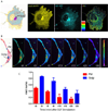
Figure 5. G proteins are active at the Golgi and on other internal membranes; such activation can be triggered in response to growth factors
A. Cos7 cell co-expressing Gαi1-YFP (internal tag) and CFP-Gβ1 and Gγ2 (untagged) were analyzed at steady-state by FRET imaging. Individual YFP and CFP channels show concentrated localization of G proteins on a perinuclear compartment, confirmed to be Golgi in a prior study [70]. FRET scale is shown as an inset. FRET is observed at the PM (yellow pixels), indicative of the formation of inactive trimers. By contrast, at steady-state, little or no FRET was seen at the Golgi, indicative of dissociated trimers. Activation of G protein and dissociation of trimers at the Golgi is abolished in GIV-depleted cells [70]. B–C. HeLa cell coexpressing Gαi1-YFP (internal tag) and CFP-Gβ1 and Gγ2 (untagged) as in A, were serum starved in 0.2% FBS and then stimulated with 50 nM EGF and analyzed by FRET imaging for dissociation of Gαβγ trimers. Briefly, ratiometric FRET imaging (FRET/CFP) was carried out using a Nikon Ti Eclipse which allows for perfect focus that locks the field and plane of image designed for long term live cell imaging. To reduce bleaching, only 5% of 405 laser was used. Upon growth factor stimulation, FRET is diminished both at the PM and at the Golgi. Bar graphs in C display the quantification of change in FRET ratio (as determined using 5–8 ROIs / cell for the PM and 1–2 ROIs for the Golgi region), in 3 cells, during 3 independent experiments.

References

    1. Lin C, Ear J, Midde K, Lopez-Sanchez I, et al. Structural basis for activation of trimeric Gi proteins by multiple growth factor receptors via GIV/Girdin. Mol Biol Cell. 2014;25:3654–3671. - PMC - PubMed
    1. Ghosh P, Garcia-Marcos M, Farquhar MG. GIV/Girdin is a rheostat that fine-tunes growth factor signals during tumor progression. Cell Adh Migr. 2011;5:237–248. - PMC - PubMed
    1. Lopez-Sanchez I, Garcia-Marcos M, Mittal Y, Aznar N, et al. Protein kinase C-theta (PKCtheta) phosphorylates and inhibits the guanine exchange factor, GIV/Girdin. Proceedings of the National Academy of Sciences of the United States of America. 2013;110:5510–5515. - PMC - PubMed
    1. Wang H, Misaki T, Taupin V, Eguchi A, et al. GIV/girdin links vascular endothelial growth factor signaling to Akt survival signaling in podocytes independent of nephrin. J Am Soc Nephrol. 2015;26:314–327. - PMC - PubMed
    1. Ghosh P, Beas AO, Bornheimer SJ, Garcia-Marcos M, et al. A G{alpha}i-GIV molecular complex binds epidermal growth factor receptor and determines whether cells migrate or proliferate. Mol Biol Cell. 2010;21:2338–2354. - PMC - PubMed

Publication types

MeSH terms

Substances