

Bone marrow-derived cells in ocular neovascularization: contribution and mechanisms
- PMID: 26880135
- PMCID: PMC4819501
- DOI: 10.1007/s10456-016-9497-6
Bone marrow-derived cells in ocular neovascularization: contribution and mechanisms
Abstract
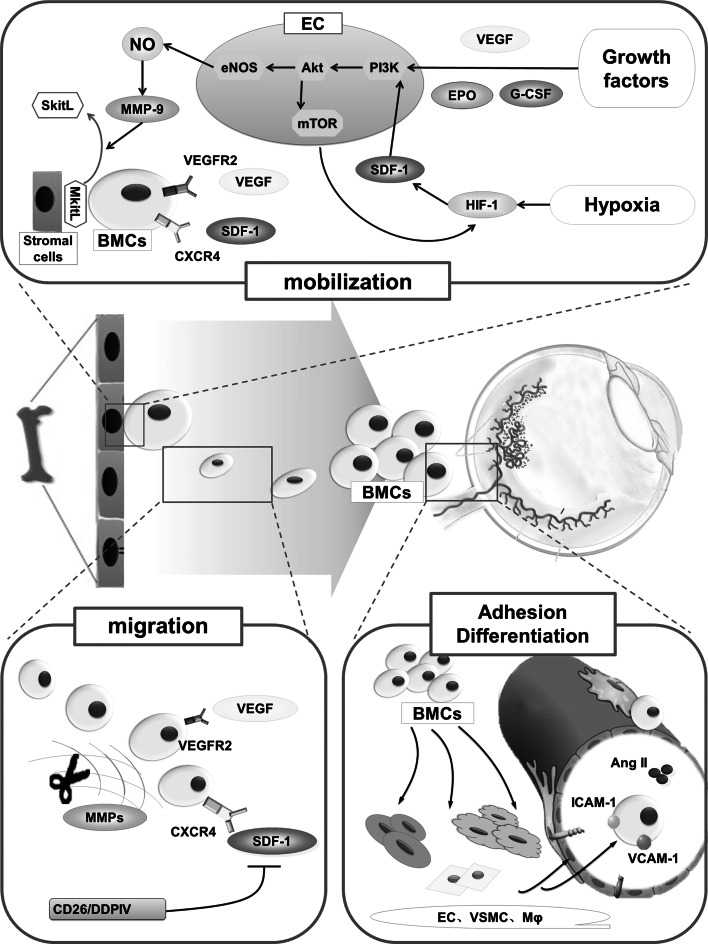
Ocular neovascularization often leads to severe vision loss. The role of bone marrow-derived cells (BMCs) in the development of ocular neovascularization, and its significance, is increasingly being recognized. In this review, we discuss their contribution and the potential mechanisms that mediate the effect of BMCs on the progression of ocular neovascularization. The sequence of events by which BMCs participate in ocular neovascularization can be roughly divided into four phases, i.e., mobilization, migration, adhesion and differentiation. This process is delicately regulated and liable to be affected by multiple factors. Cytokines such as vascular endothelial growth factor, granulocyte colony-stimulating factor and erythropoietin are involved in the mobilization of BMCs. Studies have also demonstrated a key role of cytokines such as stromal cell-derived factor-1, tumor necrosis factor-α, as well as vascular endothelial growth factor, in regulating the migration of BMCs. The adhesion of BMCs is mainly regulated by vascular cell adhesion molecule-1, intercellular adhesion molecule-1 and vascular endothelial cadherin. However, the mechanisms regulating the differentiation of BMCs are largely unknown at present. In addition, BMCs secrete cytokines that interact with the microenvironment of ocular neovascularization; their contribution to ocular neovascularization, especially choroidal neovascularization, can be aggravated by several risk factors. An extensive regulatory network is thought to modulate the role of BMCs in the development of ocular neovascularization. A comprehensive understanding of the involved mechanisms will help in the development of novel therapeutic strategies related to BMCs. In this review, we have limited the discussion to the recent progress in this field, especially the research conducted at our laboratory.
Keywords: Bone marrow-derived cells; Mechanism; Ocular neovascularization; Stem cells.
Figures
Similar articles
-
Nicotine promotes contribution of bone marrow-derived cells to experimental choroidal neovascularization in mice.Exp Eye Res. 2008 Jun;86(6):983-90. doi: 10.1016/j.exer.2008.03.018. Epub 2008 Apr 3. Exp Eye Res. 2008. PMID: 18472096
-
Integrin α5β1 promotes BMCs mobilization and differentiation to exacerbate choroidal neovascularization.Exp Eye Res. 2020 Apr;193:107991. doi: 10.1016/j.exer.2020.107991. Epub 2020 Mar 3. Exp Eye Res. 2020. PMID: 32142723
-
Hyperglycemia promotes vasculogenesis in choroidal neovascularization in diabetic mice by stimulating VEGF and SDF-1 expression in retinal pigment epithelial cells.Exp Eye Res. 2014 Jun;123:87-96. doi: 10.1016/j.exer.2014.04.012. Epub 2014 Apr 26. Exp Eye Res. 2014. PMID: 24780853
-
Endothelial progenitor cells: characterization and role in vascular biology.Circ Res. 2004 Aug 20;95(4):343-53. doi: 10.1161/01.RES.0000137877.89448.78. Circ Res. 2004. PMID: 15321944 Review.
-
Stem cell mobilisation for myocardial repair.Expert Opin Biol Ther. 2008 Nov;8(11):1675-90. doi: 10.1517/14712598.8.11.1675. Expert Opin Biol Ther. 2008. PMID: 18847304 Review.
Cited by
-
Stem Cells in the Path of Light, from Corneal to Retinal Reconstruction.Biomedicines. 2021 Jul 23;9(8):873. doi: 10.3390/biomedicines9080873. Biomedicines. 2021. PMID: 34440077 Free PMC article. Review.
-
Choroidal Neovascularization: Mechanisms of Endothelial Dysfunction.Front Pharmacol. 2019 Nov 29;10:1363. doi: 10.3389/fphar.2019.01363. eCollection 2019. Front Pharmacol. 2019. PMID: 31849644 Free PMC article. Review.
-
Amount of Mononuclear Phagocyte Infiltrate Does Not Predict Area of Experimental Choroidal Neovascularization (CNV).J Ocul Pharmacol Ther. 2018 Sep;34(7):489-499. doi: 10.1089/jop.2017.0131. J Ocul Pharmacol Ther. 2018. PMID: 30188257 Free PMC article.
-
Intriguing Roles for Endothelial ADAM10/Notch Signaling in the Development of Organ-Specific Vascular Beds.Physiol Rev. 2018 Oct 1;98(4):2025-2061. doi: 10.1152/physrev.00029.2017. Physiol Rev. 2018. PMID: 30067156 Free PMC article. Review.
-
Macrophage/microglia polarization for the treatment of diabetic retinopathy.Front Endocrinol (Lausanne). 2023 Sep 28;14:1276225. doi: 10.3389/fendo.2023.1276225. eCollection 2023. Front Endocrinol (Lausanne). 2023. PMID: 37842315 Free PMC article. Review.
References
Publication types
MeSH terms
Substances
LinkOut - more resources
Full Text Sources
Other Literature Sources
