Differential regulation of taurine biosynthesis in rainbow trout and Japanese flounder
- PMID: 26880478
- PMCID: PMC4754659
- DOI: 10.1038/srep21231
Differential regulation of taurine biosynthesis in rainbow trout and Japanese flounder
Abstract
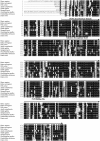
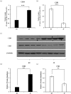
Animals have varied taurine biosynthesis capability, which was determined by activities of key enzymes including cysteine dioxygenase (CDO) and cysteine sulfinate decarboxylase (CSD). However, whether CDO and CSD are differentially regulated across species remains unexplored. In the present study, we examined the regulations of CDO and CSD in rainbow trout and Japanese flounder, the two fish species with high and low taurine biosynthesis ability respectively. Our results showed that the expression of CDO was lower in rainbow trout but more responsive to cysteine stimulation compared to that in Japanese flounder. On the other hand, both the expression and catalytic efficiency (k(cat)) of CSD were higher in rainbow trout than those of Japanese flounder. A three-residue substrate recognition motif in rainbow trout CSD with sequence of F126/S146/Y148 was identified to be responsible for high k(cat), while that with sequence of F88/N108/F110 in Japanese flounder led to low k(cat), as suggested by site-directed mutagenesis studies. In summary, our results determined new aspects of taurine biosynthesis regulation across species.
Figures


Similar articles
-
Ontogenetic taurine biosynthesis ability in rainbow trout (Oncorhynchus mykiss).Comp Biochem Physiol B Biochem Mol Biol. 2015 Jul;185:10-5. doi: 10.1016/j.cbpb.2015.03.003. Epub 2015 Mar 13. Comp Biochem Physiol B Biochem Mol Biol. 2015. PMID: 25773436
-
Enhancement of menadione stress tolerance in yeast by accumulation of hypotaurine and taurine: co-expression of cDNA clones, from Cyprinus carpio, for cysteine dioxygenase and cysteine sulfinate decarboxylase in Saccharomyces cerevisiae.Amino Acids. 2010 Apr;38(4):1173-83. doi: 10.1007/s00726-009-0328-6. Epub 2009 Jul 26. Amino Acids. 2010. PMID: 19633968
-
The synthesis and role of taurine in the Japanese eel testis.Amino Acids. 2012 Aug;43(2):773-81. doi: 10.1007/s00726-011-1128-3. Epub 2011 Nov 2. Amino Acids. 2012. PMID: 22045384
-
Taurine biosynthetic enzymes and taurine transporter: molecular identification and regulations.Neurochem Res. 2004 Jan;29(1):83-96. doi: 10.1023/b:nere.0000010436.44223.f8. Neurochem Res. 2004. PMID: 14992266 Review.
-
Taurine biosynthesis enzyme cysteine sulfinate decarboxylase (CSD) from brain: the long and tricky trail to identification.Neurochem Res. 1992 Sep;17(9):849-59. doi: 10.1007/BF00993260. Neurochem Res. 1992. PMID: 1407273 Review. No abstract available.
Cited by
-
Effects of GRP78 on Endoplasmic Reticulum Stress and Inflammatory Response in Macrophages of Large Yellow Croaker (Larimichthys crocea).Int J Mol Sci. 2023 Mar 20;24(6):5855. doi: 10.3390/ijms24065855. Int J Mol Sci. 2023. PMID: 36982929 Free PMC article.
-
Stimulatory effect of dietary taurine on growth performance, digestive enzymes activity, antioxidant capacity, and tolerance of common carp, Cyprinus carpio L., fry to salinity stress.Fish Physiol Biochem. 2018 Apr;44(2):639-649. doi: 10.1007/s10695-017-0459-8. Epub 2017 Dec 28. Fish Physiol Biochem. 2018. PMID: 29285672
-
Molecular Characterization and Directed Evolution of a Metagenome-Derived l-Cysteine Sulfinate Decarboxylase.Food Technol Biotechnol. 2018 Mar;56(1):117-123. doi: 10.17113/ftb.56.01.18.5415. Food Technol Biotechnol. 2018. PMID: 29796005 Free PMC article.
-
High-Resolution Magic Angle Spinning (HRMAS) NMR Identifies Oxidative Stress and Impairment of Energy Metabolism by Zearalenone in Embryonic Stages of Zebrafish (Danio rerio), Olive Flounder (Paralichthys olivaceus) and Yellowtail Snapper (Ocyurus chrysurus).Toxins (Basel). 2023 Jun 15;15(6):397. doi: 10.3390/toxins15060397. Toxins (Basel). 2023. PMID: 37368698 Free PMC article.
-
Dynamics of blood Taurine concentration and its correlation with nutritional and physiological status during the fattening period of Japanese black cattle.J Anim Sci. 2024 Jan 3;102:skae347. doi: 10.1093/jas/skae347. J Anim Sci. 2024. PMID: 39535934 Free PMC article.
References
-
- Jacobsen J. G. & Smith L. H. Biochemistry and physiology of taurine and taurine derivatives. Physiol Rev 48, 424–511 (1968). - PubMed
-
- Huxtable R. J. Physiological actions of taurine. Physiol Rev 72, 101–163 (1992). - PubMed
-
- Lambert I. H. Regulation of the cellular content of the organic osmolyte taurine in mammalian cells. Neurochem Res 29, 27–63 (2004). - PubMed
-
- Foos T. M. & Wu J. Y. The role of taurine in the central nervous system and the modulation of intracellular calcium homeostasis. Neurochem Res 27, 21–26 (2002). - PubMed
-
- Lima L., Obregon F., Cubillos S., Fazzino F. & Jaimes I. Taurine as a micronutrient in development and regeneration of the central nervous system. Nutr Neurosci 4, 439–443 (2001). - PubMed
Publication types
MeSH terms
Substances
LinkOut - more resources
Full Text Sources
Other Literature Sources
Miscellaneous

