

Diagnosis of adults Xp11.2 translocation renal cell carcinoma by immunohistochemistry and FISH assays: clinicopathological data from ethnic Chinese population
- PMID: 26880493
- PMCID: PMC4754949
- DOI: 10.1038/srep21677
Diagnosis of adults Xp11.2 translocation renal cell carcinoma by immunohistochemistry and FISH assays: clinicopathological data from ethnic Chinese population
Abstract
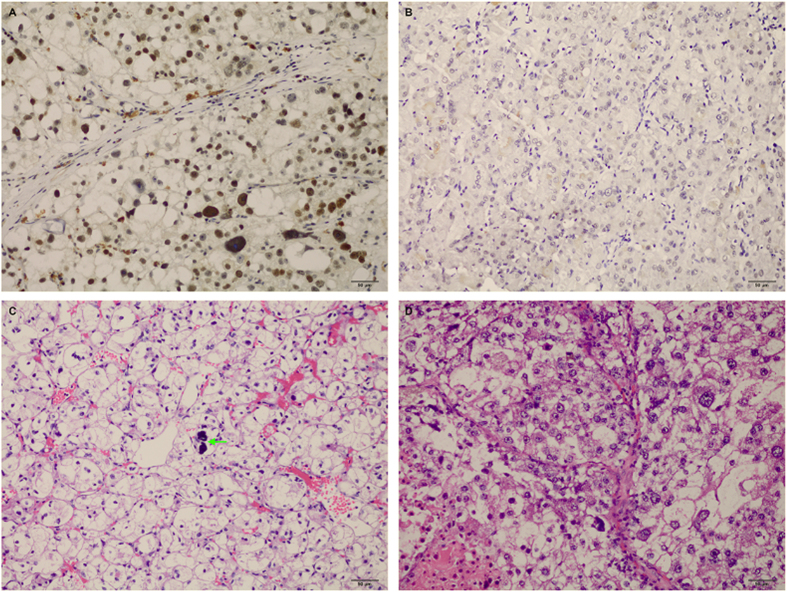
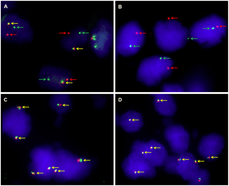
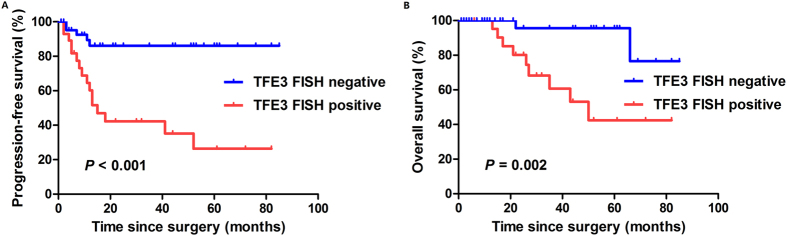
This study aimed to assess the utility of transcription factor E3 (TFE3) break-apart fluorescence in situ hybridization (FISH) assay in diagnosis of Xp11.2 translocation renal cell carcinoma (Xp11.2 RCC) and to compare the clinicopathological features between adult Xp11.2 RCC and non-Xp11.2 RCC. 76 pathologically suspected Xp11.2 RCCs were recruited from our institution. Both TFE3 immunohistochemistry (IHC) and TFE3 FISH assay were performed for the entire cohort. The progression-free survival (PFS) and overall survival (OS) curves were estimated using the Kaplan-Meier method. FISH analysis confirmed 30 Xp11.2 RCCs, including 28 cases with positive TFE3 immunostaining and 2 cases with negative immunostaining. The false-positive and false-negative rates were 6.7% (2/30) and 4.3% (2/46), respectively, for TFE3 IHC compared with FISH assay. Xp11.2 RCC was significantly associated with higher pathological stage and Fuhrman nuclear grade compared with non-Xp11.2 RCC (P < 0.05). The median PFS and OS for TFE3 FISH-positive group were 13.0 months (95% CI, 8.4-17.6 months) and 50.0 months (95% CI, 27.6-72.4 months), respectively, while the median PFS and OS had not been reached for TFE3 FISH-negative group. In conclusion, TFE3 break-apart FISH assay is a highly useful and standard diagnostic method for Xp11.2 RCC. Adult Xp11.2 RCC is clinically aggressive and often presents at advanced stage with poor prognosis.
Figures
Similar articles
-
TFE3 break-apart FISH has a higher sensitivity for Xp11.2 translocation-associated renal cell carcinoma compared with TFE3 or cathepsin K immunohistochemical staining alone: expanding the morphologic spectrum.Am J Surg Pathol. 2013 Jun;37(6):804-15. doi: 10.1097/PAS.0b013e31827e17cb. Am J Surg Pathol. 2013. PMID: 23598965
-
Utilization of a TFE3 break-apart FISH assay in a renal tumor consultation service.Am J Surg Pathol. 2013 Aug;37(8):1150-63. doi: 10.1097/PAS.0b013e31828a69ae. Am J Surg Pathol. 2013. PMID: 23715164
-
Usefulness of a break-apart FISH assay in the diagnosis of Xp11.2 translocation renal cell carcinoma.Virchows Arch. 2011 Sep;459(3):299-306. doi: 10.1007/s00428-011-1127-5. Epub 2011 Jul 20. Virchows Arch. 2011. PMID: 21773754
-
RBM10-TFE3 renal cell carcinoma characterised by paracentric inversion with consistent closely split signals in break-apart fluorescence in-situ hybridisation: study of 10 cases and a literature review.Histopathology. 2019 Aug;75(2):254-265. doi: 10.1111/his.13866. Epub 2019 Jun 25. Histopathology. 2019. PMID: 30908700 Review.
-
A case of TFE3 translocation renal cell carcinoma with rare morphological features and literature review.Indian J Pathol Microbiol. 2023 Jan-Mar;66(1):135-140. doi: 10.4103/ijpm.ijpm_755_21. Indian J Pathol Microbiol. 2023. PMID: 36656224 Review.
Cited by
-
Renal cell carcinoma associated with Xp11.2 translocation/transcription factor E3 gene fusion: an adult case report and literature review.J Int Med Res. 2020 Oct;48(10):300060520942095. doi: 10.1177/0300060520942095. J Int Med Res. 2020. PMID: 33026261 Free PMC article. Review.
-
PD-L1 expression in Xp11.2 translocation renal cell carcinoma: Indicator of tumor aggressiveness.Sci Rep. 2017 May 18;7(1):2074. doi: 10.1038/s41598-017-02005-7. Sci Rep. 2017. PMID: 28522811 Free PMC article.
-
Renal cell carcinoma associated with Xp11.2 translocation/transcription factor E3 gene fusion: A case report and literature review.Oncol Lett. 2023 Nov 21;27(1):29. doi: 10.3892/ol.2023.14162. eCollection 2024 Jan. Oncol Lett. 2023. PMID: 38073770 Free PMC article.
-
Preoperative neutrophil-to-lymphocyte ratio predicts the surgical outcome of Xp11.2 translocation/TFE3 renal cell carcinoma patients.BMC Urol. 2018 Jun 11;18(1):60. doi: 10.1186/s12894-018-0374-z. BMC Urol. 2018. PMID: 29890986 Free PMC article.
-
The suitability of NONO-TFE3 dual-fusion FISH assay as a diagnostic tool for NONO-TFE3 renal cell carcinoma.Sci Rep. 2020 Oct 1;10(1):16361. doi: 10.1038/s41598-020-73309-4. Sci Rep. 2020. PMID: 33004995 Free PMC article.
References
-
- Ramphal R., Pappo A., Zielenska M., Grant R. & Ngan B. Y. Pediatric renal cell carcinoma: clinical, pathologic, and molecular abnormalities associated with the members of the mit transcription factor family. American journal of clinical pathology 126, 349–364, doi: 10.1309/98YE9E442AR7LX2X (2006). - DOI - PubMed
-
- Armah H. B., Parwani A. V., Surti U. & Bastacky S. I. Xp11.2 translocation renal cell carcinoma occurring during pregnancy with a novel translocation involving chromosome 19: a case report with review of the literature. Diagnostic pathology 4, 15, doi: 10.1186/1746-1596-4-15 (2009). - DOI - PMC - PubMed
Publication types
MeSH terms
Substances
LinkOut - more resources
Full Text Sources
Other Literature Sources
