. 2016 Mar 7;213(3):329-36.
doi: 10.1084/jem.20151464. Epub 2016 Feb 15.

RNase H2 catalytic core Aicardi-Goutières syndrome-related mutant invokes cGAS-STING innate immune-sensing pathway in mice

Affiliations

RNase H2 catalytic core Aicardi-Goutières syndrome-related mutant invokes cGAS-STING innate immune-sensing pathway in mice

Vladislav Pokatayev et al. J Exp Med. .

Abstract

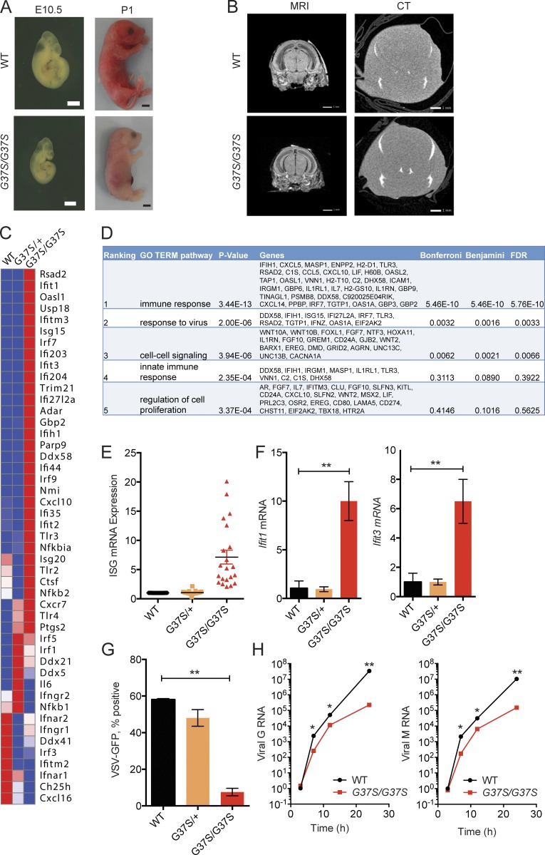
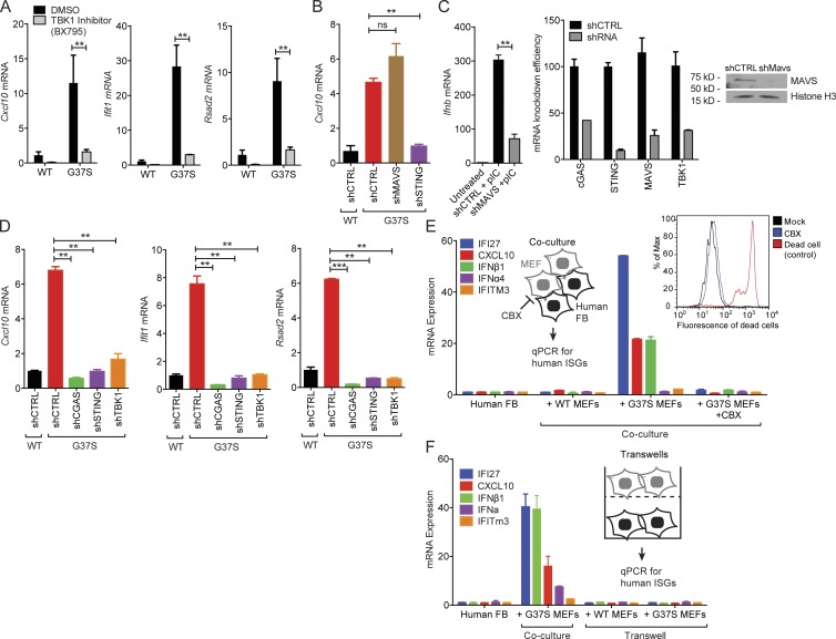
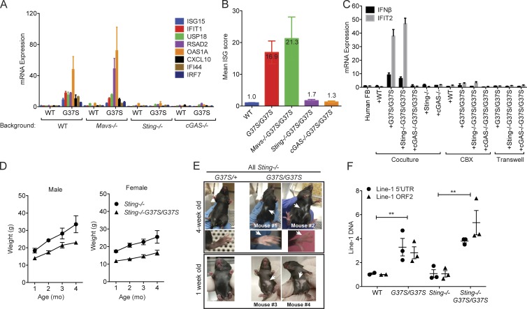
The neuroinflammatory autoimmune disease Aicardi-Goutières syndrome (AGS) develops from mutations in genes encoding several nucleotide-processing proteins, including RNase H2. Defective RNase H2 may induce accumulation of self-nucleic acid species that trigger chronic type I interferon and inflammatory responses, leading to AGS pathology. We created a knock-in mouse model with an RNase H2 AGS mutation in a highly conserved residue of the catalytic subunit, Rnaseh2a(G37S/G37S) (G37S), to understand disease pathology. G37S homozygotes are perinatal lethal, in contrast to the early embryonic lethality previously reported for Rnaseh2b- or Rnaseh2c-null mice. Importantly, we found that the G37S mutation led to increased expression of interferon-stimulated genes dependent on the cGAS-STING signaling pathway. Ablation of STING in the G37S mice results in partial rescue of the perinatal lethality, with viable mice exhibiting white spotting on their ventral surface. We believe that the G37S knock-in mouse provides an excellent animal model for studying RNASEH2-associated autoimmune diseases.


Figures

Figure 1.
Figure 1.
Primary cells from Rnaseh2aG37S/G37S embryos show increased expression in ISGs. (A) Images of WT and Rnaseh2aG37S/G37S embryos at indicated stage (bottom). Bars, 2 mm. (B) Transcranial images of ex vivo mouse E18.5 embryos WT and Rnaseh2aG37S/G37S in MRI gradient echo image and micro computed tomography scans. No appreciable calcium, other than in the bones of the cranium (top arrow) and jaw (bottom arrow), which are still under formation. Bars, 1 mm. (C) A heat map of immune gene expression in WT, Rnaseh2aG37S/+ and Rnaseh2aG37S/G37S primary MEFs. Data from RNA-seq (Table S1). (D) Gene ontology analysis of 388 genes that are increased by twofold or more in Rnaseh2aG37S/G37S compared with WT MEFs. Top five enriched pathways are shown. (E) Expression of ISGs in WT, Rnaseh2aG37S/+, and Rnaseh2aG37S/G37S primary MEFs. Each dot represents a different ISG. Data from RNA-seq. (F) Quantitative RT-PCR analysis of Ifit1 and Ifit3 mRNA (ISGs) in WT, Rnaseh2aG37S/+, and Rnaseh2aG37S/G37S primary MEFs. (G and H) VSV-GFP replication in WT, Rnaseh2aG37S/+, and Rnaseh2aG37S/G37S primary MEFs. FACS analysis measures VSV-GFP signal at 24 h after infection (G). Quantitative RT-PCR analysis of VSV G and M RNA measure viral RNA replication at indicated time after infection (H). *, P < 0.05; **, P < 0.01. Mice were compared with littermate controls. Data are representative of at least three independent experiments. Error bars represent the SEM. Unpaired Student’s t test (F–H).
Figure 2.
Figure 2.
Immune activation in Rnaseh2aG37S/G37S primary MEFs requires the cGAS–STING innate immune pathway. (A) Quantitative RT-PCR analysis of Cxcl10, Ifit1 and Rsad2 mRNA (all ISGs) in WT and Rnaseh2aG37S/G37S (G37S, same below) MEFs treated with DMSO or TBK1 inhibitor BX795 (10 µM) for 6 h. (B) Quantitative RT-PCR analysis of Cxcl10 mRNA in WT and G37S MEFs treated with shRNA against indicated genes involved in cytosolic nucleic acid-sensing. (C) shMAVS knockdown reduces poly(I:C)-induced IFN response. Knockdown efficiency is shown on the right. (D) Quantitative RT-PCR analysis of Cxcl10, Ifit1, and Rsad2 mRNA in WT and G37S MEFs treated with shRNA against indicated genes involved in DNA-sensing pathway. (E) Quantitative RT-PCR analysis of a panel of human ISGs and IFN genes in human fibroblasts (BJ-1 cells) co-cultured with WT or G37S MEFs for 18 h, with or without CBX treatment (inhibits gap junction). Left inset shows a schematic diagram of the gap junction assay. Right inset shows FACS analysis of cell death in mock- and CBX-treated cells. (F) Quantitative RT-PCR analysis of human ISGs in human fibroblasts in a trans-well assay co-cultured with WT or G37S MEFs for 18 h. Mice were compared with littermate controls. **, P < 0.01; ***, P < 0.001. ns, not significant. Data are representative of at least three independent experiments. Error bars represent the SEM. Unpaired Student’s t test (A–D).
Figure 3.
Figure 3.
Sting−/− partially rescues perinatal lethality of G37S mice. (A) Quantitative RT-PCR analysis of a panel of mouse ISGs in WT or G37S embryos on Mavs−/− or Sting−/− or cGAS−/− background. Total RNA was isolated from primary MEFs (E13.5) of indicated genotype. (B) Mean ISG score of indicated genotypes. Data from A. (C) Gap junction cGAMP bioassay. As in Fig. 2 E, MEFs of indicated genotype were co-cultured with human BJ-1 cells for 18 h, with or without CBX or direct contact (indicated on the bottom). Quantitative RT-PCR analysis of IFN-β and IFIT1 indicates cGAMP activity in MEFs. (D) Mouse body weights. n = 4. (E) White-spotting phenotype in Rnaseh2aG37S/G37S Sting−/− viable adults. (F) Quantitative PCR analysis of mouse Line-1 5′ UTR and ORF2 DNA in WT or G37S embryos (isolated from E13.5 or E15.5; n = 3). Each dot represents an individual embryo. Mice were compared with littermate controls and with age-matched knock-out mice **, P < 0.01. Data are representative of at least two independent experiments (A–C), or pooled data from multiple animals (D–F). Error bars represent the SEM. Unpaired Student’s t test (F).

Similar articles

Cited by

References

    1. Ablasser A., Schmid-Burgk J.L., Hemmerling I., Horvath G.L., Schmidt T., Latz E., and Hornung V.. 2013. Cell intrinsic immunity spreads to bystander cells via the intercellular transfer of cGAMP. Nature. 503:530–534. 10.1038/nature12640 - DOI - PMC - PubMed
    1. Ablasser A., Hemmerling I., Schmid-Burgk J.L., Behrendt R., Roers A., and Hornung V.. 2014. TREX1 deficiency triggers cell-autonomous immunity in a cGAS-dependent manner. J. Immunol. 192:5993–5997. 10.4049/jimmunol.1400737 - DOI - PubMed
    1. Cerritelli S.M., and Crouch R.J.. 2009. Ribonuclease H: the enzymes in eukaryotes. FEBS J. 276:1494–1505. 10.1111/j.1742-4658.2009.06908.x - DOI - PMC - PubMed
    1. Chon H., Vassilev A., DePamphilis M.L., Zhao Y., Zhang J., Burgers P.M., Crouch R.J., and Cerritelli S.M.. 2009. Contributions of the two accessory subunits, RNASEH2B and RNASEH2C, to the activity and properties of the human RNase H2 complex. Nucleic Acids Res. 37:96–110. 10.1093/nar/gkn913 - DOI - PMC - PubMed
    1. Chon H., Sparks J.L., Rychlik M., Nowotny M., Burgers P.M., Crouch R.J., and Cerritelli S.M.. 2013. RNase H2 roles in genome integrity revealed by unlinking its activities. Nucleic Acids Res. 41:3130–3143. 10.1093/nar/gkt027 - DOI - PMC - PubMed

Publication types

MeSH terms

Supplementary concepts