. 2016 Jun;5(6):1137-46.
doi: 10.1002/cam4.662. Epub 2016 Feb 16.

Glycosylation status of serum immunoglobulin G in patients with prostate diseases

Affiliations

Glycosylation status of serum immunoglobulin G in patients with prostate diseases

Saiko Kazuno et al. Cancer Med. 2016 Jun.

Abstract

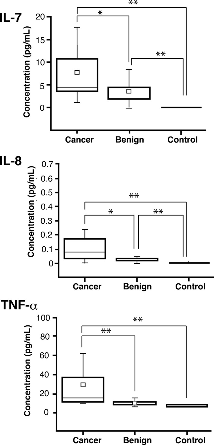
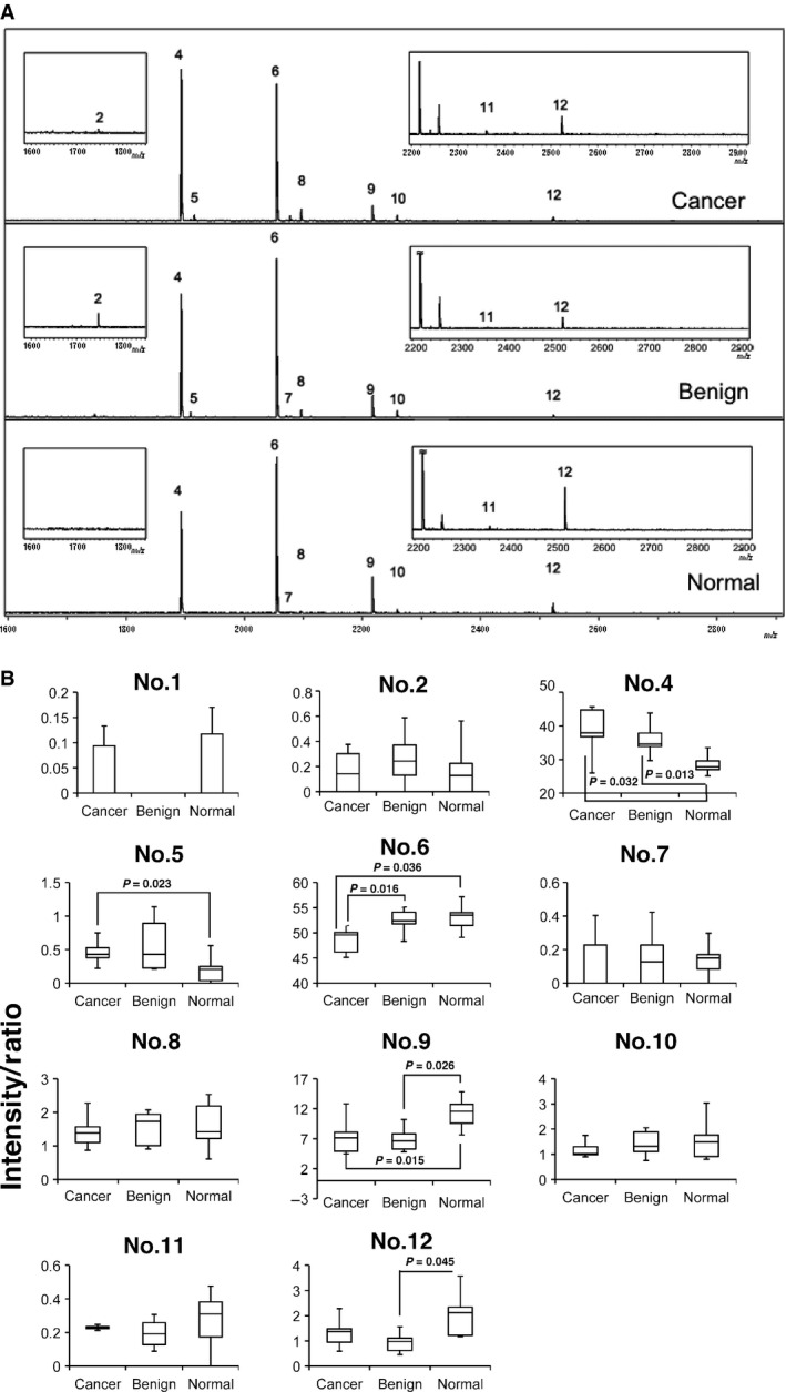
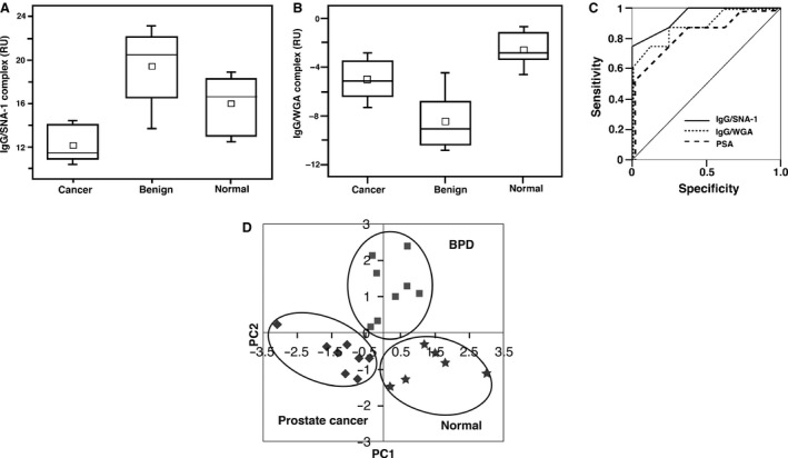
Occurrences of high values in patients with benign prostate disease and low values in patients with highly suspicious cancer have diminished the trustworthiness of prostate-specific antigen as an early diagnostic marker of prostate cancer. In the search for other complimentary markers, we focused on serum IgG from patients with prostate diseases as well as normal subjects. IgG purified from the sera of normal control subjects and patients with prostate diseases, was digested with peptide N-glycanase. Released glycans were quantified using MALDI-time of flight mass spectrometry. We report that N-linked (N-acetylhexosamine)2 (deoxyhexose)(mannose)3 (N-acetylglucosamine)2 was significantly increased in the IgG heavy chains of patients with prostate cancer compared with that of either benign prostatic disease patients or healthy subjects, whereas (hexose)(N-acetylhexosamine)2 (deoxyhexose)(mannose)3 (N-acetylglucosamine)2 was more abundant in the heavy chains of healthy subjects and benign prostatic disease patients. Thus, an absence of the terminal hexose of N-linked glycans has been closely connected to the progression of prostate cancer. Furthermore, surface plasmon resonance analyses have revealed that IgG from patients with prostate cancer has a decreased binding for Sambucus nigra lectin, compared with that from the benign prostatic disease patients or from normal subjects, suggesting lower levels of (N-acetylneuraminic acid)(α2-6)galactose/N-acetylgalactosamine groups in the N-linked glycans of patient IgG. Meanwhile, wheat germ agglutinin binding to IgG of the cancer group was significantly larger than that for the benign prostatic disease group but smaller than that for normal subjects. Our study indicates that the glycosylation changes in IgG can become useful diagnostic parameters for prostate cancer.

Keywords: IgG; N-linked glycan; PSA; mass spectrometry; prostate cancer.


Figures

Figure 1
Figure 1
Box graphs of (interleukin,IL) IL‐7, IL‐8, and tumor necrosis factor (TNF)‐α concentrations in serum samples determined by Luminex 200 multiplex beads assay. The small open square represents an average. The horizontal line in the box represents the median value (Cancer: n = 17, Benign: n = 17, Normal: n = 5). *< 0.05, **< 0.01.
Figure 2
Figure 2
Quantification of N‐linked glycans associated with serum IgG heavy chains from patients with prostate cancer, patients with benign prostatic diseases, and normal healthy subjects. (A). MALDI‐TOF mass spectra showing N‐glycan profiles of serum IgG from patient with prostate cancer (Cancer), patient with benign prostatic disease (Benign), and normal healthy subject (Normal). The data shown are representative of five separate experiments in each experimental group. (B). Quantitative analysis of MALDI‐TOF mass spectra. Quantification was performed with separated 12 peaks (= 5). Note that two glycans, No. 4 and No. 6, which were consistently found associated with IgG heavy chains, correspond to [(HexNAc)2(Deoxyhexose)1 + (Man)3(GlcNAc)2] and [(Hex)1(HexNAc)2(Deoxyhexose)1 + (Man)3(GlcNAc)2], respectively. Although peaks 1, 3, and 13 were detected mechanically, their content (intensity) was too small to quantify. The intensity/ratio as described in reference 17 was calculated for each experimental group.
Figure 3
Figure 3
The distribution of the amount of IgG bound with SNA‐1 (A) or wheat germ hemagglutinin (WGA) (B) in the sera of patients, as measured by surface plasmon resonance (SPR) analysis. The small open square represents an average. The horizontal line in the box represents the median value. In the case of IgG bound with SNA‐1 (A), there was a significant difference between the prostate cancer group and the control or BPD (benign prostatic disease) groups with P values of 0.0002 and 0.016, respectively (n = 12 for prostate cancer group, n = 13 for BPD group, n = 7 for normal control subjects). In the case of IgG bound with WGA (B), the mean RU of the cancer group was significantly larger than that of BPD (benign prostatic disease) with a P value of 0.03, but smaller than that of normal subjects with a P value of 0.003 (n = 9 for prostate cancer group, n = 9 for BPD group, n = 7 for normal control group). From the ROC curve (C), the area under the curve (AUC) value was 0.95 for IgG/SNA‐1, represented as a solid line, and was 0.88 for IgG/WGA, represented as a dotted line. Meanwhile, the AUC value for prostate‐specific antigen (PSA), a conventional diagnostic marker, represented as a broken line, was 0.84. The results of the principal components analysis (PCA) were plotted (D). The symbols represent the following: the diamond; prostate cancer patients (n = 8), the square; benign prostatic disease (BPD) patients (n = 8), the star; the normal subjects (n = 6). The four elements consisting of IgG‐SNA‐1, IgG‐WGA, haptglobin‐SNA‐1 and PSA clearly segregate the cancer patients, the BPD patients, and the normal subjects.

Similar articles

Cited by

References

    1. Candido, J. , and Hagemann T.. 2013. Cancer‐related inflammation. J. Clin. Immunol. 33 Suppl1:S79–S84. - PubMed
    1. Gabay, C. , and Kushner I.. 1999. Acute‐phase proteins and other systemic responses to inflammation. N. Engl. J. Med. 340:448–464. - PubMed
    1. Chen, Y. , Lim B. K., Peh S. C., et al. 2008. Profiling of serum and tissue high abundance acute‐phase proteins of patients with epithelial and germ line ovarian carcinoma. Proteome Sci. 6:20. - PMC - PubMed
    1. Peracaula, R. , Sarrats A., and Rudd P. M.. 2010. Liver proteins as sensor of human malignancies and inflammation. Proteomics Clin. 4:426–431. - PubMed
    1. Gornik, O. , and Lauc G.. 2008. Glycosylation of serum proteins in inflammatory diseases. Dis. Markers 28:267–278. - PMC - PubMed

Publication types

MeSH terms