. 2016 May;214(5):625.e1-625.e11.
doi: 10.1016/j.ajog.2016.01.194. Epub 2016 Feb 12.

Alterations in expression of imprinted genes from the H19/IGF2 loci in a multigenerational model of intrauterine growth restriction (IUGR)

Affiliations

Alterations in expression of imprinted genes from the H19/IGF2 loci in a multigenerational model of intrauterine growth restriction (IUGR)

Pablo Gonzalez-Rodriguez et al. Am J Obstet Gynecol. 2016 May.

Abstract

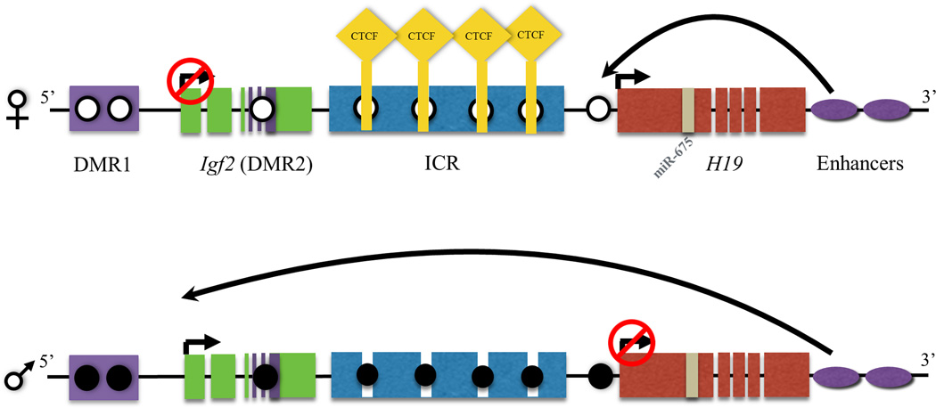
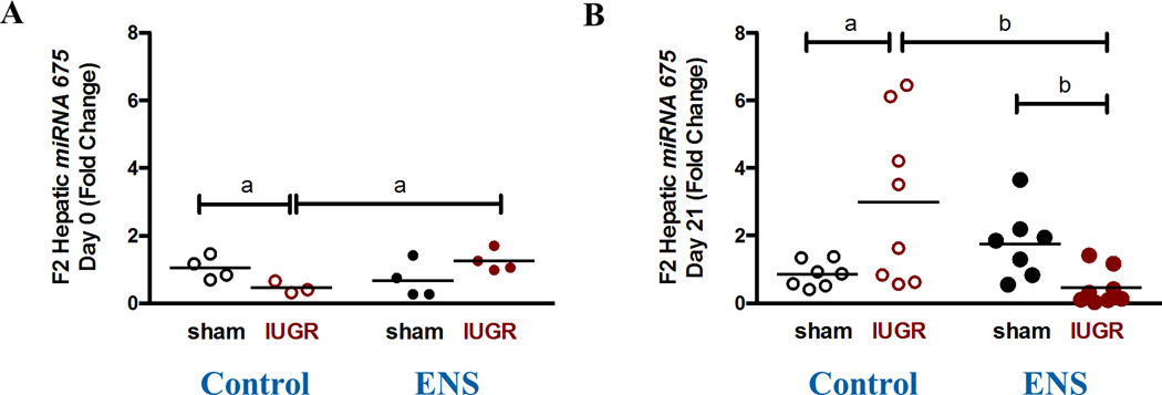
Background: The H19/IGF2 imprinted loci have attracted recent attention because of their role in cellular differentiation and proliferation, heritable gene regulation, and in utero or early postnatal growth and development. Expression from the imprinted H19/IGF2 locus involves a complex interplay of 3 means of epigenetic regulation: proper establishment of DNA methylation, promoter occupancy of CTCF, and expression of microRNA-675. We have demonstrated previously in a multigenerational rat model of intrauterine growth restriction the epigenetic heritability of adult metabolic syndrome in a F2 generation. We have further demonstrated abrogation of the F2 adult metabolic syndrome phenotype with essential nutrient supplementation of intermediates along the 1-carbon pathway and shown that alterations in the metabolome precede the adult onset of metabolic syndrome. The upstream molecular and epigenomic mediators underlying these observations, however, have yet to be elucidated fully.

Objective: In the current study, we sought to characterize the impact of the intrauterine growth-restricted lineage and essential nutrient supplementation on both levels and molecular mediators of H19 and IGF2 gene expression in the F2 generation.

Study design: F2 intrauterine growth-restricted and sham lineages were obtained by exposing P1 (grandmaternal) pregnant dams to bilateral uterine artery ligation or sham surgery at gestational day 19.5. F1 pups were allocated to the essential nutrient supplemented or control diet at postnatal day 21, and bred at 6-7 weeks of age. Hepatic tissues from the resultant F2 offspring at birth and at weaning (day 21) were obtained. Bisulfite modification and sequencing was employed for methylation analysis. H19 and IGF2 expression was measured by quantitative polymerase chain reaction. Promoter occupancy was quantified by the use of chromatin immunoprecipitation, or ChIP, against CTCF insulator proteins.

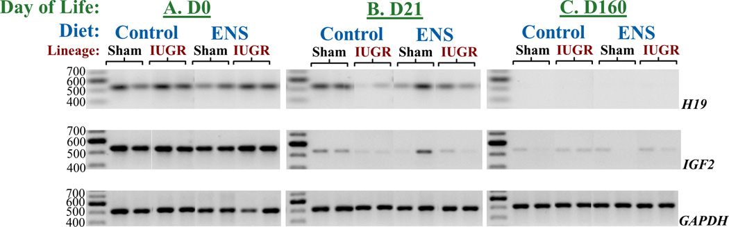
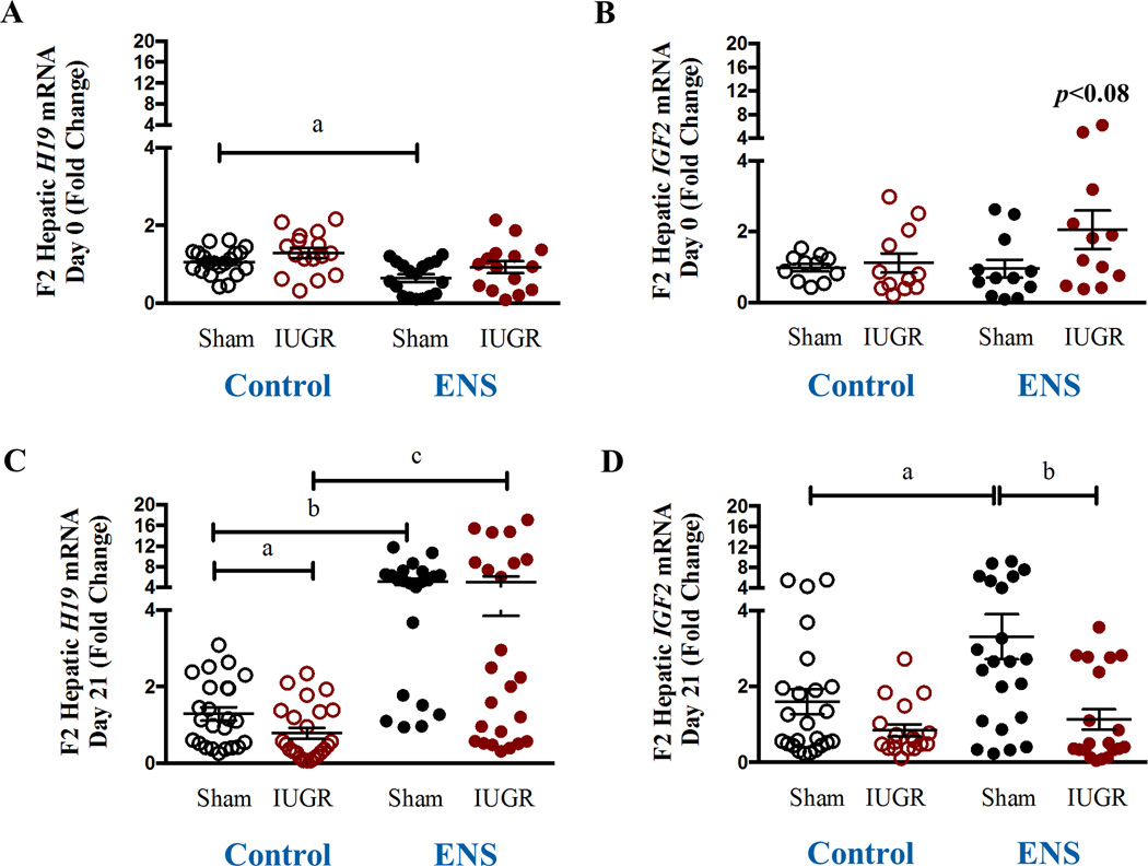
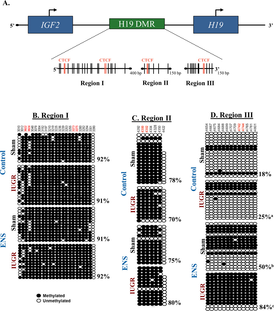
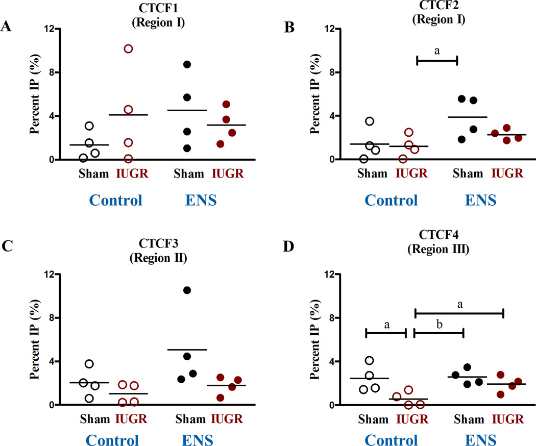
Results: Growth-restricted F2 on control diet demonstrated significant down-regulation in H19 expression compared with sham lineage (0.7831 vs 1.287; P < .05); however, essential nutrient supplementation diet abrogates this difference (4.995 vs 5.100; P > .05). Conversely, Igf2 was up-regulated by essential nutrient supplemented diet on the sham lineage (2.0 fold, P = .01), an effect that was not observed in the growth restricted offspring. A significant differential methylation was observed in the promoter region of region H19 among the intrauterine growth-restricted lineage (18% vs 25%; P < .05) on a control diet, whereas the essential nutrient supplemented diet was alternately associated with hypermethylation in both lineages (sham: 50%; intrauterine growth restriction: 84%, P < .05). Consistent with essential nutrient supplementation impacting the epigenome, a decrease of CTCF promoter occupancy was observed in CTCF4 of the growth restricted lineage (2.45% vs 0.56%; P < .05) on the control diet, an effect that was repressed with essential nutrient supplementation.

Conclusion: Heritable growth restriction is associated with changes in H19 gene expression; these changes are reversible with diet supplementation to favorably impact adult metabolic syndrome.

Keywords: CTCF; H19; IUGR; epigenomics; histone modifications; imprinting; insulin-like growth factor 2; miR-675.


Conflict of interest statement

Disclosure statement. The author(s) report(s) no conflict of interest.

Figures

Figure 1
Figure 1. Insulator protein and epigenetic regulation on Igf2/H19 gene cluster
In the mammalian genome the Igf2 and H19 are co-localized and undergo interdependent expression regulated by imprinting. Igf2 gene is transcribed from the paternal allele, while the non-coding H19 RNA is transcribed from the maternal allele. The regulation of expression from this locus involves a complex interplay of three specific epigenomic regulators, namely through DNA methylation, promoter occupancy of CTCF and expression of miR675. Epigenetic regulation is observed in this process where a demethylated (open circles) imprinted control region (ICR) binds CTCF (yellow diamond) to block the interaction of the downstream enhancers (purple ovals) with the promoter region of Igf2, thereby repressing the transcription of this gene, while the enhancers instead induce the transcription of H19. On the paternal allele, methylation (filled circle) on the ICR blocks the binding of CTCF allowing the interaction of the enhancers with the promoter of Igf2 facilitating gene transcription, while H19 is repressed due to methylation of its promoter region. miR675 is a microRNA expressed from exon one in the H19 gene.
Figure 2
Figure 2. Qualitative transcriptional changes of H19 and IGF2 by IUGR lineage and ENS supplemented diet
H19 and IGF2 cDNA expression from both sham surgery and IUGR lineages on day of life 0, 21, and 160 in animals fed either a control or ENS diet. Both H19 and IGF2 expression is strongest during development. Expression decreasing significantly by weaning (D21) and nearly absent in adulthood (D160). At day 21, animals who are destined to develop metabolic syndrome have low H19 expression, while ENS rescued this diminished expression.
Figure 3
Figure 3. Quantiative H19 and IGF2 gene expression in association with IUGR lineage and ENS supplemented diet
Expression of imprinted genes H19 and IGF2 from fetal rat liver was determined via real time PCR for (A) H19 D0 (a: 1.06±0.07 vs 0.8±0.1; p< 0.002, N=18–20) (B) IGF2 D0, (C) H19 D21 (a: 1.2±0.2 vs 0.7±0.1; p< 0.05, N=24; b: 1.2±0.2 vs 5.1±0.7; p < 0.0001, N22–24; c: 0.7±0.1 vs 5.1±0.7; p< 0.001, N=24) and (D) IGF2 D21 (a: 1.6 ± 0.3 vs 3.3 ± 0.6, N=22–24, p=0.01; b: P3.3 ± 0.6 vs 1.1 ± 0.3, N=20–22, p=0.002) in both sham and IUGR lineages on control versus ENS diet.
Figure 4
Figure 4. Altered DNA methylation of the H19 differentially methylated regions (DMR) occurs among IUGR lineage rats
(A) Three areas of the H19 DMR imprinting control region analyzed by are depicted. Vertical lines indicate CpG dinucleotides while red lines indicate the CpG falls within a CTCF finding site. (B–C) Methylation status was determined by sequencing cloned DNA following bisulfite treatment. The various regions of and their methylation status is depicted. While no changes were found in regions I and II, region III showed significant methylation changes at every site analyzed (p<0.02, Chi squared). Overall, IUGR induced a 40% increase in methylation, while IUGR fed ENS diet was increased 460%. (a, p<0.05; b, p<0.01; c, p<0.001).
Figure 5
Figure 5. CTCF promoter occupancy altered in IUGR lineage rats
Chromatin immunoprecipitation (ChIP) compared to a mock IgG was carried out for the various CTCF binding sites. A 77% decreased occupancy of CTCF at insulator site 4 (a: 2.4±0.6 vs 0.6±0.3, N=4, p<0.01) was induced by IUGR linage in D21 rats.
Figure 6
Figure 6. Expression of miRNA-675
The expression of miR-675 was determine by real time PCR for (A) D0 and (B) D21 (a: 1.7±0.4 vs 0.4±0.2, N=7–8, p=0.008). Its repression was decreased in IUGR animals fed an ENS diet at D21.

Similar articles

Cited by

References

    1. Gong L, Pan YX, Chen H. Gestational low protein diet in the rat mediates Igf2 gene expression in male offspring via altered hepatic DNA methylation. Epigenetics. 2010;5(7):619–626. - PubMed
    1. Sohi G, Marchand K, Revesz A, Arany E, Hardy DB. Maternal protein restriction elevates cholesterol in adult rat offspring due to repressive changes in histone modifications at the cholesterol 7alpha-hydroxylase promoter. Mol Endocrinol. 2011;25(5):785–798. - PMC - PubMed
    1. Suter M, Bocock P, Showalter L, Hu M, Shope C, McKnight R, Grove K, Lane R, Aagaard-Tillery K. Epigenomics: maternal high-fat diet exposure in utero disrupts peripheral circadian gene expression in nonhuman primates. FASEB J. 2011;25(2):714–726. - PMC - PubMed
    1. Zheng S, Rollet M, Pan YX. Maternal protein restriction during pregnancy induces CCAAT/enhancer-binding protein (C/EBPbeta) expression through the regulation of histone modification at its promoter region in female offspring rat skeletal muscle. Epigenetics. 2011;6(2):161–170. - PubMed
    1. Suter MA, Takahashi D, Grove KL, Aagaard KM. Postweaning exposure to a high-fat diet is associated with alterations to the hepatic histone code in Japanese macaques. Pediatr Res. 2013;74(3):252–258. - PMC - PubMed

Publication types

MeSH terms