. 2016 Apr 15;76(8):2125-36.
doi: 10.1158/0008-5472.CAN-15-1490. Epub 2016 Feb 15.

Myeloid-Derived Suppressor Cells Express Bruton's Tyrosine Kinase and Can Be Depleted in Tumor-Bearing Hosts by Ibrutinib Treatment

Affiliations

Myeloid-Derived Suppressor Cells Express Bruton's Tyrosine Kinase and Can Be Depleted in Tumor-Bearing Hosts by Ibrutinib Treatment

Andrew Stiff et al. Cancer Res. .

Abstract

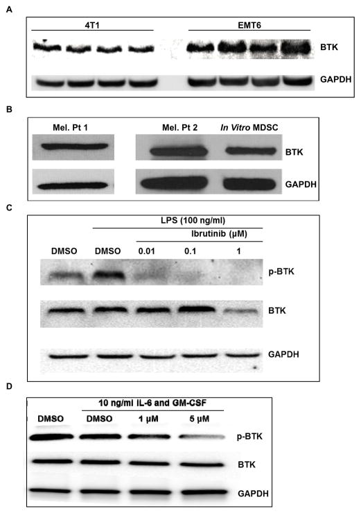
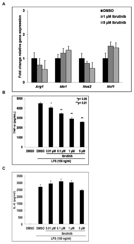
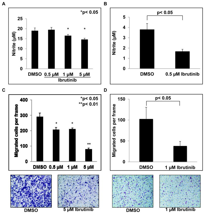
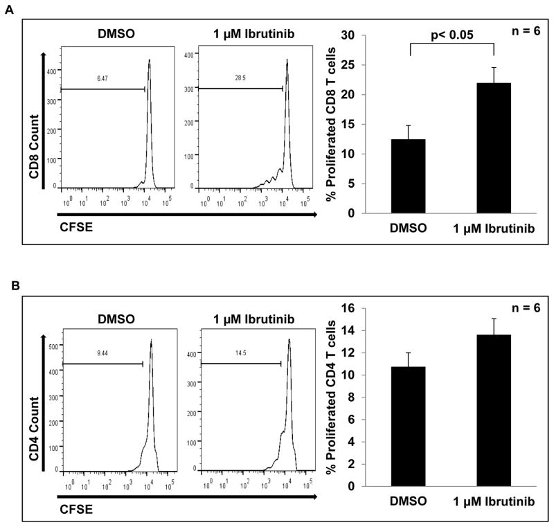
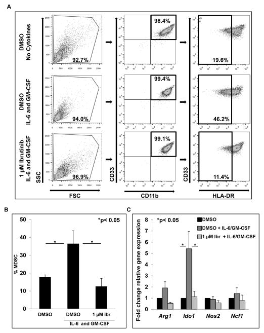
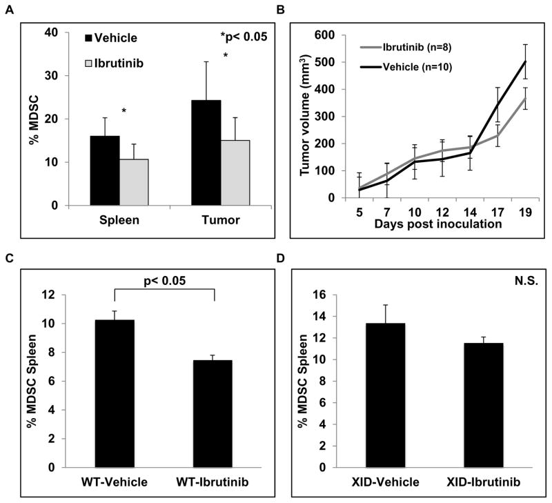
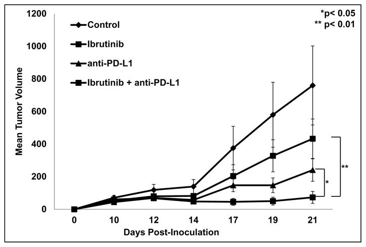
Myeloid-derived suppressor cells (MDSC) are a heterogeneous group of immature myeloid cells that expand in tumor-bearing hosts in response to soluble factors produced by tumor and stromal cells. MDSC expansion has been linked to loss of immune effector cell function and reduced efficacy of immune-based cancer therapies, highlighting the MDSC population as an attractive therapeutic target. Ibrutinib, an irreversible inhibitor of Bruton's tyrosine kinase (BTK) and IL2-inducible T-cell kinase (ITK), is in clinical use for the treatment of B-cell malignancies. Here, we report that BTK is expressed by murine and human MDSCs, and that ibrutinib is able to inhibit BTK phosphorylation in these cells. Treatment of MDSCs with ibrutinib significantly impaired nitric oxide production and cell migration. In addition, ibrutinib inhibited in vitro generation of human MDSCs and reduced mRNA expression of indolamine 2,3-dioxygenase, an immunosuppressive factor. Treatment of mice bearing EMT6 mammary tumors with ibrutinib resulted in reduced frequency of MDSCs in both the spleen and tumor. Ibrutinib treatment also resulted in a significant reduction of MDSCs in wild-type mice bearing B16F10 melanoma tumors, but not in X-linked immunodeficiency mice (XID) harboring a BTK mutation, suggesting that BTK inhibition plays an important role in the observed reduction of MDSCs in vivo Finally, ibrutinib significantly enhanced the efficacy of anti-PD-L1 (CD274) therapy in a murine breast cancer model. Together, these results demonstrate that ibrutinib modulates MDSC function and generation, revealing a potential strategy for enhancing immune-based therapies in solid malignancies. Cancer Res; 76(8); 2125-36. ©2016 AACR.


Conflict of interest statement

The authors have no conflicts of interest to disclose.

Figures

Figure 1
Figure 1. Murine and human MDSC express BTK and its phosphorylation is inhibited by ibrutinib
(A) Immunoblot showing BTK and GAPDH expression in MDSC isolated from the spleen of mice bearing 4T1 and EMT6 mammary carcinoma tumors. (B) Immunoblot of BTK and GAPDH expression in human MDSC isolated from patients with metastatic melanoma and in vitro generated MDSC. Results displayed are from two separate immunoblots. (C) Immunoblot showing ibrutinib reduces the level of phosphorylated BTK (p-BTK) in LPS stimulated MSC2 cells. (D) In vitro generated MDSC were treated with DMSO or ibrutinib for 1 hour followed by stimulation with IL-6 and GM-CSF. Lysates were collected 15 minutes after stimulation and probed for expression of p-BTK, BTK, and GAPDH.
Figure 2
Figure 2. Effect of ibrutinib on gene expression and cytokine production by activated MSC2 cells
(A) Effect of ibrutinib on expression of Arg1, Ido1, Nos2, and Ncf1 by LPS activated MSC2 cells. Total RNA was collected 24 hours later and expression of Arg1, Ido1, Nos2, and Ncf1 determined by qRT-PCR. Values displayed are means ± SE from three independent experiments. (B and C) After 24 hours the supernatants of cells treated as in Fig. 2A were collected and the levels of TNF-α and IL-6 were measured by flow cytometry using a cytokine bead assay. Values displayed are means ± SE from three independent experiments.
Figure 3
Figure 3. Ibrutinib reduces MDSC NO production and migraton
(A) MSC2 cells were treated with DMSO or ibrutinib followed by stimulation with LPS. Supernatants were collected after 24 hrs and nitrite levels were measured using Griess reagent. Values displayed are means ± SE from three independent experiments, *p< 0.05. (B) MDSC isolated from metastatic melanoma patients were treated with DMSO or ibrutinib for 1 hour. After treatment, MDSC were cultured with 10 ng/ml IL-6 and 10 ng/ml GM-CSF for 48 hours and nitrite measured as above. Values represent mean ± SE from three patients, p< 0.05. (C) MSC2 cells were treated with DMSO or ibrutinib for one hour. Media conditioned by the EMT6 cell line served to stimulate migration. Values represent mean ± SE from three independent experiments. (D) MDSC isolated from patients with melanoma were treated with DMSO or ibrutinib. 200 ng/ml GM-CSF was used to stimulate chemotaxis and PBS supplemented media served as a negative control. Migration proceeded for 12 hrs. Values displayed are means ± SE from 3 patients. Representative images are given below migration results. Migrated cells are stained purple against the light blue transwell insert.
Figure 4
Figure 4. Ibrutinib reduces MDSC suppression of T cell proliferation
DMSO or ibrutinib treated MDSC were co-cultured with anti-CD3/CD28 bead activated T cells labelled with CFSE at a 2:1 ratio. After three days cells were collected and stained with anti-CD8 (A) and anti-CD4 (B) antibodies and proliferation assessed by flow cytometry. Histograms show results from one representative experiment. Bar graphs represent means ± SE from six independent experiments, p< 0.05 for CD8 T cells.
Figure 5
Figure 5. Ibrutinib inhibits in vitro generation of human MDSC
Monocytes were cultured for 6 days in the three different conditions. (A) Gating strategy and flow panels from a representative experiment. (B) Percent MDSC in different culture conditions after 6 days. Values represented are means ± SE from three independent experiments. MDSC were identified by a CD33+/CD11b+/HLA-DRlow phenotype, *p<0.05. (C) Total RNA was collected on day 6 of monocyte cultures and expression of Arg1, Ido1, Nos2, and Ncf1 was determined by qRT-PCR. Values represent mean ± SE from three independent experiments, *p< 0.05 for Ido1.
Figure 6
Figure 6. Ibrutinib reduces MDSC frequency in vivo
Eight mice were included in the ibrutinib treatment group and ten mice in the vehicle control group. (A) Splenocytes and single cell suspensions of the tumors were stained with GR-1 and CD11b antibodies. Values are the mean ± SE of GR-1+/CD11b+ MDSC in the spleen and tumor, *p< 0.05. (B) Tumor volumes were measured three times weekly. Values are the mean ± SE of tumor volumes at each time point. (C) Wild type C57BL/6 mice were inoculated with 1×10⁵ B16F10 melanoma cells subcutaneously. After two weeks of treatment mice were sacrificed and the frequency of GR-1+/CD11b+ MDSC in spleen was measured by flow cytometry. Values represented are means ± SE of MDSC from 5 mice for each treatment group, p< 0.05. (D) BTK mutant C57BL/6 XID mice were inoculated with 1×10⁵ B16F10 melanoma cells subcutaneously. After two weeks of treatment mice were sacrificed and the frequency of GR-1+/CD11b+ MDSC in spleen was measured by flow cytometry. Values represented are means ± SE of MDSC from 5 mice for each treatment group, p = 0.73.
Figure 7
Figure 7. Ibrutinib improves anti-PD-L1 immune checkpoint blockade
Once tumors were palpable (5mm diameter) mice were divided into 4 treatment groups: vehicle/IgG control, ibrutinib (25 mg/kg daily), anti-PD-L1 (100 μg per mouse MWF), or the combination of ibrutinib and anti-PD-L1 therapy. 9–10 mice were included in each treatment group. Tumor volumes were measured three times weekly with digital calipers. Treatment with the combination of ibrutinib and anti-PD-L1 resulted in a significant reduction in tumor growth compared to treatment with ibrutinib (**p< 0.01) or anti-PD-L1 (*p<0.05) alone.

References

    1. Lechner MG, Liebertz DJ, Epstein AL. Characterization of cytokine-induced myeloid-derived suppressor cells from normal human peripheral blood mononuclear cells. Journal of immunology (Baltimore, Md : 1950) 2010;185(4):2273–84. - PMC - PubMed
    1. Melani C, Chiodoni C, Forni G, Colombo MP. Myeloid cell expansion elicited by the progression of spontaneous mammary carcinomas in c-erbB-2 transgenic BALB/c mice suppresses immune reactivity. Blood. 2003;102(6):2138–45. - PubMed
    1. Gabrilovich DI, Nagaraj S. Myeloid-derived suppressor cells as regulators of the immune system. Nature reviews Immunology. 2009;9(3):162–74. - PMC - PubMed
    1. Gabrilovich DI, Ostrand-Rosenberg S, Bronte V. Coordinated regulation of myeloid cells by tumours. Nature reviews Immunology. 2012;12(4):253–68. - PMC - PubMed
    1. Trikha P, Carson WE., 3rd Signaling pathways involved in MDSC regulation. Biochimica et biophysica acta. 2014;1846(1):55–65. - PMC - PubMed

Publication types