

Review
doi: 10.1155/2016/7651047.
Epub 2016 Jan 3.
Chalcone Scaffold in Anticancer Armamentarium: A Molecular Insight
Affiliations
- PMID: 26880913
- PMCID: PMC4735904
- DOI: 10.1155/2016/7651047
Item in Clipboard
Review
Chalcone Scaffold in Anticancer Armamentarium: A Molecular Insight
J Toxicol.
2016.
Abstract
Cancer is an inevitable matter of concern in the medicinal chemistry era. Chalcone is the well exploited scaffold in the anticancer domain. The molecular mechanism of chalcone at cellular level was explored in past decades. This mini review provides the most recent updates on anticancer potential of chalcones.
Figures
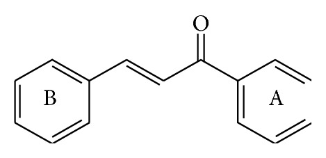
Chemical structure of chalcone.
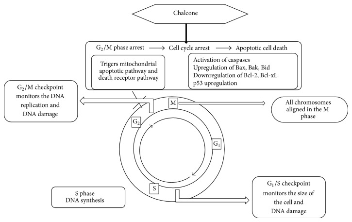
Action of chalcone on cell cycle.

Chalcones against leukemia cell lines. (1) CC50 of 30 μM, (2) CC50 of 40 μM, and (3) CC50 of 40 μM: increases proapoptotic proteins Bax, Bid, and Bak (only chalcone 2) and decreases antiapoptotic Bcl-2 expression. Caspase-9 and caspase-12 activation increases CHOP expression resulting in induction of ER stress. (4) IC50 values (4 to 8 μM) and (5) IC50 values (4 to 8 μM): induction of apoptosis through the reduction of the mitochondrial membrane potential, reduction in Bcl-2 expression, increase in Bax expression, and increase in active caspase-3. ((6a) to (6c)) IC50s between 12 and 85 μM for HDAC isoenzymes, inhibiting total HDAC activity by 20–50% at 100 μM: histone deacetylases (HDACs) inhibitor. (7) IC50 of 13.7 ± 0.8 μM: caspase-3 and caspase-7 activation.
Chalcones against colon cancer cell lines. (8) IC50 of 14.5 ± 1.1 μM: increases proapoptotic proteins Bax, Bid, and Bak (only chalcone 2) and decreases antiapoptotic Bcl-2 expression. Caspase 9 and caspase 12 activation increases CHOP expression resulting in induction of ER stress. (9) IC50 of 4.39 μM: increases the death receptors expression (TRAIL-R1 and TRAIL-R2) (Tumor necrosis factor- (TNF-) related apoptosis-inducing ligand) and proapoptotic markers (p21, Bad, Bim, Bid, Bax, Smac, caspase-3, and caspase-8) as well as reducing the antiapoptotic markers (livin, XIAP, and HSP27).
Chalcones against cervix adenocarcinoma cell lines. ((10a) to (10c)) IC50 values ranging from 2.36 to 2.73 μM: increases p18 Bax, accumulation of cells in S, and G2/M phases. (11) IC50 of 4.7 to 7.6 μM: induction of caspase-dependent intrinsic pathway p53 and its transcriptional target PUMA upregulation.
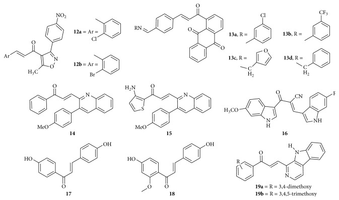
Chalcones against lung cancer and breast cancer cell lines. (12a) IC50 values of 1.35–2.07 μM and (12b) IC50 values of 7.27–11.07 μM: upregulation of the DR5; extrinsic pathway of apoptosis. ((13a) to (13d)) IC50 values ranging from 1.76 to 6.11 μM: inhibited tubulogenesis, caspase-3 and caspase-8 activation, and inhibitory potential against matrix metalloproteinases (MMP-2) secretion. (14) IC50 values of 1.41 and 0.70 μM: cell cycle arrest at G2/M phase, activation of caspase-3, and cleavage of PARP. (15) IC50 value of less than 0.10 μM: cell cycle arrest at G2/M phase, activation of caspase-3, and cleavage of PARP. (16) IC50 of 0.8 μM: enhances tubulin polymerization suggesting the activity as microtubule stabilizing agents. (17) IC50 of 0.16 to 0.44 μM and (18) IC50 of 0.56 to 0.65 μM: suppress the activation of NF-κB pathway. (19a) IC50 of 2.25 μM and (19b) IC50 of 3.29 μM: induced DNA fragmentation and apoptosis.
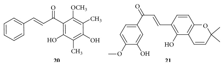
Chalcones against hepatocellular carcinoma cell lines. (20) IC50 of 6.25 μM: mitochondria dependent pathway involving inhibition of Bcl-2 expression leading to disintegration of the outer mitochondrial membrane. Caspase-3 and caspase-9 activities. (21) IC50 of 10 μM: hypoxia inducible factor-1 (HIF-1) inhibitor.
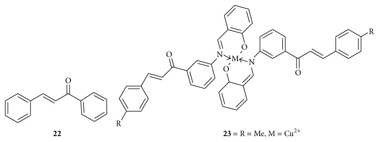
Chalcones against bladder and prostate cancer cell lines. (22) Concentration of 6 μg/mL: reduction in the expression of cyclin A and cyclin B1, decrease in the expression of Cdc2 and an increase in the expression of p21 and p27, increase in the expression of proapoptotic proteins Bax and Bak, and decrease in the expression of antiapoptotic proteins Bcl-2, Bcl-xL proteins; NF-κB activation by increasing the expression of IκBα in cytoplasm leads to apoptosis. (23) IC50 value of 5.95 μM: copper ions might act as penetration enhancers of chalcones into cancer cells and might act as inhibitors of drug efflux proteins.
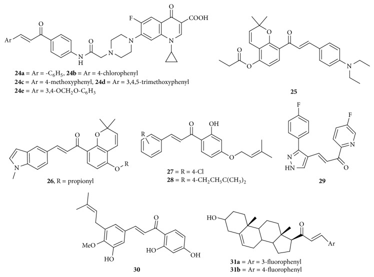
Chalcones against multiple cancer cell lines. ((24a) to (24c)) GI50 of 0.21 to 57.6 μM: topoisomerase-I and topoisomerase-II inhibition. (25) IC50 of 0.15 to 0.52 μM: arrest cells in the G2/M phase of the cell cycle and inhibit the polymerization of tubulin. Binds into the colchicine binding site of tubulin (molecular docking study). (26) IC50 values of 0.22 to 1.80 μM: induced cell cycle arrest in G2/M phase and inhibited the polymerization of tubulin; binds at the colchicine binding site of tubulin (from molecular docking analysis). (27) IC50 value of 0.89 μmol/L: cell division cycle 25 (CDC25) inhibitor. (28) IC50 values of 1.76 μmol/L: cell division cycle 25 (CDC25) inhibitor. (29) IC50 value of 0.01 μg/mL and 0.04 μg/mL: molecular mechanism not studied. (30) IC50 values of 6.4 μM and 8.5 μM: cytotoxic, molecular mechanism not studied. (31a) IC50 value of 1.02 μM and 0.79 μM and (31b) IC50 value of 0.81 μM and 1 μM: cytotoxic.
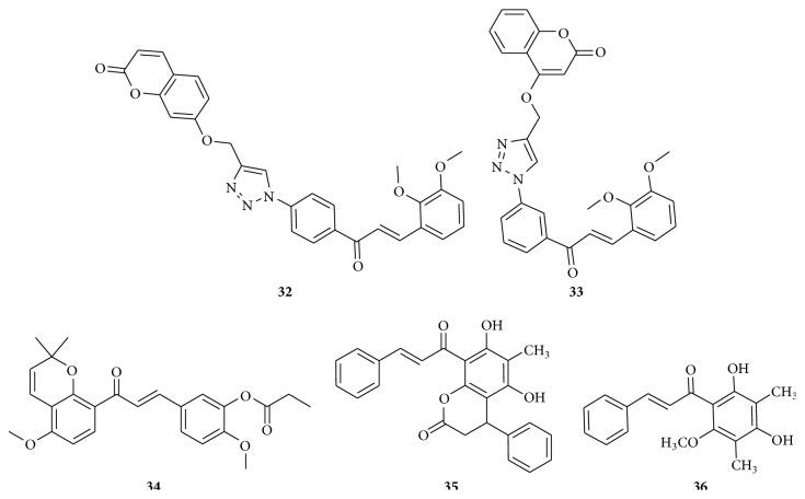
Chalcones against multiple cancer cell lines. (32) IC50 = 0.53 μM and (33) IC50 value of 8.18 μM: could snugly occupy the colchicine binding site of β-tubulin (molecular docking study). (34) Concentration of 0.2 μM: cell cycle arrest in G2/M phase promoted tubulin polymerization into microtubules and caused microtubule stabilization similar to paclitaxel. (35) IC50 values of 1.60 μM and (36) IC50 values of 2.82 μM: inhibition of clonogenicity.
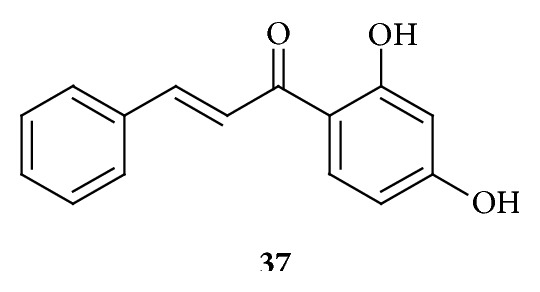
Chalcone obtained by using a fission yeast bioassay. (37) Concentration of 8 μM to 20 μM induces DNA damage which blocks cell cycle progression at the G2/M transition in a Rad3-dependent and Chk1-dependent manner.
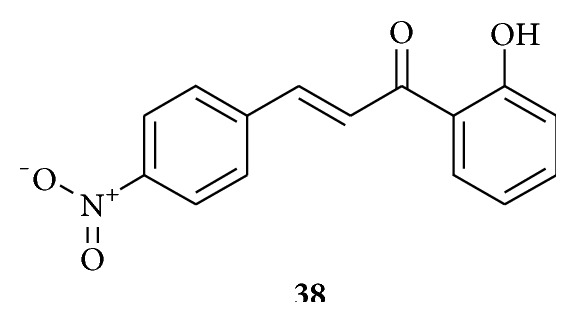
Chalcone against cathepsin B and cathepsin H obtained from goat liver. (38) K
i values of 6.18 × 10⁻⁸ M for cathepsin B. K
i values of 2.8 × 10⁻⁷ M for cathepsin H: cathepsin B and cathepsin H inhibitor.
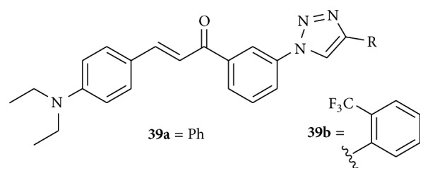
Chalcones against brain cancer cell line. ((39a) and (39b)) cotreatment of 10 μM with TRAIL (25 ng/mL): TRAIL sensitizers.
Chalcone against multidrug resistant cancer cell lines. (40) IC50 value of 2.54 μM: arresting the cell cycle between G0/G1 phases, which is via the strong induction of apoptosis by disrupting mitochondrial membrane potential (MMP) and an increased production of reactive oxygen species (ROS). (41) Combined effects of 36 and Tipifarnib reduce the IC50 value of Tipifarnib from 9.0 μM (applied alone) to 3.8 μM: increased the expression of HIF-1α. (42) GI50 of 3.87 μM and (43) GI50 of 3.95 μM: trigger cell cycle arrest at the G2/M phase and apoptotic cell death, aurora A kinase inhibitor.
Similar articles
-
Heterocyclic chalcone analogues as potential anticancer agents.Anticancer Agents Med Chem. 2013 Mar;13(3):422-32. Anticancer Agents Med Chem. 2013. PMID: 22721390 Review.
-
Recent developments in biological activities of chalcones: a mini review.Eur J Med Chem. 2014 Oct 6;85:758-77. doi: 10.1016/j.ejmech.2014.08.033. Epub 2014 Aug 12. Eur J Med Chem. 2014. PMID: 25137491 Review.
-
Advances in chalcones with anticancer activities.Recent Pat Anticancer Drug Discov. 2015;10(1):97-115. doi: 10.2174/1574892809666140819153902. Recent Pat Anticancer Drug Discov. 2015. PMID: 25138130 Review.
-
Targeting triple negative breast cancer heterogeneity with chalcones: a molecular insight.J Drug Target. 2019 Sep;27(8):830-838. doi: 10.1080/1061186X.2018.1561889. Epub 2019 Feb 11. J Drug Target. 2019. PMID: 30582377 Review.
-
Exploring pharmacological significance of chalcone scaffold: a review.Curr Med Chem. 2012;19(2):209-25. doi: 10.2174/092986712803414132. Curr Med Chem. 2012. PMID: 22320299 Review.
Cited by
-
Multienzymatic biotransformation of flavokawain B by entomopathogenic filamentous fungi: structural modifications and pharmacological predictions.Microb Cell Fact. 2024 Feb 24;23(1):65. doi: 10.1186/s12934-024-02338-9. Microb Cell Fact. 2024. PMID: 38402203 Free PMC article.
-
The effect of novel nitrogen-based chalcone analogs on colorectal cancer cells: Insight into the molecular pathways.Heliyon. 2024 Feb 27;10(5):e27002. doi: 10.1016/j.heliyon.2024.e27002. eCollection 2024 Mar 15. Heliyon. 2024. PMID: 38463818 Free PMC article.
-
Chalcone: A Privileged Structure in Medicinal Chemistry.Chem Rev. 2017 Jun 28;117(12):7762-7810. doi: 10.1021/acs.chemrev.7b00020. Epub 2017 May 10. Chem Rev. 2017. PMID: 28488435 Free PMC article. Review.
-
Molecular targets and anticancer activity of quinoline-chalcone hybrids: literature review.RSC Adv. 2020 Aug 21;10(52):31139-31155. doi: 10.1039/d0ra05594h. eCollection 2020 Aug 21. RSC Adv. 2020. PMID: 35520674 Free PMC article. Review.
-
Excavating medicinal virtues of chalcones to illuminate a new scope in cancer chemotherapy.RSC Adv. 2025 Apr 14;15(15):11617-11638. doi: 10.1039/d5ra01280e. eCollection 2025 Apr 9. RSC Adv. 2025. PMID: 40230627 Free PMC article. Review.
References
-
- American Cancer Society. Global Cancer Facts & Figures. 2nd. Atlanta, Ga, USA: American Cancer Society; 2011.
-
- Alison M. R. Cancer. Encyclopedia of life sciences, Nature Publishing Group, 2011, http://www.els.net.
-
- Dhar D. N. The Chemistry of Chalcones and Related Compounds. New York, NY, USA: John Wiley & Sons; 1981.
Publication types
LinkOut - more resources
Full Text Sources
Other Literature Sources
Miscellaneous
