Longitudinal Cell Tracking and Simultaneous Monitoring of Tissue Regeneration after Cell Treatment of Natural Tendon Disease by Low-Field Magnetic Resonance Imaging
- PMID: 26880932
- PMCID: PMC4736965
- DOI: 10.1155/2016/1207190
Longitudinal Cell Tracking and Simultaneous Monitoring of Tissue Regeneration after Cell Treatment of Natural Tendon Disease by Low-Field Magnetic Resonance Imaging
Abstract
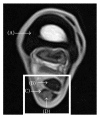
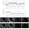
Treatment of tendon disease with multipotent mesenchymal stromal cells (MSC) is a promising option to improve tissue regeneration. To elucidate the mechanisms by which MSC support regeneration, longitudinal tracking of MSC labelled with superparamagnetic iron oxide (SPIO) by magnetic resonance imaging (MRI) could provide important insight. Nine equine patients suffering from tendon disease were treated with SPIO-labelled or nonlabelled allogeneic umbilical cord-derived MSC by local injection. Labelling of MSC was confirmed by microscopy and MRI. All animals were subjected to clinical, ultrasonographical, and low-field MRI examinations before and directly after MSC application as well as 2, 4, and 8 weeks after MSC application. Hypointense artefacts with characteristically low signal intensity were identified at the site of injection of SPIO-MSC in T1- and T2 (∗) -weighted gradient echo MRI sequences. They were visible in all 7 cases treated with SPIO-MSC directly after injection, but not in the control cases treated with nonlabelled MSC. Furthermore, hypointense artefacts remained traceable within the damaged tendon tissue during the whole follow-up period in 5 out of 7 cases. Tendon healing could be monitored at the same time. Clinical and ultrasonographical findings as well as T2-weighted MRI series indicated a gradual improvement of tendon function and structure.
Figures


Similar articles
-
In Vivo Magic Angle Magnetic Resonance Imaging for Cell Tracking in Equine Low-Field MRI.Stem Cells Int. 2019 Dec 17;2019:5670106. doi: 10.1155/2019/5670106. eCollection 2019. Stem Cells Int. 2019. PMID: 31933650 Free PMC article.
-
Long-Term Cell Tracking Following Local Injection of Mesenchymal Stromal Cells in the Equine Model of Induced Tendon Disease.Cell Transplant. 2016 Dec 13;25(12):2199-2211. doi: 10.3727/096368916X692104. Epub 2016 Jul 7. Cell Transplant. 2016. PMID: 27392888
-
Tracking of autologous adipose tissue-derived mesenchymal stromal cells with in vivo magnetic resonance imaging and histology after intralesional treatment of artificial equine tendon lesions--a pilot study.Stem Cell Res Ther. 2016 Feb 1;7:21. doi: 10.1186/s13287-016-0281-8. Stem Cell Res Ther. 2016. PMID: 26830812 Free PMC article.
-
Tracking mesenchymal stem cells using magnetic resonance imaging.Brain Circ. 2016 Jul-Sep;2(3):108-113. doi: 10.4103/2394-8108.192521. Epub 2016 Oct 18. Brain Circ. 2016. PMID: 30276283 Free PMC article. Review.
-
In vivo tracking of stem cells in brain and spinal cord injury.Prog Brain Res. 2007;161:367-83. doi: 10.1016/S0079-6123(06)61026-1. Prog Brain Res. 2007. PMID: 17618991 Review.
Cited by
-
Approaches to standardising the magnetic resonance image analysis of equine tendon lesions.Vet Rec Open. 2023 Feb 23;10(1):e257. doi: 10.1002/vro2.57. eCollection 2023 Jun. Vet Rec Open. 2023. PMID: 36846276 Free PMC article.
-
MSC in Tendon and Joint Disease: The Context-Sensitive Link Between Targets and Therapeutic Mechanisms.Front Bioeng Biotechnol. 2022 Apr 4;10:855095. doi: 10.3389/fbioe.2022.855095. eCollection 2022. Front Bioeng Biotechnol. 2022. PMID: 35445006 Free PMC article.
-
In Vivo Magic Angle Magnetic Resonance Imaging for Cell Tracking in Equine Low-Field MRI.Stem Cells Int. 2019 Dec 17;2019:5670106. doi: 10.1155/2019/5670106. eCollection 2019. Stem Cells Int. 2019. PMID: 31933650 Free PMC article.
-
Characterization of Equine Chronic Tendon Lesions in Low- and High-Field Magnetic Resonance Imaging.Vet Sci. 2022 Jun 15;9(6):297. doi: 10.3390/vetsci9060297. Vet Sci. 2022. PMID: 35737349 Free PMC article.
-
Tenogenic Properties of Mesenchymal Progenitor Cells Are Compromised in an Inflammatory Environment.Int J Mol Sci. 2018 Aug 28;19(9):2549. doi: 10.3390/ijms19092549. Int J Mol Sci. 2018. PMID: 30154348 Free PMC article.
References
-
- Dowling B. A., Dart A. J., Hodgson D. R., Smith R. K. W. Superficial digital flexor tendonitis in the horse. Equine Veterinary Journal. 2000;32(5):369–378. - PubMed
-
- Godwin E. E., Young N. J., Dudhia J., Beamish I. C., Smith R. K. W. Implantation of bone marrow-derived mesenchymal stem cells demonstrates improved outcome in horses with overstrain injury of the superficial digital flexor tendon. Equine Veterinary Journal. 2012;44(1):25–32. doi: 10.1111/j.2042-3306.2011.00363.x. - DOI - PubMed
-
- Smith R. K. W., Korda M., Blunn G. W., Goodship A. E. Isolation and implantation of autologous equine mesenchymal stem cells from bone marrow into the superficial digital flexor tendon as a potential novel treatment. Equine Veterinary Journal. 2003;35(1):99–102. - PubMed
LinkOut - more resources
Full Text Sources
Other Literature Sources

