

Mimicking Neural Stem Cell Niche by Biocompatible Substrates
- PMID: 26880934
- PMCID: PMC4736764
- DOI: 10.1155/2016/1513285
Mimicking Neural Stem Cell Niche by Biocompatible Substrates
Abstract
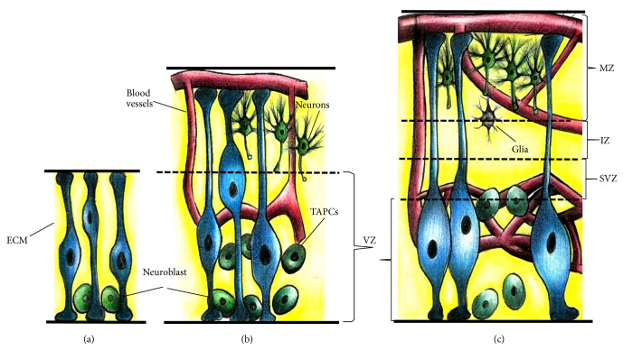
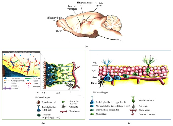
Neural stem cells (NSCs) participate in the maintenance, repair, and regeneration of the central nervous system. During development, the primary NSCs are distributed along the ventricular zone of the neural tube, while, in adults, NSCs are mainly restricted to the subependymal layer of the subventricular zone of the lateral ventricles and the subgranular zone of the dentate gyrus in the hippocampus. The circumscribed areas where the NSCs are located contain the secreted proteins and extracellular matrix components that conform their niche. The interplay among the niche elements and NSCs determines the balance between stemness and differentiation, quiescence, and proliferation. The understanding of niche characteristics and how they regulate NSCs activity is critical to building in vitro models that include the relevant components of the in vivo niche and to developing neuroregenerative approaches that consider the extracellular environment of NSCs. This review aims to examine both the current knowledge on neurogenic niche and how it is being used to develop biocompatible substrates for the in vitro and in vivo mimicking of extracellular NSCs conditions.
Figures
Similar articles
-
3D Reconstitution of the Neural Stem Cell Niche: Connecting the Dots.Front Bioeng Biotechnol. 2021 Oct 28;9:705470. doi: 10.3389/fbioe.2021.705470. eCollection 2021. Front Bioeng Biotechnol. 2021. PMID: 34778223 Free PMC article. Review.
-
Persistent Cyfip1 Expression Is Required to Maintain the Adult Subventricular Zone Neurogenic Niche.J Neurosci. 2020 Mar 4;40(10):2015-2024. doi: 10.1523/JNEUROSCI.2249-19.2020. Epub 2020 Jan 27. J Neurosci. 2020. PMID: 31988061 Free PMC article.
-
Grafted Subventricular Zone Neural Stem Cells Display Robust Engraftment and Similar Differentiation Properties and Form New Neurogenic Niches in the Young and Aged Hippocampus.Stem Cells Transl Med. 2016 Sep;5(9):1204-15. doi: 10.5966/sctm.2015-0270. Epub 2016 May 18. Stem Cells Transl Med. 2016. PMID: 27194744 Free PMC article.
-
Ontogeny of adult neural stem cells in the mammalian brain.Curr Top Dev Biol. 2021;142:67-98. doi: 10.1016/bs.ctdb.2020.11.002. Epub 2020 Dec 17. Curr Top Dev Biol. 2021. PMID: 33706926 Free PMC article. Review.
-
Area-Specific Regulation of Quiescent Neural Stem Cells by Notch3 in the Adult Mouse Subependymal Zone.J Neurosci. 2017 Dec 6;37(49):11867-11880. doi: 10.1523/JNEUROSCI.0001-17.2017. Epub 2017 Nov 3. J Neurosci. 2017. PMID: 29101245 Free PMC article.
Cited by
-
Ascorbic Acid Promotes the Stemness of Corneal Epithelial Stem/Progenitor Cells and Accelerates Epithelial Wound Healing in the Cornea.Stem Cells Transl Med. 2017 May;6(5):1356-1365. doi: 10.1002/sctm.16-0441. Epub 2017 Mar 9. Stem Cells Transl Med. 2017. PMID: 28276172 Free PMC article.
-
Microfluidic systems for stem cell-based neural tissue engineering.Lab Chip. 2016 Jul 5;16(14):2551-71. doi: 10.1039/c6lc00489j. Lab Chip. 2016. PMID: 27296463 Free PMC article. Review.
-
Fractone Stem Cell Niche Components Provide Intuitive Clues in the Design of New Therapeutic Procedures/Biomatrices for Neural Repair.Int J Mol Sci. 2022 May 5;23(9):5148. doi: 10.3390/ijms23095148. Int J Mol Sci. 2022. PMID: 35563536 Free PMC article. Review.
-
Engineering hydrogels with affinity-bound laminin as 3D neural stem cell culture systems.Biomater Sci. 2019 Nov 19;7(12):5338-5349. doi: 10.1039/c9bm00348g. Biomater Sci. 2019. PMID: 31620727 Free PMC article.
-
Microfluidic engineering of neural stem cell niches for fate determination.Biomicrofluidics. 2017 Jan 25;11(1):014106. doi: 10.1063/1.4974902. eCollection 2017 Jan. Biomicrofluidics. 2017. PMID: 28798841 Free PMC article.
References
Publication types
LinkOut - more resources
Full Text Sources
Other Literature Sources
