. 2016:2016:1894782.
doi: 10.1155/2016/1894782. Epub 2016 Jan 6.

Cancer Stem Cell Signaling during Repopulation in Head and Neck Cancer

Affiliations

Cancer Stem Cell Signaling during Repopulation in Head and Neck Cancer

George D Wilson et al. Stem Cells Int. 2016.

Abstract

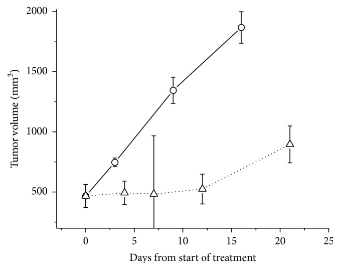
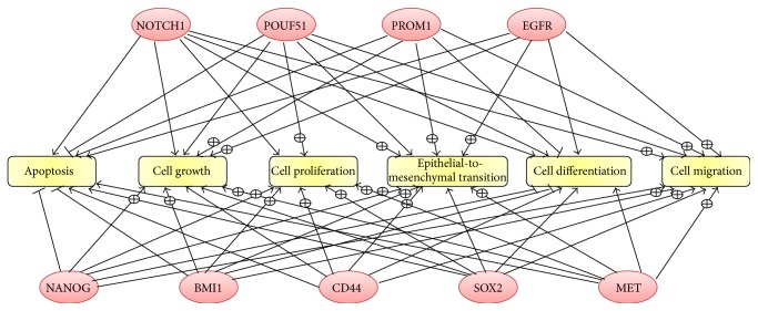
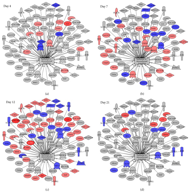
The aim of the study was to investigate cancer stem signaling during the repopulation response of a head and neck squamous cell cancer (HNSCC) xenograft after radiation treatment. Xenografts were generated from low passage HNSCC cells and were treated with either sham radiation or 15 Gy in one fraction. At different time points, days 0, 3, and 10 for controls and days 4, 7, 12, and 21, after irradiation, 3 tumors per group were harvested for global gene expression, pathway analysis, and immunohistochemical evaluation. 316 genes were identified that were associated with a series of stem cell-related genes and were differentially expressed (p ≤ 0.01 and 1.5-fold) at a minimum of one time point in UT-SCC-14 xenografts after radiation. The largest network of genes that showed significant changes after irradiation was associated with CD44, NOTCH1, and MET. c-MET and ALDH1A3 staining correlated with the changes in gene expression. A clear pattern emerged that was consistent with the growth inhibition data in that genes associated with stem cell pathways were most active at day 7 and day 12 after irradiation. The MET/CD44 axis seemed to be an important component of the repopulation response.


Figures

Figure 1
Figure 1
The tumor growth response of UT-SCC-14 xenografts to a dose of 15 Gy. Flank tumors were either irradiated with 15 Gy (∆) or sham-irradiated (O) and their growth was monitored over time with calipers.
Figure 2
Figure 2
Immunohistochemical staining for CD44, ALDH1A3, and c-MET (a) and image analysis quantification of protein expression (b) after radiation (RT) treatment. CD44 was mainly associated with the cell surface and tended to stain cells in the outer layers of the tumor islands; its expression decreased after radiation but recovered between days 12 and 21. ALDH1A3 staining was cytoplasmic and showed a diffuse pattern throughout the tumors. After irradiation ALDH1A3 increased to a maximum on day 7 and remained elevated for the period of observation. In some areas cells became more intensely stained with ALDH1A3 after irradiation. c-MET showed a staining pattern which was similar to ALDH1A3 being diffuse and cytoplasmic. The levels of c-MET increased at day 4 and reached a maximum at day 12 after which they declined rapidly to control levels (×20).
Figure 3
Figure 3
Cellular processes common to all nine cancer stem cell- (CSC-) associated genes. Pathway Studio was used to identify known relationships and cellular processes that are common between the CSC-associated genes. The ⨁ symbol on the arrow represents positive regulation of the process whilst a – through the arrow represents negative regulation.
Figure 4
Figure 4
Changes in genes associated with epithelial-to-mesenchymal transition after radiation. Using Pathway Studio, genes known to be associated with epithelial-to-mesenchymal transition and differentially expressed at a minimum of one time point after radiation with a p value ≤ 0.01 and fold change (negative or positive) of 1.5 or greater were identified. Red represents genes upregulated after irradiation whilst blue represents downregulated genes.

Similar articles

Cited by

References

    1. Pignon J.-P., Maître A. L., Maillard E., Bourhis J. Meta-analysis of chemotherapy in head and neck cancer (MACH-NC): an update on 93 randomised trials and 17,346 patients. Radiotherapy and Oncology. 2009;92(1):4–14. doi: 10.1016/j.radonc.2009.04.014. - DOI - PubMed
    1. Bryne M., Boysen M., Alfsen C. G., et al. The invasive front of carcinomas. The most important area for tumour prognosis? Anticancer Research. 1998;18(6):4757–4764. - PubMed
    1. Tremmel S. C., Götte K., Popp S., et al. Intratumoral genomic heterogeneity in advanced head and neck cancer detected by comparative genomic hybridization. Cancer Genetics and Cytogenetics. 2003;144(2):165–174. doi: 10.1016/s0165-4608(03)00011-6. - DOI - PubMed
    1. Tabor M. P., Brakenhoff R. H., Van Houten V. M. M., et al. Persistence of genetically altered fields in head and neck cancer patients: biological and clinical implications. Clinical Cancer Research. 2001;7(6):1523–1532. - PubMed
    1. Reya T., Morrison S. J., Clarke M. F., Weissman I. L. Stem cells, cancer, and cancer stem cells. Nature. 2001;414(6859):105–111. doi: 10.1038/35102167. - DOI - PubMed