

Revisiting Epithelial-to-Mesenchymal Transition in Liver Fibrosis: Clues for a Better Understanding of the "Reactive" Biliary Epithelial Phenotype
- PMID: 26880950
- PMCID: PMC4736590
- DOI: 10.1155/2016/2953727
Revisiting Epithelial-to-Mesenchymal Transition in Liver Fibrosis: Clues for a Better Understanding of the "Reactive" Biliary Epithelial Phenotype
Abstract
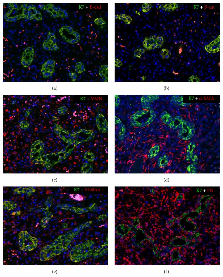
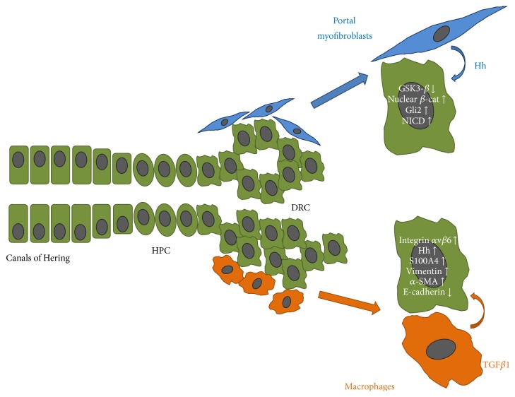
Whether liver epithelial cells contribute to the development of hepatic scarring by undergoing epithelial-to-mesenchymal transition (EMT) is a controversial issue. Herein, we revisit the concept of EMT in cholangiopathies, a group of severe hepatic disorders primarily targeting the bile duct epithelial cell (cholangiocyte), leading to progressive portal fibrosis, the main determinant of liver disease progression. Unfortunately, therapies able to halt this process are currently lacking. In cholangiopathies, fibrogenesis is part of ductular reaction, a reparative complex involving epithelial, mesenchymal, and inflammatory cells. Ductular reactive cells (DRC) are cholangiocytes derived from the activation of the hepatic progenitor cell compartment. These cells are arranged into irregular strings and express a "reactive" phenotype, which enables them to extensively crosstalk with the other components of ductular reaction. We will first discuss EMT in liver morphogenesis and then highlight how some of these developmental programs are partly reactivated in DRC. Evidence for "bona fide" EMT changes in cholangiocytes is lacking, but expression of some mesenchymal markers represents a fundamental repair mechanism in response to chronic biliary damage with potential harmful fibrogenetic effects. Understanding microenvironmental cues and signaling perturbations promoting these changes in DRC may help to identify potential targets for new antifibrotic therapies in cholangiopathies.
Figures
Similar articles
-
The crucial role of cholangiocytes in cholangiopathies.Gut Liver. 2012 Jul;6(3):295-304. doi: 10.5009/gnl.2012.6.3.295. Epub 2012 May 2. Gut Liver. 2012. PMID: 22844556 Free PMC article.
-
Emerging concepts in biliary repair and fibrosis.Am J Physiol Gastrointest Liver Physiol. 2017 Aug 1;313(2):G102-G116. doi: 10.1152/ajpgi.00452.2016. Epub 2017 May 19. Am J Physiol Gastrointest Liver Physiol. 2017. PMID: 28526690 Free PMC article. Review.
-
Knockdown of vimentin reduces mesenchymal phenotype of cholangiocytes in the Mdr2-/- mouse model of primary sclerosing cholangitis (PSC).EBioMedicine. 2019 Oct;48:130-142. doi: 10.1016/j.ebiom.2019.09.013. Epub 2019 Sep 12. EBioMedicine. 2019. PMID: 31522982 Free PMC article.
-
The Emerging Role of Macrophages in Chronic Cholangiopathies Featuring Biliary Fibrosis: An Attractive Therapeutic Target for Orphan Diseases.Front Med (Lausanne). 2020 Apr 21;7:115. doi: 10.3389/fmed.2020.00115. eCollection 2020. Front Med (Lausanne). 2020. PMID: 32373615 Free PMC article. Review.
-
Pathogenesis of Type 2 Epithelial to Mesenchymal Transition (EMT) in Renal and Hepatic Fibrosis.J Clin Med. 2015 Dec 30;5(1):4. doi: 10.3390/jcm5010004. J Clin Med. 2015. PMID: 26729181 Free PMC article. Review.
Cited by
-
miR-219-3p regulates the occurrence of hepatic fibrosis by targeting Smad2.Exp Ther Med. 2019 Jun;17(6):4635-4642. doi: 10.3892/etm.2019.7480. Epub 2019 Apr 11. Exp Ther Med. 2019. PMID: 31086594 Free PMC article.
-
The transcriptional regulators GATA6 and TET1 regulate the TGF-β pathway in cancer-associated fibroblasts to promote breast cancer progression.Cell Death Discov. 2025 Apr 11;11(1):164. doi: 10.1038/s41420-025-02438-4. Cell Death Discov. 2025. PMID: 40216762 Free PMC article.
-
The Potentiality of Herbal Remedies in Primary Sclerosing Cholangitis: From In Vitro to Clinical Studies.Front Pharmacol. 2020 Jun 10;11:813. doi: 10.3389/fphar.2020.00813. eCollection 2020. Front Pharmacol. 2020. PMID: 32587513 Free PMC article. Review.
-
Ultrastructural Characteristics of Rat Hepatic Oval Cells and Their Intercellular Contacts in the Model of Biliary Fibrosis: New Insights into Experimental Liver Fibrogenesis.Gastroenterol Res Pract. 2017;2017:2721547. doi: 10.1155/2017/2721547. Epub 2017 Jul 9. Gastroenterol Res Pract. 2017. PMID: 28769978 Free PMC article.
-
Dual Pharmacological Targeting of HDACs and PDE5 Inhibits Liver Disease Progression in a Mouse Model of Biliary Inflammation and Fibrosis.Cancers (Basel). 2020 Dec 13;12(12):3748. doi: 10.3390/cancers12123748. Cancers (Basel). 2020. PMID: 33322158 Free PMC article.
References
Publication types
LinkOut - more resources
Full Text Sources
Other Literature Sources
