. 2016:2016:3516574.
doi: 10.1155/2016/3516574. Epub 2016 Jan 10.

Comparative Analysis of Human Mesenchymal Stem Cells from Umbilical Cord, Dental Pulp, and Menstrual Blood as Sources for Cell Therapy

Affiliations

Comparative Analysis of Human Mesenchymal Stem Cells from Umbilical Cord, Dental Pulp, and Menstrual Blood as Sources for Cell Therapy

Huaijuan Ren et al. Stem Cells Int. 2016.

Abstract

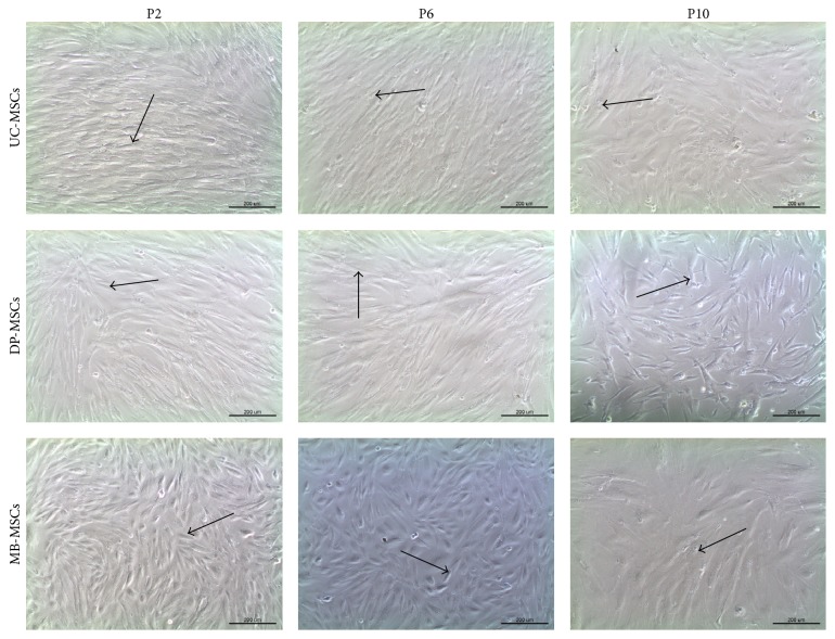
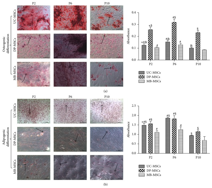
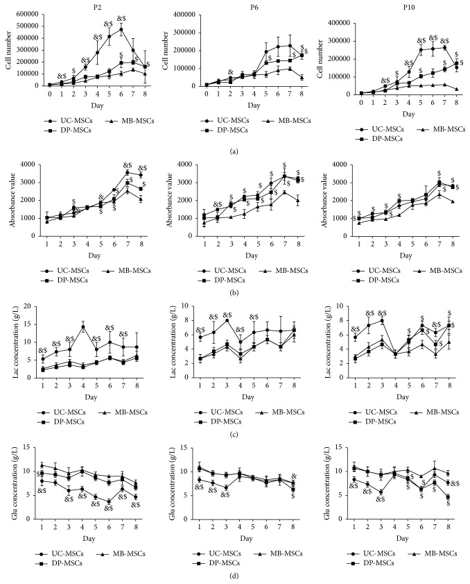
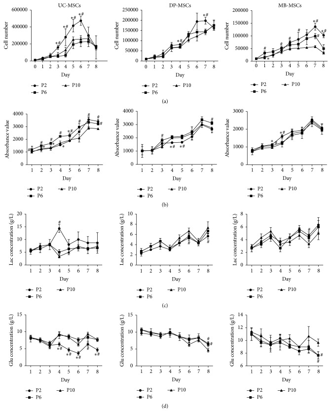
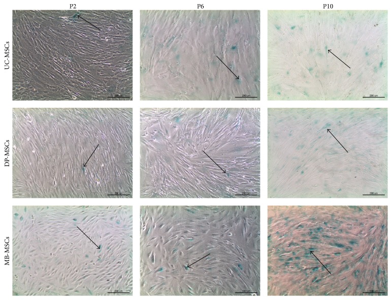
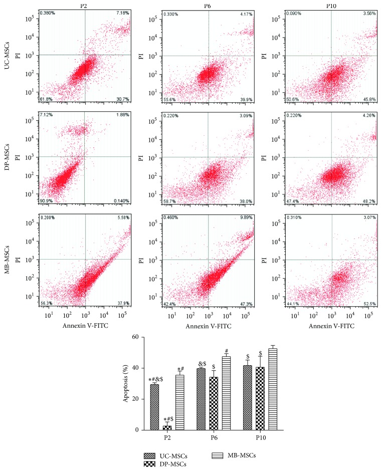
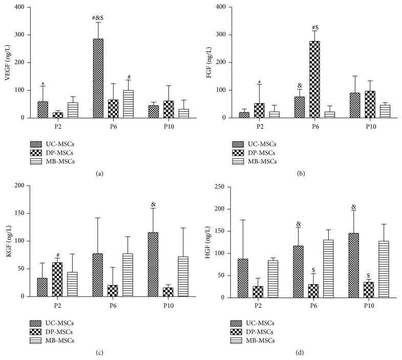
Although mesenchymal stem cells (MSCs) based therapy has been considered as a promising tool for tissue repair and regeneration, the optimal cell source remains unknown. Umbilical cord (UC), dental pulp (DP), and menstrual blood (MB) are easily accessible sources, which make them attractive candidates for MSCs. The goal of this study was to compare the biological characteristics, including morphology, proliferation, antiapoptosis, multilineage differentiation capacity, and immunophenotype of UC-, DP-, and MB-MSCs in order to provide a theoretical basis for clinical selection and application of these cells. As a result, all UC-, DP-, and MB-MSCs have self-renewal capacity and multipotentiality. However, the UC-MSCs seemed to have higher cell proliferation ability, while DP-MSCs may have significant advantages for osteogenic differentiation, lower cell apoptosis, and senescence. These differences may be associated with the different expression level of cytokines, including vascular endothelial growth factor, fibroblast growth factor, keratinocyte growth factor, and hepatocyte growth factor in each of the MSCs. Comprehensively, our results suggest DP-MSCs may be a desired source for clinical applications of cell therapy.


Figures

Figure 1
Figure 1
Morphology of UC-, DP-, and MB-MSCs (P2, P6, and P10) (100x). All MSCs exhibited a spindle-shaped morphology at P2 (arrow). However, MB-MSCs gradually became flatted and fragmented at P6 and P10 (arrow); a polygonal shape and cytoplasmic granulations were observed in UC-MSCs at P10 (arrow). A fibroblast-like morphology was maintained in DP-MSCs even at P10 (arrow). UC-MSCs: umbilical cord mesenchymal stem cells; DP-MSCs: dental pulp mesenchymal stem cells; MB-MSCs: menstrual blood mesenchymal stem cells; P: passage.
Figure 2
Figure 2
Multidifferentiation of UC-, DP-, and MB-MSCs. (a) Osteogenic differentiation, in which the cells were stained with Alizarin red (100x); (b) adipogenic differentiation in which the cells were stained with Oil Red O (100x). The differentiation capacity was semiquantified by enzyme immunoassay analyzer. Each experiment was performed in triplicate: compared with P6 of corresponding MSCs; #compared with P10 of corresponding MSCs; &compared with DP-MSCs at corresponding passage; $compared with MB-MSCs at corresponding passage, p < 0.05. UC-MSCs: umbilical cord mesenchymal stem cells; DP-MSCs: dental pulp mesenchymal stem cells; MB-MSCs: menstrual blood mesenchymal stem cells; P: passage.
Figure 3
Figure 3
Growth kinetics of UC-, DP-, and MB-MSCs. (a) The cell number determined on an automatic cell counter; (b) cell viability determined by Alamar Blue staining; (c-d) the glucose (Glu) and lactate (Lac) levels monitored using the glucose and lactate biosensors. Each experiment was performed in triplicate: &compared with DP-MSCs at corresponding day; $compared with MB-MSCs at corresponding day, p < 0.05. UC-MSCs: umbilical cord mesenchymal stem cells; DP-MSCs: dental pulp mesenchymal stem cells; MB-MSCs: menstrual blood mesenchymal stem cells; P: passage.
Figure 4
Figure 4
Growth kinetics of UC-, DP-, and MB-MSCs. (a) The cell number determined on an automatic cell counter; (b) cell viability determined by Alamar Blue staining; (c-d) the glucose (Glu) and lactate (Lac) levels monitored using the glucose and lactate biosensors. Each experiment was performed in triplicate: compared with P6 of corresponding MSCs at corresponding day; #compared with P10 of corresponding MSCs at corresponding day, p < 0.05. UC-MSCs: umbilical cord mesenchymal stem cells; DP-MSCs: dental pulp mesenchymal stem cells; MB-MSCs: menstrual blood mesenchymal stem cells; P: passage.
Figure 5
Figure 5
Senescence of UC-, DP-, and MB-MSCs. Senescence was assayed by SA-β-gal activity with positive green staining (100x, arrow). UC-MSCs: umbilical cord mesenchymal stem cells; DP-MSCs: dental pulp mesenchymal stem cells; MB-MSCs: menstrual blood mesenchymal stem cells; P: passage.
Figure 6
Figure 6
Apoptosis of UC-, DP-, and MB-MSCs. Apoptosis was detected by Annexin V-FITC/PI apoptosis kit and flow cytometry. Each experiment was performed in triplicate: compared with DP-MSCs at corresponding passage; #compared with MB-MSCs at corresponding passage; &compared with P6 of corresponding MSCs; $compared with P10 of corresponding MSCs, p < 0.05. UC-MSCs: umbilical cord mesenchymal stem cells; DP-MSCs: dental pulp mesenchymal stem cells; MB-MSCs: menstrual blood mesenchymal stem cells; P: passage.
Figure 7
Figure 7
Cytokines of UC-, DP-, and MB-MSCs. (a) VEGF expression; (b) FGF expression; (c) KGF expression; (d) HGF expression. Each experiment was performed in triplicate: compared with DP-MSCs at corresponding passage; #compared with MB-MSCs at corresponding passage; &compared with P6 of corresponding MSCs; $compared with P10 of corresponding MSCs, p < 0.05. UC-MSCs: umbilical cord mesenchymal stem cells; DP-MSCs: dental pulp mesenchymal stem cells; MB-MSCs: menstrual blood mesenchymal stem cells; P: passage; VEGF: vascular endothelial growth factor; FGF: fibroblast growth factor; KGF: keratinocyte growth factor; HGF: hepatocyte growth factor.

Similar articles

Cited by

References

    1. Hilfiker A., Kasper C., Hass R., Haverich A. Mesenchymal stem cells and progenitor cells in connective tissue engineering and regenerative medicine: is there a future for transplantation? Langenbeck's Archives of Surgery. 2011;396(4):489–497. doi: 10.1007/s00423-011-0762-2. - DOI - PubMed
    1. Jadalannagari S., Aljitawi O. S. Ectodermal differentiation of wharton's jelly mesenchymal stem cells for tissue engineering and regenerative medicine applications. Tissue Engineering B: Reviews. 2015;21(3):314–322. doi: 10.1089/ten.teb.2014.0404. - DOI - PubMed
    1. Li Y., Charif N., Mainard D., Bensoussan D., Stoltz J.-F., de Isla N. Donor's age dependent proliferation decrease of human bone marrow mesenchymal stem cells is linked to diminished clonogenicity. Bio-Medical Materials and Engineering. 2014;24(1, supplement):47–52. doi: 10.3233/BME-140973. - DOI - PubMed
    1. Wu L. W., Wang Y.-L., Christensen J. M., et al. Donor age negatively affects the immunoregulatory properties of both adipose and bone marrow derived mesenchymal stem cells. Transplant Immunology. 2014;30(4):122–127. doi: 10.1016/j.trim.2014.03.001. - DOI - PubMed
    1. Sanberg P. R., Eve D. J., Willing A. E., et al. The treatment of neurodegenerative disorders using umbilical cord blood and menstrual blood-derived stem cells. Cell Transplantation. 2011;20(1):85–94. doi: 10.3727/096368910X532855. - DOI - PubMed