. 2016:2016:4095072.
doi: 10.1155/2016/4095072. Epub 2016 Jan 6.

Monitoring the Bystander Killing Effect of Human Multipotent Stem Cells for Treatment of Malignant Brain Tumors

Affiliations

Monitoring the Bystander Killing Effect of Human Multipotent Stem Cells for Treatment of Malignant Brain Tumors

Cindy Leten et al. Stem Cells Int. 2016.

Abstract

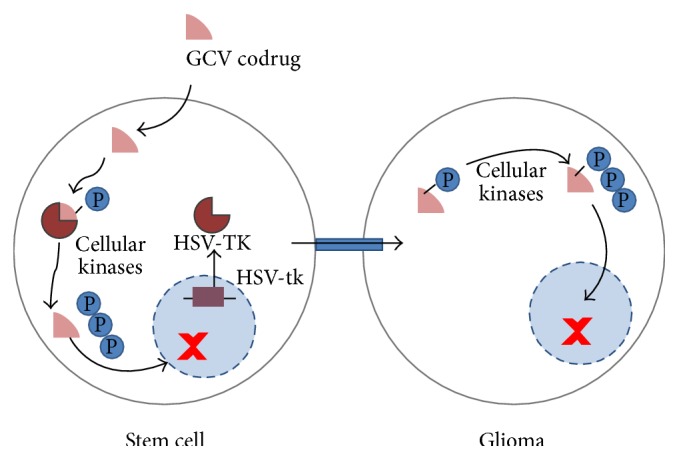
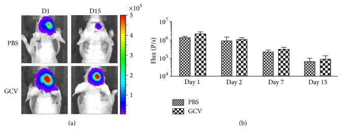
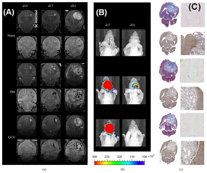
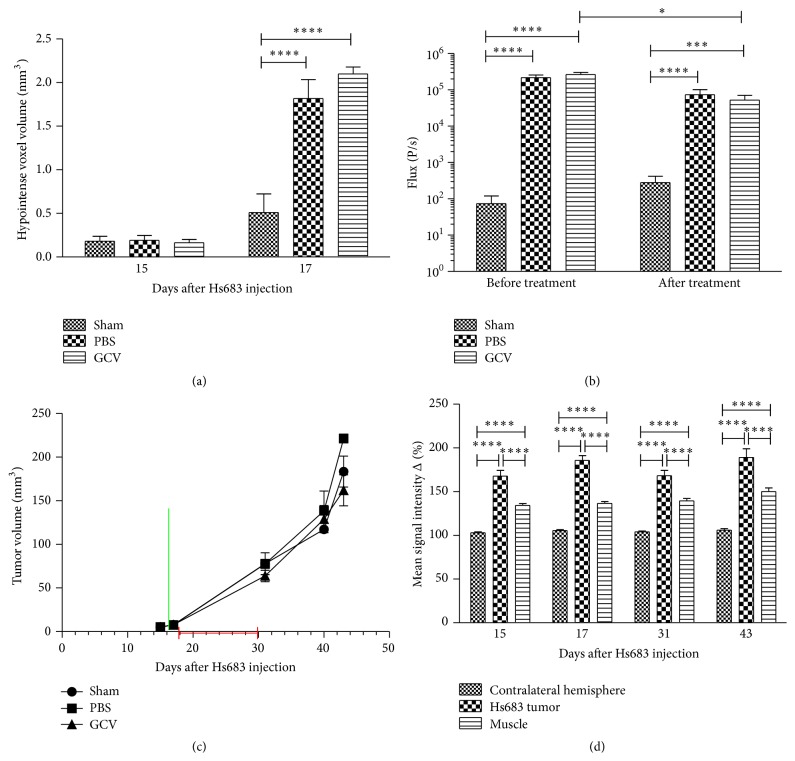
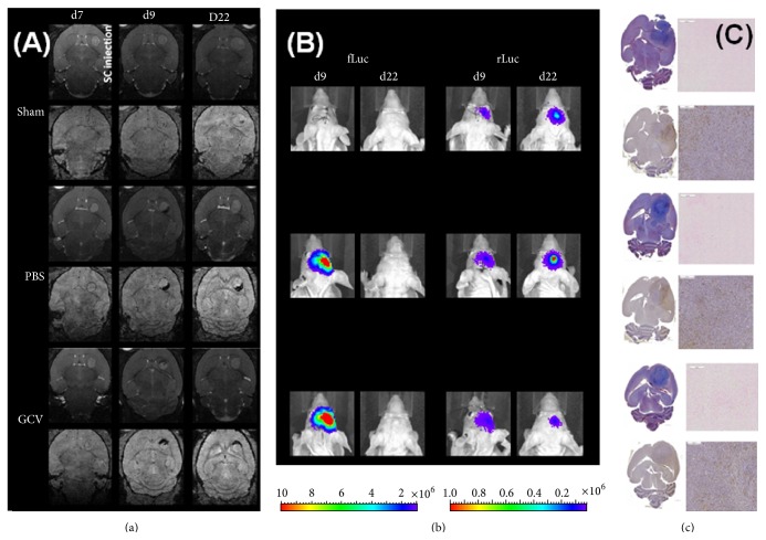
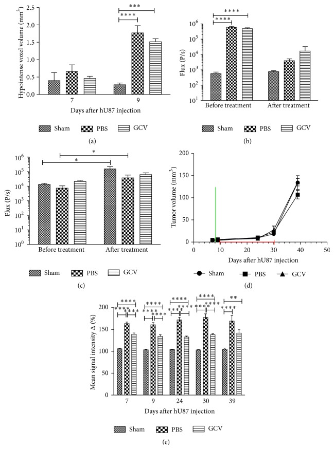
Tumor infiltrating stem cells have been suggested as a vehicle for the delivery of a suicide gene towards otherwise difficult to treat tumors like glioma. We have used herpes simplex virus thymidine kinase expressing human multipotent adult progenitor cells in two brain tumor models (hU87 and Hs683) in immune-compromised mice. In order to determine the best time point for the administration of the codrug ganciclovir, the stem cell distribution and viability were monitored in vivo using bioluminescence (BLI) and magnetic resonance imaging (MRI). Treatment was assessed by in vivo BLI and MRI of the tumors. We were able to show that suicide gene therapy using HSV-tk expressing stem cells can be followed in vivo by MRI and BLI. This has the advantage that (1) outliers can be detected earlier, (2) GCV treatment can be initiated based on stem cell distribution rather than on empirical time points, and (3) a more thorough follow-up can be provided prior to and after treatment of these animals. In contrast to rodent stem cell and tumor models, treatment success was limited in our model using human cell lines. This was most likely due to the lack of immune components in the immune-compromised rodents.


Figures

Figure 1
Figure 1
Concept of tumor therapy by using suicide gene expressing stem cells that are able to track tumor cells. It has been shown that certain stem cells are able to track infiltrating tumor cells [, –17, 22]. In addition, the therapeutic cells must carry a suicide gene, in this case the herpes simplex virus thymidine kinase (HSV-TK). When a substrate for the HSV-TK enzyme, ganciclovir (GCV), is provided, it enters the cell and is converted by HSV-TK into GCV-monophosphate. The HSV-TK displays a 1000-fold higher affinity for GCV than the mammalian thymidine kinase so that systemic toxicity is limited while the increased affinity boosts tumor therapy capabilities [5]. Cellular kinases will phosphorylate the GCV-monophosphate further to GCV-triphosphate, a guanine nucleoside analogue which inhibits cellular DNA polymerase and results in chain termination with subsequent cell death. While this would erase the therapeutic cell but not the targeted tumor cell, a means for transferring the cytotoxic compound to the tumor cell is required. GCV-monophosphate can passively diffuse into neighboring cells after the formation of gap junctions between adjacent cells, which results mostly in tumor and therapeutic cell killing as normal adult brain cells usually do not replicate [36]. This is also known as “the bystander killing effect” [18, 37]. This approach can in theory terminate both primary and infiltrating tumor cells, thus eliminating sources of possible recurrent tumors [5].
Figure 2
Figure 2
In vitro validation of hMultistem transduction. (a) Assessment of firefly luciferase (fLuc) expression via BLI showed a significant difference between transduced (eGFP-fLuc as well as eGFP-fLuc-HSV-tk) and wt hMultistem. (b) GCV killing experiment for validation of HSV-tk expression. Left: BLI signal was significantly decreased in eGFP-fLuc-HSV-tk expressing cells but not in eGFP-fLuc expressing control cells for all GCV concentrations compared to cells without GCV treatment. Right: the BCA protein assay confirmed BLI results and showed a dose-dependent cell killing in eGFP-fLuc-HSV-tk expressing cells but not in eGFP-fLuc expressing cells. Levels of significance are expressed as p < 0.05, ∗∗ p < 0.01, ∗∗∗ p < 0.001, and ∗∗∗∗ p < 0.0001.
Figure 3
Figure 3
In vitro validation of SPIO labeling of hMultistem cells. (a) ICP-OES experiments confirmed iron internalization after SPIO labeling (10 ± 1 pg Fe cell−1) but not for unlabeled control cells. (b) Multiecho T2 map (MRI) confirms higher relaxation rats for SPIO labeled cells compared to controls. (c) BLI signal intensity was not significantly affected by SPIO labeling of cells (∗∗∗ p < 0.001).
Figure 4
Figure 4
In vitro and in vivo validation of hU87 transduction with a mCherry-rLuc encoding vector. (a) In vitro analysis of mCherry-rLuc expressing hU87 cells showing sufficient expression of rLuc for in vitro detection by BLI using coelenterazine-h as a substrate. Furthermore, results show no statistically significant signal intensity differences after exposure of the rLuc positive hU87 cells to D-luciferin. (b) MRI-based tumor volume measurements from hU87 tumor bearing animals showed rapid growth with tumors reaching an average tumor size of 27.6 ± 2.4 mm3 at day 21 after injection. (c) BLI measurements on mCherry-rLuc positive hU87 tumor bearing mice also indicated tumor growth over time. Levels of significance are expressed as p < 0.05, ∗∗ p < 0.01, ∗∗∗ p < 0.001, and ∗∗∗∗ p < 0.0001.
Figure 5
Figure 5
In vivo validation of BLI signal intensity and hMultistem survival in Hsd:Athymic-FoxN1 nu mice. (a) Bioluminescence images of a representative animal that received 5 × 10⁵ hMultistems (day 1) and was treated from day 2 to day 15 with either PBS or GCV (50 mg/kg). (b) Quantification of BLI signal intensities shows that (1) the stem cells were detectable by BLI, (2) stem cell survival was reduced in the environment of the host's brain, and (3) no statistically significant difference in stem cell viability between the PBS and GCV receiving animals was detected. Number of animals was 6 per group.
Figure 6
Figure 6
Follow-up of suicide gene therapy using hMultistem as cellular vehicles in the Hs683 oligodendroglioma model by using multimodal in vivo imaging and histology. (a) MR images of a representative animal from the sham, PBS, and GCV treated groups show a comparable tumor growth prior to hMultistem injection on T2-weighted MR images (upper row for each group). While there is little hypointense contrast visible on 3D T2 -weighted MR images (lower row for each group), the distribution of SPIO labeled hMultistem cells could clearly be detected due to their hypointense contrast in the PBS and GCV treated group. When tumors grew larger, the hypointense voxels got more dispersed over time. (b) BLI of representative animals. The fLuc expression of the stem cells was clearly detectable after engraftment and only diminished after 14 days. (c) One week after the end of therapy, animals developed symptoms after which histological analysis was performed. Masson's trichrome staining (upper left) of all animals showed very large tumors. Prussian blue (upper right) staining was also performed which confirmed the presence of iron in PBS and GCV treated animals. Finally, Iba1 staining (bottom) was performed which showed a predominant absence of activated microglia in all treatment groups. Some minor microglial activation was however detectable around the tumor.
Figure 7
Figure 7
Quantification of in vivo imaging data to monitor suicide gene therapy using hMultistem as cellular vehicles in the Hs683 oligodendroglioma model. (a) Quantification of the volume of hypointense voxels in animals receiving SPIO labeled hMultistem cells indicates sufficient contrast to detect the therapeutic cells in the Hs683 tumor model (sham (no cells): N = 3, PBS treated: N = 6, and GCV treated: N = 6). (b) Bioluminescent imaging data showed that the stem cells were detectable through BLI, but no statistically significant difference in stem cell viability between the PBS and GCV receiving animals could be detected following treatment. However, there was a small decrease in the BLI signal before and after treatment in the GCV treated group. (c) Tumor volume measurements proved no statistical differences between sham operated, PBS treated, or GCV treated animals (green bar: hMultistem injection; red bar: duration of GCV/PBS treatment). (d) T1-weighted MRI pre- and postcontrast injection showed BBB integrity loss in the Hs683 tumor compared to the contralateral hemisphere and the extracranial muscle at all time points (N = 15). Levels of significance are expressed as p < 0.05, ∗∗ p < 0.01, ∗∗∗ p < 0.001, and ∗∗∗∗ p < 0.0001.
Figure 8
Figure 8
Follow-up of suicide gene therapy using hMultistem as cellular vehicles in the hU87 glioma model by using multimodal in vivo imaging and histology. (a) MR images of a representative animal from the sham, PBS, and GCV treated groups show a comparable tumor growth prior to hMultistem injection on T2-weighted MR images (upper row for each group). While there is little hypointense contrast visible on 3D T2 -weighted MR images (lower row for each group), the distribution of SPIO labeled hMultistem cells could clearly be detected due to their hypointense contrast in the PBS and GCV treated group (one day after engraftment). The contrast distribution changed only very little until the end of treatment. When tumors grew larger, the hypointense voxels got more dispersed over time. (b) Left: bioluminescent imaging using D-luciferin as a substrate for fLuc expressing hMultistem cells. Stem cells were detectable after engraftment but not after the end of treatment for both, the PBS and GCV receiving groups. Right: bioluminescence imaging using coelenterazine-h as a substrate for rLuc expressing hU87 tumor cells. The signal intensity increased for the sham and PBS group but not for the GCV group, indicating some inhibition of tumor growth. (c) Two weeks after the end of therapy, animals developed symptoms after which histological analysis was performed. Masson's trichrome staining (upper left) of all animals showed large, dense tumors. Prussian blue (upper right) staining was also performed which confirmed the presence of iron in PBS and GCV treated animals. Finally, Iba1 staining (bottom) was performed which showed a predominant absence of activated microglia in all treatment groups.
Figure 9
Figure 9
Quantification of in vivo imaging data to monitor suicide gene therapy using hMultistem as cellular vehicles in the hU87 glioblastoma model. (a) Quantification of the volume of hypointense voxels in animals receiving SPIO labeled eGFP-fLuc-HSV-tk+ hMultistem cells indicates sufficient contrast to detect the therapeutic cells in the hU87 tumor model (sham (no cells): N = 3, PBS treated: N = 5, and GCV treated: N = 6). (b) Bioluminescent imaging data using D-luciferin as a substrate for fLuc expressing hMultistem cells showed that the stem cells were detectable through BLI. A decrease in stem cell viability was detected for both PBS and GCV treated animals, which indicates that the stem cells are unable to survive well in the tumor microenvironment. (c) Bioluminescent imaging data using coelenterazine-h as a substrate for rLuc to investigate tumor viability indicated tumor growth in the sham operated and PBS treated group but not in the GCV treated group. (d) Tumor volume measurements proved no statistical differences between sham operated, PBS treated, or GCV treated animals (green bar: hMultistem injection; red bar: duration of GCV/PBS treatment). (e) T1-weighted MRI pre- and postcontrast injection showed BBB integrity loss in the hU87 tumor compared to the contralateral hemisphere and the extracranial muscle at all time points (N = 11). Levels of significance are expressed as p < 0.05, ∗∗ p < 0.01, ∗∗∗ p < 0.001, and ∗∗∗∗ p < 0.0001.

Similar articles

Cited by

References

    1. Louis D. N., Ohgaki H., Wiestler O. D., et al. The 2007 WHO classification of tumours of the central nervous system. Acta Neuropathologica. 2007;114(2):97–109. doi: 10.1007/s00401-007-0243-4. - DOI - PMC - PubMed
    1. Stupp R., Mason W. P., Van Den Bent M. J., et al. Radiotherapy plus concomitant and adjuvant temozolomide for glioblastoma. The New England Journal of Medicine. 2005;352(10):987–996. doi: 10.1056/nejmoa043330. - DOI - PubMed
    1. Thomas A. A., Brennan C. W., DeAngelis L. M., Omuro A. M. Emerging therapies for glioblastoma. JAMA Neurology. 2014;71(11):1437–1444. doi: 10.1001/jamaneurol.2014.1701. - DOI - PubMed
    1. Auffinger B., Spencer D., Pytel P., Ahmed A. U., Lesniak M. S. The role of glioma stem cells in chemotherapy resistance and glioblastoma multiforme recurrence. Expert Review of Neurotherapeutics. 2015;15(7):741–752. doi: 10.1586/14737175.2015.1051968. - DOI - PMC - PubMed
    1. Miletic H., Fischer Y., Litwak S., et al. Bystander killing of malignant glioma by bone marrow-derived tumor-infiltrating progenitor cells expressing a suicide gene. Molecular Therapy. 2007;15(7):1373–1381. doi: 10.1038/sj.mt.6300155. - DOI - PubMed

LinkOut - more resources