

Stromal Derived Factor-1/CXCR4 Axis Involved in Bone Marrow Mesenchymal Stem Cells Recruitment to Injured Liver
- PMID: 26880995
- PMCID: PMC4737461
- DOI: 10.1155/2016/8906945
Stromal Derived Factor-1/CXCR4 Axis Involved in Bone Marrow Mesenchymal Stem Cells Recruitment to Injured Liver
Abstract
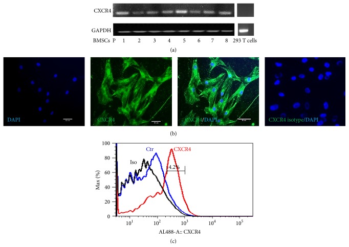
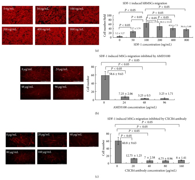
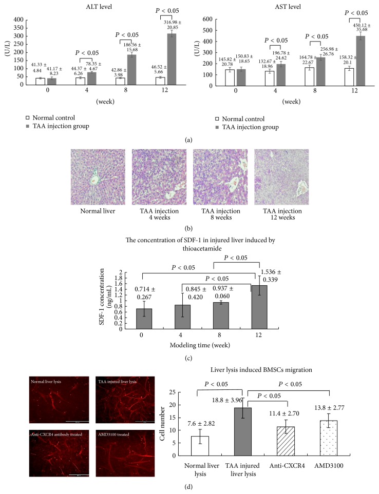
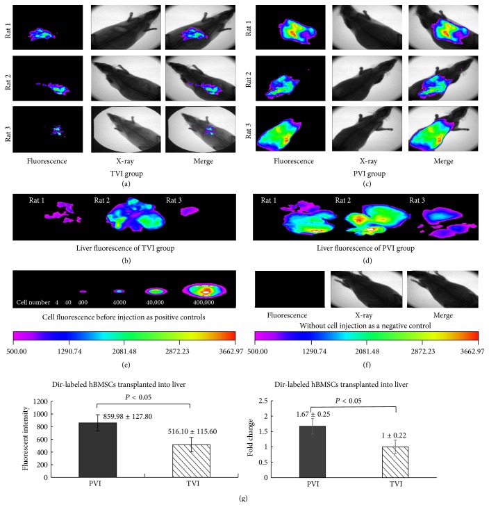
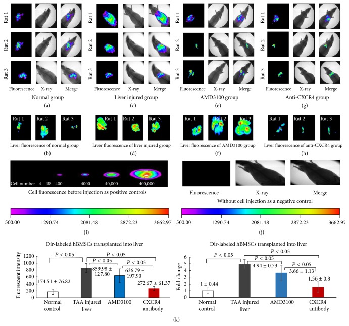
The molecular mechanism of bone marrow mesenchymal stromal stem cells (BMSCs) mobilization and migration to the liver was poorly understood. Stromal cell-derived factor-1 (SDF-1) participates in BMSCs homing and migration into injury organs. We try to investigate the role of SDF-1 signaling in BMSCs migration towards injured liver. The expression of CXCR4 in BMSCs at mRNA level and protein level was confirmed by RT-PCR, flow cytometry, and immunocytochemistry. The SDF-1 or liver lysates induced BMSCs migration was detected by transwell inserts. CXCR4 antagonist, AMD3100, and anti-CXCR4 antibody were used to inhibit the migration. The Sprague-Dawley rat liver injury model was established by intraperitoneal injection of thioacetamide. The concentration of SDF-1 increased as modeling time extended, which was determined by ELISA method. The Dir-labeled BMSCs were injected into the liver of the rats through portal vein. The cell migration in the liver was tracked by in vivo imaging system and the fluorescent intensity was measured. In vivo, BMSCs migrated into injured liver which was partially blocked by AMD3100 or anti-CXCR4 antibody. Taken together, the results demonstrated that the migration of BMSCs was regulated by SDF-1/CXCR4 signaling which involved in BMSCs recruitment to injured liver.
Figures
Similar articles
-
[Electroacupuncture combined with bone marrow mesenchymal stem cell transplantation promotes repair of thin endometrium by regulating SDF-1/CXCR4 signaling].Zhen Ci Yan Jiu. 2023 Sep 25;48(9):870-80. doi: 10.13702/j.1000-0607.20220942. Zhen Ci Yan Jiu. 2023. PMID: 37730257 Chinese.
-
Effect of SDF-1/CXCR4 axis on the migration of transplanted bone mesenchymal stem cells mobilized by erythropoietin toward lesion sites following spinal cord injury.Int J Mol Med. 2015 Nov;36(5):1205-14. doi: 10.3892/ijmm.2015.2344. Epub 2015 Sep 14. Int J Mol Med. 2015. PMID: 26398409 Free PMC article.
-
The SDF-1/CXCR4 axis regulates migration of transplanted bone marrow mesenchymal stem cells towards the pancreas in rats with acute pancreatitis.Mol Med Rep. 2014 May;9(5):1575-82. doi: 10.3892/mmr.2014.2053. Epub 2014 Mar 14. Mol Med Rep. 2014. PMID: 24626964 Free PMC article.
-
SDF-1/CXCR4 Augments the Therapeutic Effect of Bone Marrow Mesenchymal Stem Cells in the Treatment of Lipopolysaccharide-Induced Liver Injury by Promoting Their Migration Through PI3K/Akt Signaling Pathway.Cell Transplant. 2020 Jan-Dec;29:963689720929992. doi: 10.1177/0963689720929992. Cell Transplant. 2020. PMID: 32452221 Free PMC article.
-
Role of SDF-1 as a regulatory chemokine in renal regeneration after acute kidney injury.Kidney Int Suppl (2011). 2011 Sep;1(3):87-89. doi: 10.1038/kisup.2011.20. Kidney Int Suppl (2011). 2011. PMID: 25018907 Free PMC article. Review.
Cited by
-
Enzyme-Cleaved Bone Marrow Transplantation Improves the Engraftment of Bone Marrow Mesenchymal Stem Cells.JBMR Plus. 2023 Feb 11;7(3):e10722. doi: 10.1002/jbm4.10722. eCollection 2023 Mar. JBMR Plus. 2023. PMID: 36936364 Free PMC article.
-
Correlation between SDF-1α, CD34 positive hematopoietic stem cells and CXCR4 expression with liver fibrosis in CCl4 rat model.BMC Gastroenterol. 2023 Sep 21;23(1):323. doi: 10.1186/s12876-023-02932-y. BMC Gastroenterol. 2023. PMID: 37730560 Free PMC article.
-
High-mobility group box 1 fragment ameliorates chronic pancreatitis induced by caerulein in mice.J Gastroenterol. 2024 Aug;59(8):744-757. doi: 10.1007/s00535-024-02112-z. Epub 2024 May 10. J Gastroenterol. 2024. PMID: 38727823
-
Sequential SDF-1/CGRP-releasing smart composite hydrogel promotes osteoporotic fracture healing by targeting sensory nerve-regulated bone remodeling.Mater Today Bio. 2025 Apr 17;32:101750. doi: 10.1016/j.mtbio.2025.101750. eCollection 2025 Jun. Mater Today Bio. 2025. PMID: 40331153 Free PMC article.
-
Osteogenesis of bone marrow mesenchymal stem cell in hyperglycemia.Front Endocrinol (Lausanne). 2023 Jun 21;14:1150068. doi: 10.3389/fendo.2023.1150068. eCollection 2023. Front Endocrinol (Lausanne). 2023. PMID: 37415664 Free PMC article. Review.
References
LinkOut - more resources
Full Text Sources
Other Literature Sources
