Review
. 2016:2016:8373020.
doi: 10.1155/2016/8373020. Epub 2016 Jan 6.

Endogenous and Synthetic Cannabinoids as Therapeutics in Retinal Disease

Affiliations
Review

Endogenous and Synthetic Cannabinoids as Therapeutics in Retinal Disease

Despina Kokona et al. Neural Plast. 2016.

Abstract

The functional significance of cannabinoids in ocular physiology and disease has been reported some decades ago. In the early 1970s, subjects who smoked Cannabis sativa developed lower intraocular pressure (IOP). This led to the isolation of phytocannabinoids from this plant and the study of their therapeutic effects in glaucoma. The main treatment of this disease to date involves the administration of drugs mediating either the decrease of aqueous humour synthesis or the increase of its outflow and thus reduces IOP. However, the reduction of IOP is not sufficient to prevent visual field loss. Retinal diseases, such as glaucoma and diabetic retinopathy, have been defined as neurodegenerative diseases and characterized by ischemia-induced excitotoxicity and loss of retinal neurons. Therefore, new therapeutic strategies must be applied in order to target retinal cell death, reduction of visual acuity, and blindness. The aim of the present review is to address the neuroprotective and therapeutic potential of cannabinoids in retinal disease.


Figures

Figure 1
Figure 1
Effect of HU-210 on somatostatin's release in rat retina. (a) HU-210 at the low dose of 10−16 M had no effect on the release of somatostatin (1.7 ± 0.15 pg/μg, n = 4) compared to the control tissue (CTRL, 1.5 ± 0.04 pg/μg, n = 12). HU-210 at 10−14 M increased the release of somatostatin in the retina (2.4 ± 0.17 pg/μg, n = 5, ∗∗∗ p < 0.001 compared to CTRL), whereas higher concentrations caused a statistically significant decrease in somatostatin's release (10−12 M, 0.6 ± 0.06 pg/μg, n = 5, ∗∗∗ p < 0.001 compared to CTRL; 10−10 M, 0.63 ± 0.09 pg/μg, n = 5, ∗∗∗ p < 0.001 compared to CTRL; 10−8 M, 0.68 ± 0.06 pg/μg, n = 6, ∗∗∗ p < 0.001 compared to CTRL; 10−7 M, 0.7 ± 0.07 pg/μg, n = 6, ∗∗∗ p < 0.001 compared to CTRL). Dunnett's Multiple Comparison Test was used for the statistical analysis of the data. All values represent the mean ± SEM. (b) Effect of the CB1 preferred antagonist AM251 in the actions of HU-210 (10−7 M) on somatostatin's release. AM251 (10−7 M) reversed the attenuation of somatostatin release by HU-210 (10−7 M) (1.42 ± 0.22 pg/μg, ### p < 0.001 compared to HU-210, n = 5). One-way ANOVA with Tukey's post hoc analysis test was used for the statistical analysis of the data. All values represent the mean ± SEM.
Figure 2
Figure 2
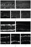
Effect of the synthetic CB1/CB2 cannabinoid HU-210 on ChAT and PKC immunoreactivity. (a) ChAT immunoreactivity. ChAT immunoreactivity in control tissue ((A) n = 12) is localized in cholinergic amacrine cell somata in the INL and GCL, as well as in their processes in the IPL. (B) Chemical ischemia mixture (n = 12) caused a reduction of ChAT immunoreactivity as revealed by loss of cholinergic cell somata and less intense signal in cell processes. HU-210 afforded neuroprotection at the concentrations of 10−6 M ((C) n = 5), 10−5 M ((D) n = 5), and 10−4 M ((E) n = 5). Arrows depict ChAT-immunoreactive amacrine cells. (b) PKC immunoreactivity. PKC immunoreactivity in control tissue ((A) n = 3) is localized in rod bipolar cells in the OPL and in their axons extending toward the IPL. Reduced immunoreactivity is observed in the presence of the chemical ischemia mixture ((B) n = 3). HU-210 afforded neuroprotection at all of concentrations used (10−6 M ((C) n = 3), 10−5 M ((D) n = 3), and 10−4 M ((E) n = 3)). Scale bar: 50 μm. OPL: outer plexiform layer; INL: inner nuclear layer; IPL: inner plexiform layer; GCL: ganglion cell layer.
Figure 3
Figure 3
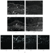
Involvement of the CB1 receptor on the neuroprotective actions of HU-210. (a) ChAT immunoreactivity. The selective inverse agonist AM251 attenuated the neuroprotective actions of HU-210 on cholinergic amacrine cells ((A) control, n = 12; (B) chemical ischemia mixture, n = 12; (C) HU-210 (10−5 M), n = 3; (D) HU-210 (10−5 M) + AM251 (10−5 M)). Arrows depict ChAT-immunoreactive amacrine cells. Scale bar: 50 μm. (b) Effect of cannabinoids on retinal cell death. TUNEL staining was prominent in the chemical ischemia incubated tissues ((B) n = 2) while less staining was observed in the control tissues ((A) n = 2). TUNEL staining substantiates the neuroprotective effects of HU-210 ((C) 10−5 M, n = 2) and the reduced neuroprotection (increased TUNEL staining) in the presence of AM251 ((D) 10−5 M, n = 3). ONL: outer nuclear layer; INL: inner nuclear layer; IPL: inner plexiform layer; GCL: ganglion cell layer.
Figure 4
Figure 4
Involvement of the TRPV1 channel on the neuroprotective actions of endogenous and synthetic cannabinoids on bNOS expressing amacrine cells. (a) Intravitreal coinjection of the TRPV1 antagonist capsazepine (10−6 M) with AMPA (42 nmol/eye) + AEA (10−7 M, n = 6) or 2-AG (b) (10−7 M, n = 3) or HU-210 (c) (10−7 M, n = 3) attenuated the neuroprotective effects of the cannabinoid ligands (∗∗∗ p < 0.001 compared to CTRL, ## p < 0.01, ### p < 0.001 compared to AMPA, and + p < 0.05, ++ p < 0.01 compared to AEA (10−7 M) or 2-AG (10−7 M) or HU-210 (10−7 M), resp.). (d) The TRPV1 channel agonist capsaicin (10−6–10−4 M, n = 3) coinjected with AMPA had no neuroprotective effect at any of the doses tested (∗∗∗ p < 0.001, compared to CTRL). One-way ANOVA with Tukey's post hoc analysis was used for the statistical analysis of all data presented in this figure.

References

    1. Yazulla S. Endocannabinoids in the retina: from marijuana to neuroprotection. Progress in Retinal and Eye Research. 2008;27(5):501–526. doi: 10.1016/j.preteyeres.2008.07.002. - DOI - PMC - PubMed
    1. Schwitzer T., Schwan R., Angioi-Duprez K., et al. The cannabinoid system and visual processing: a review on experimental findings and clinical presumptions. European Neuropsychopharmacology. 2014;25(1):100–112. doi: 10.1016/j.euroneuro.2014.11.002. - DOI - PubMed
    1. Hepler R. S., Frank I. R. Marihuana smoking and intraocular pressure. Journal of the American Medical Association. 1971;217(10):p. 1392. doi: 10.1001/jama.217.10.1392. - DOI - PubMed
    1. Merritt J. C., Perry D. D., Russell D. N., Jones B. F. Topical delta 9-tetrahydrocannabinol and aqueous dynamics in glaucoma. Journal of Clinical Pharmacology. 1981;21(8-9, supplement):467S–471S. - PubMed
    1. Järvinen T., Pate D. W., Laine K. Cannabinoids in the treatment of glaucoma. Pharmacology & Therapeutics. 2002;95(2):203–220. doi: 10.1016/s0163-7258(02)00259-0. - DOI - PubMed

Publication types