. 2016 Feb 17:16:115.
doi: 10.1186/s12885-016-2170-z.

MB3W1 is an orthotopic xenograft model for anaplastic medulloblastoma displaying cancer stem cell- and Group 3-properties

Affiliations

MB3W1 is an orthotopic xenograft model for anaplastic medulloblastoma displaying cancer stem cell- and Group 3-properties

Sebastian Dietl et al. BMC Cancer. .

Abstract

Background: Medulloblastoma is the most common malignant brain tumor in children and can be divided in different molecular subgroups. Patients whose tumor is classified as a Group 3 tumor have a dismal prognosis. However only very few tumor models are available for this subgroup.

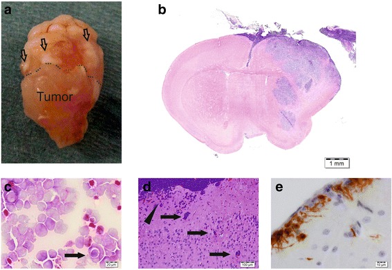
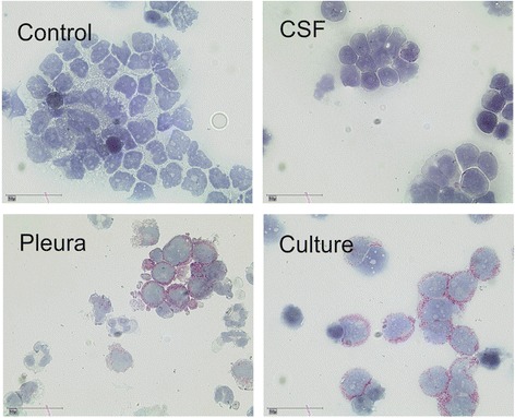
Methods: We established a robust orthotopic xenograft model with a cell line derived from the malignant pleural effusions of a child suffering from a Group 3 medulloblastoma.

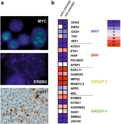
Results: Besides classical characteristics of this tumor subgroup, the cells display cancer stem cell characteristics including neurosphere formation, multilineage differentiation, CD133/CD15 expression, high ALDH-activity and high tumorigenicity in immunocompromised mice with xenografts exactly recapitulating the original tumor architecture.

Conclusions: This model using unmanipulated, human medulloblastoma cells will enable translational research, specifically focused on Group 3 medulloblastoma.


Figures

Fig. 1
Fig. 1
Illustration of the clinical case. a. Sagittal cranial MRI of the 22-month-old boy showing the initial tumor in the 4th ventricle (left, single arrow) and signs of massive meningeosis 3 weeks later (right, multiple arrows). b. HE staining of the initial tumor showing typical anaplastic morphology of the tumor cells. Insert: medulloblastoma cells from the CSF (Pappenheim-staining)
Fig. 2
Fig. 2
Characteristics of medulloblastoma cells recovered from the pleural effusions after progression of disease. a. Representative flow cytometry plots outlining the expression of CD133 and CD15 in MB3W1 cells. b. Light-microscopy of MB3W1 cells in culture in serum-free medium showing sphere forming growth (left) and partly adherent growth in serum-containing medium (right). c. Growth characteristics of MB3W1 cells grown in serum-free medium (left, purple columns), compared to growth in serum-containing medium (right, light blue columns = adherent fraction, dark blue columns = spheroid fraction). d. Expression of CD133 on cells grown as spheres in serum-free medium at in vitro passage 1 (blue full line) and passage 30 (blue broken line). Black line = isotype control. e. ALDH expression as measured by substrate conversion using the ALDEFLUOR assay. Different cell lines were incubated with ALDH-substrate either without (green lines) or with a specific enzyme inhibitor to block ALDH-activity (black lines). The degree of fluorescence correlates with ALDH-activity. R28 and R11 are glioblastoma cell lines recently characterized as cells with CSC activity. U251 is a glioblastoma cell line with no known CSC activity
Fig. 3
Fig. 3
In vivo growth characteristics of MB3W1. a. 5 × 104 luciferase-transduced MB3W1 cells were xenografted either infra- (open circles) or supratentorially (closed circles), and tumor growth was monitored by BLI. b. Survival curves after infra- (pink, n = 6) versus supratentorial inoculation (blue, n = 5; one animal lost during surgery (censored)); grey: untreated controls, surgery only (n = 2). c. Survival curves of mice inoculated with untransduced tumor cells: comparison of MB3W1 cells previously grown in different culture conditions: supratentorial inoculation of cells previously grown as spheres in serum-free medium (pink curves, n = 5) and compared to inoculation of cells previously grown in serum-containing medium – cells were only derived from the non-proliferative adherent fraction - (blue curves, n = 5). Circles: 5 × 105 cells, squares: 5 × 104 cells, triangles: 5 × 103 cells. d. Growth characteristics of CD133+ (closed triangles, n = 5) and CD133 (open triangles, n = 5) MB3W1 cells after supratentorial inoculation of 5000 tumor cells
Fig. 4
Fig. 4
Immunohistological evaluation of the original tumor and xenografted infratentorial tumor. Selected markers are shown in comparison. For some markers, there was not sufficient material left from the primary tumor. All specimens were formaldehyde fixed and paraffin embedded prior to histopathological evaluation
Fig. 5
Fig. 5
Xenografts show signs of metastasis. a. Macroscopically subarachnoidal tumor dissemination is seen frequently at tumor progression and b. tumor cells can then constantly be found in the CSF (HE stained gross section). c. Xenograft cytospin of subarachnoidal disseminated tumor cells at the humane endpoint reveal blue-cell tumor cells with for LCA histology typical cell wrapping (arrow). d. Histology (HE-staining) indicates aggressive, infiltrative behavior with xenografted tumor cells invading from the brain surface (arrow head) and the Virchow-Robin spaces (arrows). e. Vimentin-staining showing tumor cells with pseudopodia reaching from the brain surface into the brain tissue
Fig. 6
Fig. 6
Evaluation of CD133 expression in MB3W1 cells directly after cell isolation and during cell culture. Cytospins from different cell preparations were evaluated for CD133. Preferred expression of CD133 on cells directly derived from the pleural effusions (lower left) and on cultivated MB3W1 cells at day 4 after isolation (lower right). In comparison cytospins from the CSF at diagnosis showed little (if any) staining for CD133 (upper right). (Upper left: staining control)
Fig. 7
Fig. 7
MB3W1 cells comprise a faithful Group 3 model. a. Upper panel: FISH analysis of MB3W1 cells revealed a clear amplification of the MYC oncogene (green signal) as a hallmark of Group 3 tumors (in red: CEP8). Middle panel: FISH on chromosome 17 shows (consistent with the patient’s karyogram) tetraploidy of the ERBB2 gen (red signal) and an unbalanced gain of chromosome 17q (green signal of the chromosome enumeration probe 17p11.1-17q11.1). Lower panel: Immunohistochemical staining of xenotransplanted tumor specimen revealing accumulation of p53 protein. b. Heatmap, illustrating analysis of a set of 22 marker genes previously validated for MB sub-grouping. Strong clustering of genes regulated in Group 3 MB is observed. No significant differences in gene expression were detected in RNA derived from early passages (<5 passages) or late passages (>20 passages)

References

    1. Rickert CH, Paulus W. Epidemiology of central nervous system tumors in childhood and adolescence based on the new WHO classification. Childs Nerv. Syst. 2001;17(9):503–511. doi: 10.1007/s003810100496. - DOI - PubMed
    1. Pizer BL, Clifford SC. The potential impact of tumour biology on improved clinical practice for medulloblastoma: progress towards biologically driven clinical trials. Br J Neurosurg. 2009;23(4):364–375. doi: 10.1080/02688690903121807. - DOI - PubMed
    1. Eberhart CG, Kratz J, Wang Y, Summers K, Stearns D, Cohen K, Dang CV, Burger PC. Histopathological and molecular prognostic markers in medulloblastoma: c-myc, N-myc, TrkC, and anaplasia. J Neuropathol Exp Neurol. 2004;63(5):441–449. doi: 10.1093/jnen/63.5.441. - DOI - PubMed
    1. von Hoff K, Hartmann W, von Bueren AO, Gerber NU, Grotzer MA, Pietsch T, Rutkowski S. Large cell/anaplastic medulloblastoma: outcome according to myc status, histopathological, and clinical risk factors. Pediatr Blood Cancer. 2010;54(3):369–376. doi: 10.1002/pbc.22339. - DOI - PubMed
    1. Ellison DW, Kocak M, Dalton J, Megahed H, Lusher ME, Ryan SL, Zhao W, Nicholson SL, Taylor RE, Bailey S, et al. Definition of disease-risk stratification groups in childhood medulloblastoma using combined clinical, pathologic, and molecular variables. J. Clin. Oncol. Off. J. Am. Soc. Clin. Oncol. 2011;29(11):1400–1407. doi: 10.1200/JCO.2010.30.2810. - DOI - PMC - PubMed

Publication types

Substances

LinkOut - more resources