Review
. 2016 Feb 15;30(4):355-85.
doi: 10.1101/gad.275776.115.

Genetics and biology of pancreatic ductal adenocarcinoma

Affiliations
Review

Genetics and biology of pancreatic ductal adenocarcinoma

Haoqiang Ying et al. Genes Dev. .

Abstract

With 5-year survival rates remaining constant at 6% and rising incidences associated with an epidemic in obesity and metabolic syndrome, pancreatic ductal adenocarcinoma (PDAC) is on track to become the second most common cause of cancer-related deaths by 2030. The high mortality rate of PDAC stems primarily from the lack of early diagnosis and ineffective treatment for advanced tumors. During the past decade, the comprehensive atlas of genomic alterations, the prominence of specific pathways, the preclinical validation of such emerging targets, sophisticated preclinical model systems, and the molecular classification of PDAC into specific disease subtypes have all converged to illuminate drug discovery programs with clearer clinical path hypotheses. A deeper understanding of cancer cell biology, particularly altered cancer cell metabolism and impaired DNA repair processes, is providing novel therapeutic strategies that show strong preclinical activity. Elucidation of tumor biology principles, most notably a deeper understanding of the complexity of immune regulation in the tumor microenvironment, has provided an exciting framework to reawaken the immune system to attack PDAC cancer cells. While the long road of translation lies ahead, the path to meaningful clinical progress has never been clearer to improve PDAC patient survival.

Keywords: KRAS; cancer metabolism; pancreatic cancer; tumor immune modulation.


Figures

Figure 1.
Figure 1.
Metabolism reprogramming in PDAC. KRAS-driven PDAC is characterized by enhanced glycolysis, including increased glucose uptake and lactate production. Oncogenic KRAS also promotes the efflux of lactate to mitigate the toxic effect of intracellular lactate accumulation due to elevated glycolysis. The flux of glycolysis intermediates was directed by oncogenic KRAS into biosynthetic pathways, including the nonoxidative pentose phosphate pathway (PPP) for nucleotide biosynthesis and the hexosamine biosynthesis pathway (HBP) to support glycosylation. The reprogramming of glucose metabolism in PDAC cells by oncogenic KRAS is mediated by up-regulation of multiple enzymes in a MYC-dependent manner. Oncogenic KRAS also induces a noncanonical glutamine metabolism pathway to maintain redox homeostasis in PDAC cells through the induction of aspartate transaminase 1 (GOT1) expression. The redox balance is also maintained by KRAS-mediated activation of NRF2, which is a master transcription factor for antioxidant genes. Another feature of PDAC metabolism is the activation of nutrient salvage pathways to fuel tumor growth. Oncogenic KRAS induces macropinocytosis and uptake of protein from extracellular space, which leads to lysosomal degradation and the release of nutrients to support the tricarboxylic acid (TCA) cycle. In addition, KRAS-driven PDAC cells are also characterized by increased autophagy, which leads to the degradation of organelles and proteins and the production of amino acids and other components that support metabolism. The activation of autophagy is achieved through the MiT/TFE-mediated expression of autophagy and lysosome genes. Enzymes whose expression is induced in oncogenic KRAS are indicated in red. (GFPT1) Glucosamine-fructose-6-phosphate aminotransferase 1; (ME1) malic enzyme 1; (ROS) reactive oxygen species; (RPE) ribulose-5-phosphate-3-epimerase; (RPIA) ribulose-5-phosphate isomerase.
Figure 2.
Figure 2.
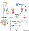
Immune network, prospective targets, and therapies in PDAC. PDAC cancer cells secrete cytokines such as TGFβ, CXCL5, and GM-CSF (granulocyte-macrophage colony-stimulating factor), which mobilizes bone marrow-derived, immune-suppressive immune cells such as myeloid-derived suppressor cells (MDSCs) and regulatory T (Treg) cells. The secretion of these protumorigenic cytokines by the cancer cells is tightly regulated by oncogenic KRAS-dependent pathways. These tumor-infiltrated MDSCs and Treg cells create an immune-suppressive environment by suppressing the activity and functions of CD8+ cytotoxic T cells and blocking the M1 phenotype of tumor-associated macrophages (TAMs). PDAC cells also secrete metabolites such as lactate, which suppresses CD8+ cytotoxic T-cell activity. Furthermore, the paracrine network between PDAC cancer cells and myofibroblasts such as pancreatic stellate cells (PSCs) creates a desmoplastic response leading to fibrosis and immune suppression. Also, cancer cells secrete factors such as Sonic hedgehog (SHH) ligands that activate PSCs to produce matrix metalloproetinases (MMPs) that promote invasion and extracellular matrix (ECM)—mostly collagens. In addition, PDAC cancer cells and the infiltrating MDSCs secrete proinflammatory cytokines such as IL6, which further promotes JAK/STAT-mediated pathways, leading to cancer cell survival, proliferation, and diminished tumor antigen presentation to the dendritic cells. Numerous strategies are being pursued to manipulate the immunosuppressive environment in PDAC and reduce immune evasion by cancer cells. Unlike melanoma, the immune modulation strategies such as anti-CTLA4 (cytotoxic T-lymphocyte-associated protein 4) and anti-PD1/PD-L1 (programmed cell death 1 ligand 1) have yet to show any promising outcome as monotherapies, although, in a αSMA-Tk-KPC GEMM, the depletion of stroma creates a therapeutic opportunity for checkpoint blockers such as PD1. Clinical trials involving GVAX (allogeneic pancreatic cancer cells modified to express GM-CSF) alone and/or in combination with CRS-207 (live attenuated Listeria monocytogenes expressing mesothelin) have shown positive outcomes and generated excitement among the immunotherapy community. Other immune-modulating therapies currently being tested and showing some efficacy include the adaptive T-cell therapy such as the mesothelin chimeric antigen receptor (CAR) T-cell therapy. (αSMA) α-Smooth muscle actin.

Comment in

Similar articles

Cited by

References

    1. Aguirre AJ, Bardeesy N, Sinha M, Lopez L, Tuveson DA, Horner J, Redston MS, DePinho RA. 2003. Activated Kras and Ink4a/Arf deficiency cooperate to produce metastatic pancreatic ductal adenocarcinoma. Genes Dev 17: 3112–3126. - PMC - PubMed
    1. Aguirre AJ, Brennan C, Bailey G, Sinha R, Feng B, Leo C, Zhang Y, Zhang J, Gans JD, Bardeesy N, et al. 2004. High-resolution characterization of the pancreatic adenocarcinoma genome. Proc Natl Acad Sci 101: 9067–9072. - PMC - PubMed
    1. Alagesan B, Contino G, Guimaraes AR, Corcoran RB, Deshpande V, Wojtkiewicz GR, Hezel AF, Wong KK, Loda M, Weissleder R, et al. 2015. Combined MEK and PI3K inhibition in a mouse model of pancreatic cancer. Clin Cancer Res 21: 396–404. - PMC - PubMed
    1. Alexandrov LB, Nik-Zainal S, Wedge DC, Aparicio SA, Behjati S, Biankin AV, Bignell GR, Bolli N, Borg A, Borresen-Dale AL, et al. 2013. Signatures of mutational processes in human cancer. Nature 500: 415–421. - PMC - PubMed
    1. Alvarez-Calderon F, Gregory MA, Pham-Danis C, DeRyckere D, Stevens BM, Zaberezhnyy V, Hill AA, Gemta L, Kumar A, Kumar V, et al. 2015. Tyrosine kinase inhibition in leukemia induces an altered metabolic state sensitive to mitochondrial perturbations. Clin Cancer Res 21: 1360–1372. - PMC - PubMed

Publication types

MeSH terms

Substances