. 2016 Apr:39:349-364.
doi: 10.1016/j.meegid.2016.02.015. Epub 2016 Feb 12.

S1 gene-based phylogeny of infectious bronchitis virus: An attempt to harmonize virus classification

Affiliations

S1 gene-based phylogeny of infectious bronchitis virus: An attempt to harmonize virus classification

Viviana Valastro et al. Infect Genet Evol. 2016 Apr.

Abstract

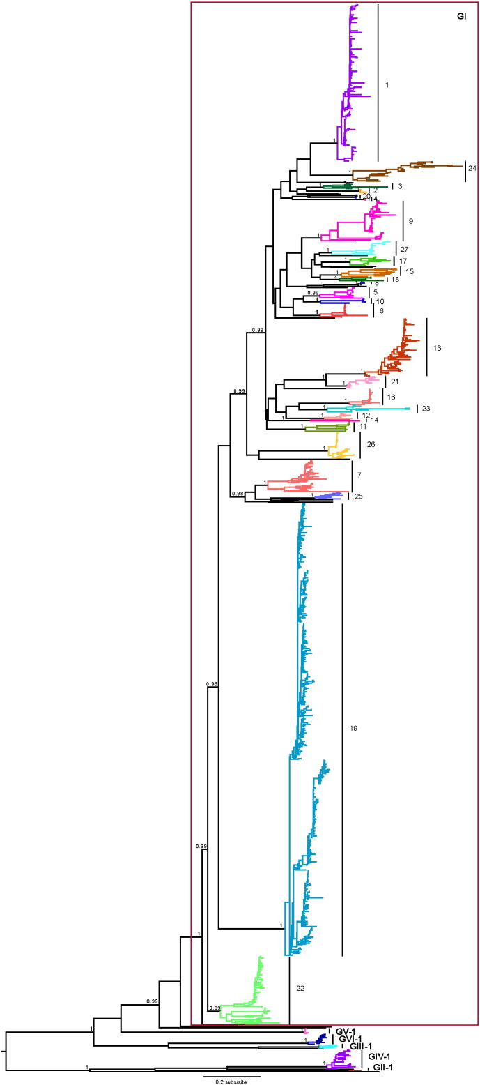
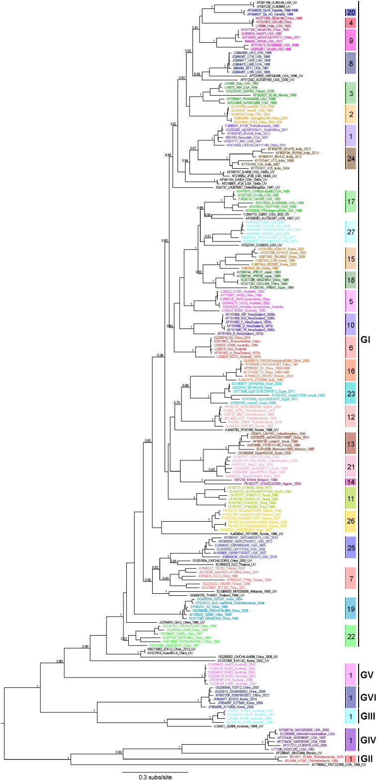
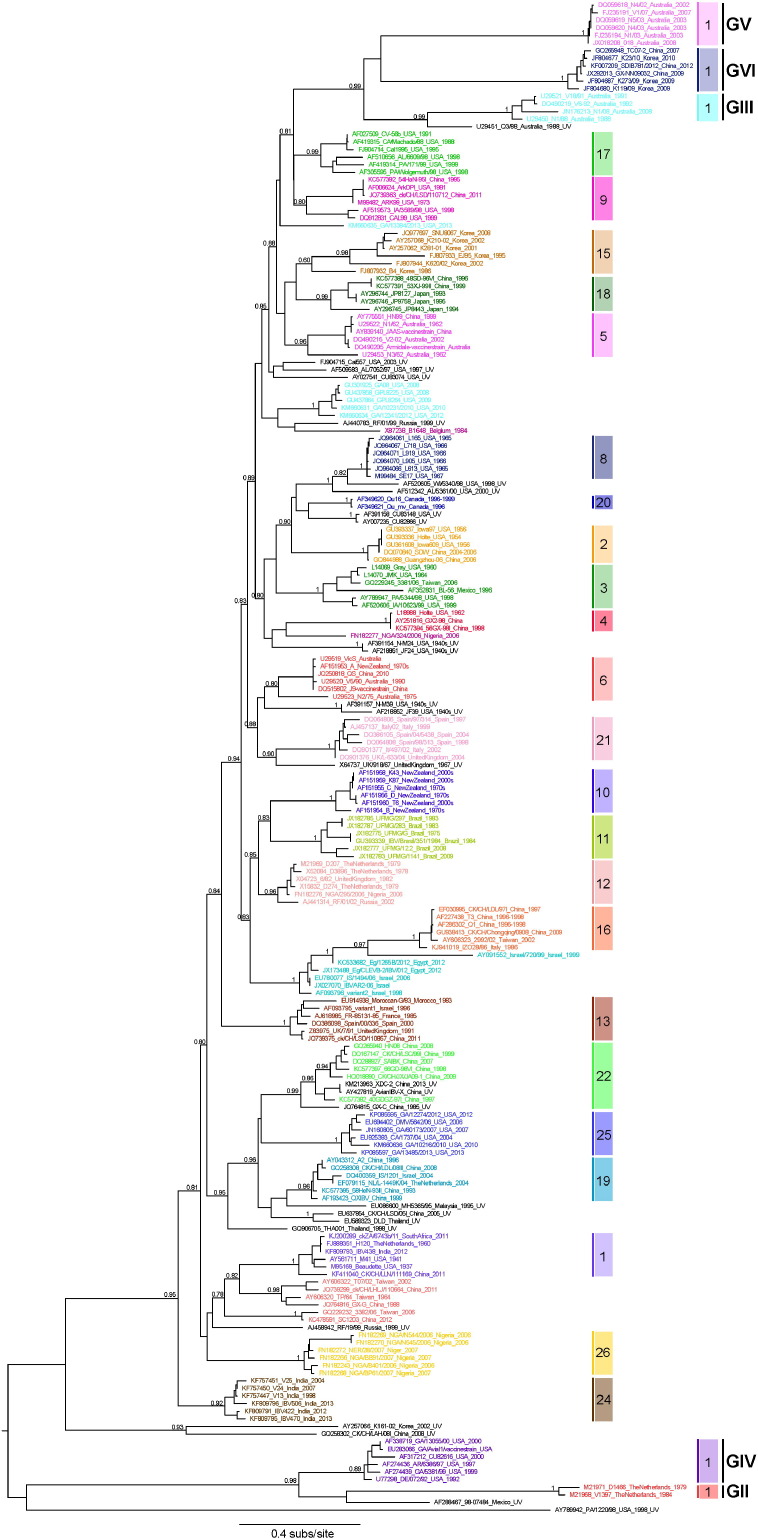
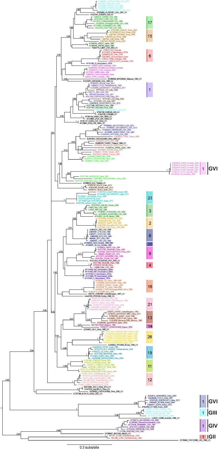
Infectious bronchitis virus (IBV) is the causative agent of a highly contagious disease that results in severe economic losses to the global poultry industry. The virus exists in a wide variety of genetically distinct viral types, and both phylogenetic analysis and measures of pairwise similarity among nucleotide or amino acid sequences have been used to classify IBV strains. However, there is currently no consensus on the method by which IBV sequences should be compared, and heterogeneous genetic group designations that are inconsistent with phylogenetic history have been adopted, leading to the confusing coexistence of multiple genotyping schemes. Herein, we propose a simple and repeatable phylogeny-based classification system combined with an unambiguous and rationale lineage nomenclature for the assignment of IBV strains. By using complete nucleotide sequences of the S1 gene we determined the phylogenetic structure of IBV, which in turn allowed us to define 6 genotypes that together comprise 32 distinct viral lineages and a number of inter-lineage recombinants. Because of extensive rate variation among IBVs, we suggest that the inference of phylogenetic relationships alone represents a more appropriate criterion for sequence classification than pairwise sequence comparisons. The adoption of an internationally accepted viral nomenclature is crucial for future studies of IBV epidemiology and evolution, and the classification scheme presented here can be updated and revised novel S1 sequences should become available.

Keywords: Classification; Evolution; IBV; Phylogeny.


Figures

Fig. 1
Fig. 1
Phylogenetic tree of complete S1 nucleotide sequences (1456 nt) of 1286 IBVs. The phylogeny shows the evolutionary relationships among all IBV genotypes and lineages proposed here. Each lineage is color-coded and its corresponding designation is reported. Unique variants (UVs) are marked in black. The red box designates the 27 lineages within GI. SH-like branch supports are shown for key nodes. The scale bar represents the number of nucleotide substitutions per site, and the tree is mid-point rooted for clarity only.
Fig. 2
Fig. 2
Phylogenetic tree of complete S1 nucleotide sequences. The phylogeny contains a total of 199 IBV strains, including 6 representative sequences of each lineage detected and 26 strains recognized as unique variants. Each lineage is color-coded and its corresponding designation is reported. Bars reporting the genotypes in which the lineages fall are shown. GenBank accession number, isolate number or name, country of origin and collection date is given for each strain. The designation “UV” indicates unique variants, here marked in black. A complete list of the 199 sequences used is provided in Table S1. SH-like branch supports are shown for key nodes. The scale bar represents the number of nucleotide substitutions per site, and the tree is mid-point rooted for clarity only. Phylogenetic tree of complete S1 nucleotide sequences. The phylogeny contains a total of 199 IBV strains, including 6 representative sequences of each lineage detected and 26 strains recognized as unique variants. Each lineage is color-coded and its corresponding designation is reported. Bars reporting the genotypes in which the lineages fall are shown. GenBank accession number, isolate number or name, country of origin and collection date is given for each strain. The designation “UV” indicates unique variants, here marked in black. A complete list of the 199 sequences used is provided in Table S1. SH-like branch supports are shown for key nodes. The scale bar represents the number of nucleotide substitutions per site, and the tree is mid-point rooted for clarity only.
Fig. 3
Fig. 3
Phylogenetic tree of partial S1 nucleotide sequences including HVRs1 and 2. The phylogeny contains a total of 199 IBV strains, including 6 representative sequences of each lineage detected and 26 strains recognized as unique variants. All strains belonging to the same lineage, assessed on the basis of the complete full-length sequences, are labeled with a unique color code as in Fig. 1, Fig. 2. The color-coded boxes reporting the lineage designations are only shown for those lineages correctly identified. GenBank accession number, isolate number or name, country of origin and collection date is given for each strain. The designation “UV” indicates unique variants, here marked in black. SH-like branch supports are shown for key nodes. The scale bar represents the number of nucleotide substitutions per site, and the tree is mid-point rooted for clarity only.
Fig. 4
Fig. 4
Phylogenetic tree of partial S1 nucleotide sequences including HVR3. The phylogeny contains a total of 199 IBV strains, including 6 representative sequences of each lineage detected and 26 strains recognized as unique variants. All strains belonging to the same lineage, assessed on the basis of the complete full-length sequences, are labeled with a unique color code as in Fig. 1, Fig. 2. The color-coded boxes reporting the lineage designations are shown only for those lineages correctly identified. GenBank accession number, isolate number or name, country of origin and collection date is given for each strain. The designation “UV” indicates unique variants, here marked in black. SH-like branch supports are shown for key nodes. The scale bar represents the number of nucleotide substitutions per site, and the tree is mid-point rooted for clarity only.

References

    1. Ababneh M., Dalab A.E., Alsaad S., Al-Zghoul M. Presence of infectious bronchitis virus strain CK/CH/LDL/97I in the Middle East. ISRN Vet. Sci. 2012;2012:201721. - PMC - PubMed
    1. Abdel-Moneim A.S., Madbouly H.M., Gelb J., Jr., Ladman B.S. Isolation and identification of Egypt/Beni-Suef/01 a novel genotype of infectious bronchitis virus. Vet. Med. J. Giza. 2002;50:1065–1078.
    1. Abdel-Moneim A.S., El-Kady M.F., Ladman B.S., Gelb J., Jr. S1 gene sequence analysis of a nephropathogenic strain of avian infectious bronchitis virus in Egypt. Virol. J. 2006;3:78. - PMC - PubMed
    1. Abdel-Moneim A.S., Afifi M.A., El-Kady M.F. Emergence of a novel genotype of avian infectious bronchitis virus in Egypt. Arch. Virol. 2012;157(12):2453–2457. - PubMed
    1. Abro S.H., Renström L.H., Ullman K., Isaksson M., Zohari S., Jansson D.S., Belák S., Baule C. Emergence of novel strains of avian infectious bronchitis virus in Sweden. Vet. Microbiol. 2011;155(2–4):237–246. - PMC - PubMed

Publication types

Substances