. 2016 Oct;36(7):1169-78.
doi: 10.1007/s10571-015-0313-4. Epub 2016 Feb 16.

Inhibition of Epidermal Growth Factor Receptor Improves Myelination and Attenuates Tissue Damage of Spinal Cord Injury

Affiliations

Inhibition of Epidermal Growth Factor Receptor Improves Myelination and Attenuates Tissue Damage of Spinal Cord Injury

Si Zhang et al. Cell Mol Neurobiol. 2016 Oct.

Abstract

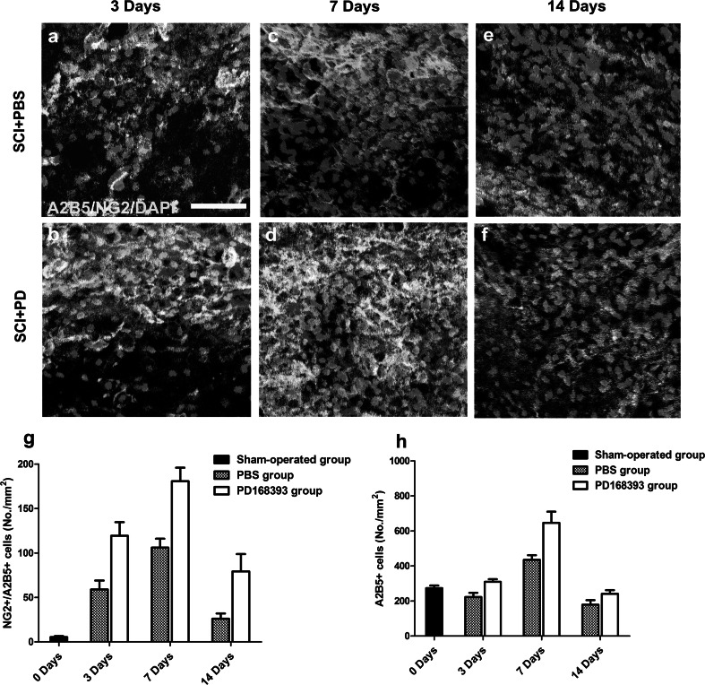
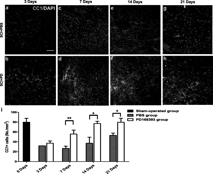
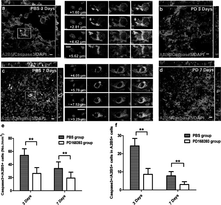
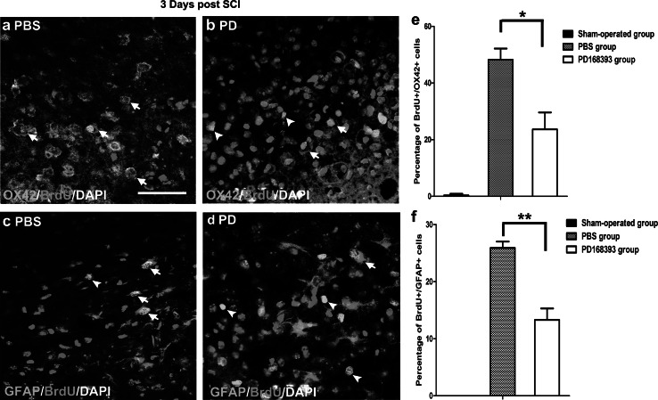
Preventing demyelination and promoting remyelination of denuded axons are promising therapeutic strategies for spinal cord injury (SCI). Epidermal growth factor receptor (EGFR) inhibition was reported to benefit the neural functional recovery and the axon regeneration after SCI. However, its role in de- and remyelination of axons in injured spinal cord is unclear. In the present study, we evaluated the effects of EGFR inhibitor, PD168393 (PD), on the myelination in mouse contusive SCI model. We found that expression of myelin basic protein (MBP) in the injured spinal cords of PD treated mice was remarkably elevated. The density of glial precursor cells and oligodendrocytes (OLs) was increased and the cell apoptosis in lesions was attenuated after PD168393 treatment. Moreover, PD168393 treatment reduced both the numbers of OX42 + microglial cells and glial fibrillary acidic protein + astrocytes in damaged area of spinal cords. We thus conclude that the therapeutic effects of EGFR inhibition after SCI involves facilitating remyelination of the injured spinal cord, increasing of oligodendrocyte precursor cells and OLs, as well as suppressing the activation of astrocytes and microglia/macrophages.

Keywords: EGFR inhibition; Remyelination; Spinal cord injury.


Conflict of interest statement

All authors declare that there are no conflicts of interest.

Figures

Fig. 1
Fig. 1
Effects of EGFR inhibition on gross histological change and myelin sparing post SCI. In control and PD168393 (PD)-treated group at 3 weeks post injury, histological changes were shown by HE staining (sagittal sections). The injured segment showed distinct cell loss, cell aggregation and scar formation at the injury epicenter (ac). Immunostaining with anti-MBP antibody of spinal cords (coronal sections) in both control (left panel) and PD-treated group (right panel) at 3 weeks post injury (d, e). MBP immunofluorescent staining of the injury site at 3, 7 and 14 days post SCI. Nuclei were counterstained with DAPI in all images (sagittal sections) (fm). Quantitative analysis of MBP signals in the injured areas per mm2 shown in fm (n). Scale bars 500 µm (ae); 25 µm (fm)
Fig. 2
Fig. 2
A2B5 +/NG2 + OPCs at 3, 7 and 14 days of SCI. Immunostaining of A2B5 (red) and NG2 proteoglycan (green) in both control group (a, c, e) and PD-treated group (b, d, f) at 3, 7 and 14 days post SCI. Nuclei were counterstained with DAPI in all images. The number of NG2 +/A2B5 + cells was quantitatively compared between control and PD-treated groups in 3, 7 and 14 days post injury (g). Quantitative analysis of A2B5 immunopositive cells between control and PD-treated groups in 3, 7 and 14 days post injury (h). Scale bars 50 µm
Fig. 3
Fig. 3
The CC1 + oligodendrocytes are increased by PD treatment. Immunostaining of CC1 (red) in both control group (a, c, e, g) and PD-treated group (b, d, f, h) at 3, 7, 14 and 21 days post SCI. Nuclei were counterstained with DAPI in all images. Quantification of CC1 + cells in above experiments (i). Scale bars 50 µm
Fig. 4
Fig. 4
PD alleviates OPC apoptosis in SCI. Z-stack confocal images of tissue harvested from control and PD-treated mice at 3 (a, c) and 7 days (b, d) post injury was stained with antibodies against caspase-3 (green) and A2B5 (red). Nuclei were counterstained with DAPI in all images. Quantification of apoptotic A2B5 + oligodendrocyte progenitors within the 6 mm along rostral-caudal axis of spinal cord (e) (n = 5, **P < 0.01). The proportion of caspase3 +/A2B5 + cells in A2B5 + cells per mm2 was determined by dividing the number of cells double-labeled for caspase-3 and A2B5 by the total number of A2B5 + cells in control and PD-treated group respectively (f) (n = 5, **P < 0.01). Images showed z-axis projections of 23 × 0.40 µm (a), 22 × 0.55 µm (b), 25 × 0.40 µm (c), 21 × 0.40 µm (d). Scale bars 10 µm
Fig. 5
Fig. 5
EGFR inhibition reduces the proliferative OX42 + macrophages and GFAP + astrocytes at 3 days post injury. Immunofluorescent images showed the BrdU +/OX42 + (a, b), BrdU/GFAP + (c, d) cells in the injury epicenter in both control and PD-treated groups. Nuclei were counterstained with DAPI. Examples of double-labeled OX42 +/BrdU + cells and GFAP +/BrdU + cells were indicated by arrow. Examples of BrdU+ only cells were indicated by arrowhead. The results were confirmed by quantitative analysis of number counting in (e) and (f) (n = 5, *P < 0.05, **P < 0.01). Scale bars 50 µm
Fig. 6
Fig. 6
PD treatment down-regulates the number of OX42 + cells. The panoramic images of sections at 3 and 21 days post SCI were shown in control (a, c) and PD-treated (b, d) group. Quantification OX42 + cells in control (black columns) and PD-treated injured segment (white columns) at 3 or 21 days of injury were shown in (e) and (f). Scale bars 500 µm

References

    1. Aguirre A, Gallo V (2007) Reduced EGFR signaling in progenitor cells of the adult subventricular zone attenuates oligodendrogenesis after demyelination. Neuron Glia Biol 3(3):209–220 - PMC - PubMed
    1. Aguirre A, Dupree JL, Mangin JM, Gallo V (2007) A functional role for EGFR signaling in myelination and remyelination. Nat Neurosci 10(8):990–1002 - PubMed
    1. Ahmed Z, Jacques SJ, Berry M, Logan A (2009) Epidermal growth factor receptor inhibitors promote CNS axon growth through off-target effects on glia. Neurobiol Dis 36(1):142–150 - PubMed
    1. Berry M, Ahmed Z, Douglas MR, Logan A (2011) Epidermal growth factor receptor antagonists and CNS axon regeneration: mechanisms and controversies. Brain Res Bull 84(4–5):289–299 - PubMed
    1. Bezzi P, Volterra A (2001) A neuron-glia signalling network in the active brain. Curr Opin Neurobiol 11(3):387–394 - PubMed

MeSH terms

LinkOut - more resources