. 2016 Feb;41(1-2):431-49.
doi: 10.1007/s11064-016-1850-z. Epub 2016 Feb 17.

Proof-of Concept that an Acute Trophic Factors Intervention After Spinal Cord Injury Provides an Adequate Niche for Neuroprotection, Recruitment of Nestin-Expressing Progenitors and Regeneration

Affiliations

Proof-of Concept that an Acute Trophic Factors Intervention After Spinal Cord Injury Provides an Adequate Niche for Neuroprotection, Recruitment of Nestin-Expressing Progenitors and Regeneration

Warin Krityakiarana et al. Neurochem Res. 2016 Feb.

Erratum in

Abstract

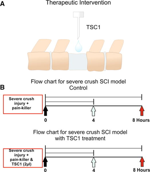
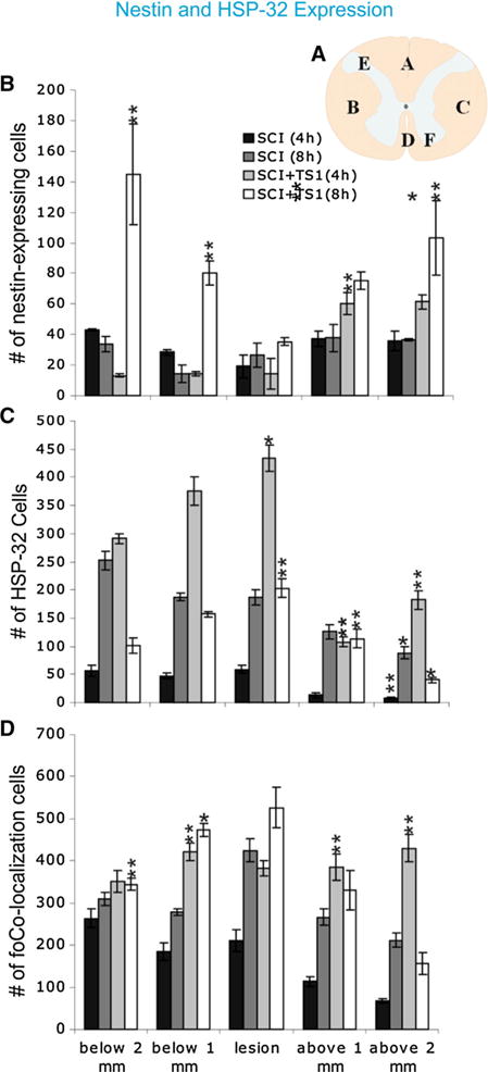
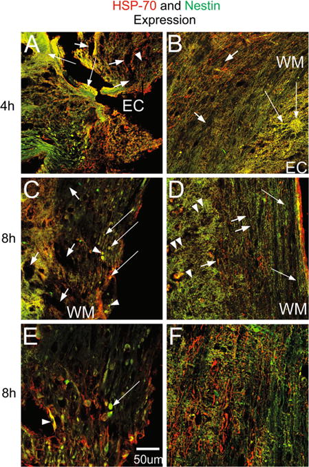
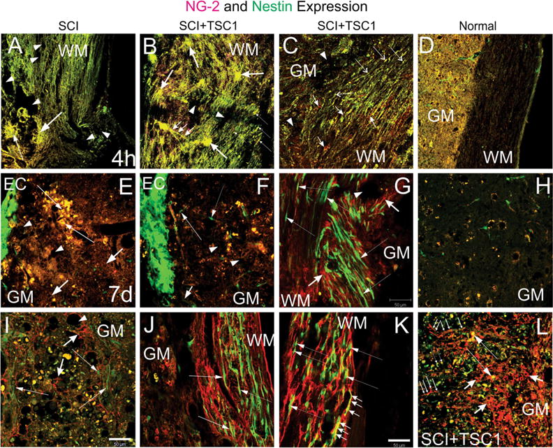
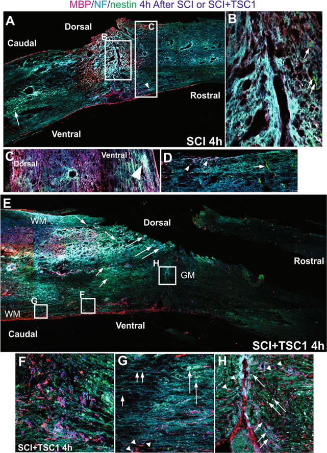
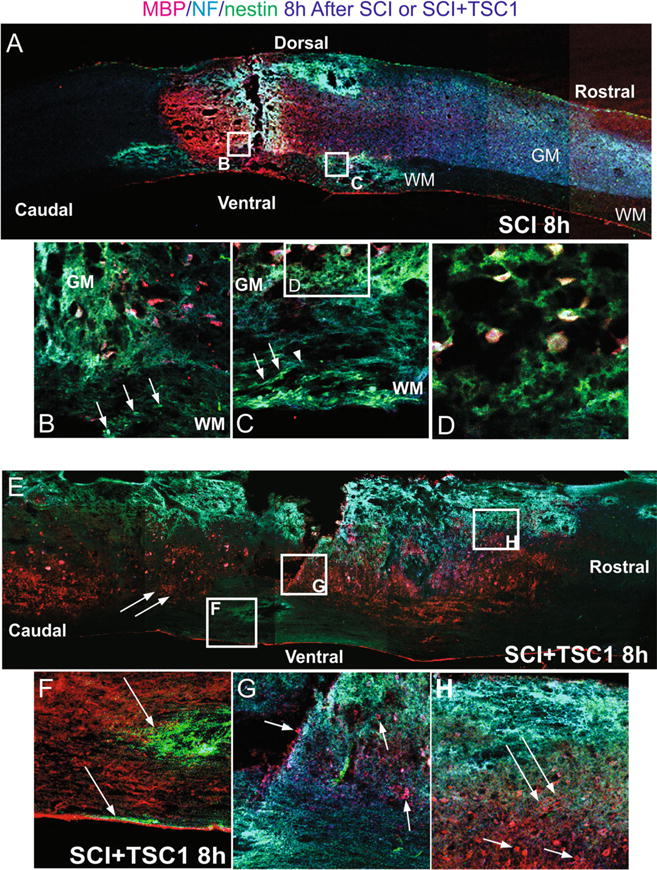
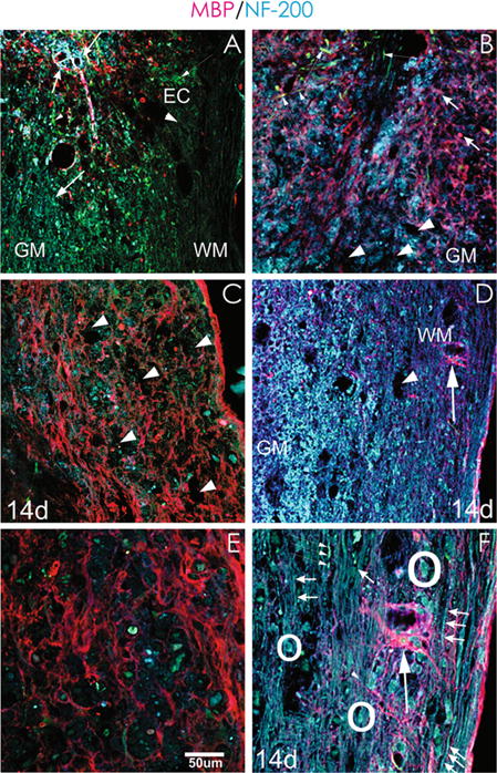
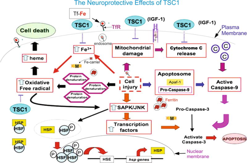
Trophic factor treatment has been shown to improve the recovery of brain and spinal cord injury (SCI). In this study, we examined the effects of TSC1 (a combination of insulin-like growth factor 1 and transferrin) 4 and 8 h after SCI at the thoracic segment level (T12) in nestin-GFP transgenic mice. TSC1 treatment for 4 and 8 h increased the number of nestin-expressing cells around the lesion site and prevented Wallerian degeneration. Treatment with TSC1 for 4 h significantly increased heat shock protein (HSP)-32 and HSP-70 expression 1 and 2 mm from lesion site (both, caudal and rostral). Conversely, the number of HSP-32 positive cells decreased after an 8-h TSC1 treatment, although it was still higher than in both, non-treated SCI and intact spinal cord animals. Furthermore, TSC1 increased NG2 expressing cell numbers and preserved most axons intact, facilitating remyelination and repair. These results support our hypothesis that TSC1 is an effective treatment for cell and tissue neuroprotection after SCI. An early intervention is crucial to prevent secondary damage of the injured SC and, in particular, to prevent Wallerian degeneration.

Keywords: Heat shock proteins; IGF-1; Myelin; Oligodendrocytes; Transferrin; Wallerian degeneration.


Figures

Fig. 1
Fig. 1
a Schematic representation on TSC1 mode of administration as drops over the lesion at the thoracic level (T12). Nestin-GFP transgenic mice generated by Yamaguchi and co-workers [34] (2000) 6 months of age were used in this study. The surgery included laminectomy and after laminectomy, mice received a severe crush at the lumbar level (L1–L2) followed by the administration of a 2 μl drop of TSC1 as shown in Fig. 1B. This approach allowed immediate contact with the spinal cord tissue and rapid absorption of TSC1. Because the volume is small the full amount of TSC1 reaches the dorsal portion of the spinal cord tissue. b Time line showing when the severe crush was performed as well as, the TSC1 administration
Fig. 2
Fig. 2
TSC1 increased the number of nestin-expressing cells 4 and 8 h after treatment. a Schematic representation of a transverse section of the spinal cord showing the six different areas used to count cells. The bar graph represents the total number of cells from three separate groups that were counted: nestin-GFP expressing cells (b), HSP-32 positive cells (c), and, co-expression of both markers by the same cell (d). Quantification was performed from coronal spinal cord sections of control (SCI) mice and SCI + TSC1 mice. HSP-32 positive cell numbers reached their highest at the level of the lesion and below (caudal to) the lesion after TSC1 treatment. Nestin-GFP expressing cells in SCI mice were increased after been treated with TSC1 for 4 and 8 h. The number of cells co-expressing nestin-GFP and HSP-32 was increased after treatment with TSC1. Note that the total number of nestin or HSP-32 expressing cells would be the sum of the corresponding bars for each marker and the number represented in the colocalization bar. Bars represent the mean ± SD; *p < 0.05, **p < 0.01 versus respective control
Fig. 3
Fig. 3
Sagittal views of the spinal cord at the lesion level of SCI and SCI + TSC1 mice at 4 and 8 h after injury. a HSP-70 positive cells and nestin-GFP expressing cells were present at the epicenter (EC) where the tissue that showed sponginess (EC), massive cell loss and cavitation were already visible (short arrows). In most regions nestin expressing cells weren’t distinguishable. HSP-70 labeled the soma of some cells as well as, some thin and hair-like fibers. These cells seemed to migrate in rows towards the lesion in the caudal semi-stump and they would stop at the lesion edge as if there was an imaginary border. A real colocalization of nestin and HSP-70 was not observed. b Mice treated with TSC1 after SCI displayed HSP-70 in the WM both in their thin fibers and in their cell body (long arrows). Numerous HSP +/nestin negative cells were present in the gray matter. Certain large multipolar cells in the WM co-expressed HSP-70 and nestin. c Eight hours after injury caudal and adjacent to the epicenter of the lesion cavitation was considerably reduced and very few cells expressed nestin intensely (arrowheads) while others faintly co-expressed nestin and HSP70 (long arrows). There were also small HSP-70 + positive nestin-negative cells in the white and gray matter. d Eight hours after lesion and TSC1 administration, the cytoarchitecture of both WM and GM were very well preserved. Nestin labeled hairy structures were organized as fishing net over the extent of the gray matter (arrowheads) while, in the WM nestin labeled fibers were parallel to axons and perpendicular to the nestin positive structures in the gray matter (arrows). Hairy fibers were labeled for HSP-70 mainly in the WM and they were devoid of nestin expression. e Eight hours after injury and 1 mm from the lesion epicenter, enlarged green cells were present, and almost no HSP-70 was found. Beyond 1 mm and caudal to de lesion, colocalization of both markers and more HSP-70 were observed. f Eight hours after mice were treated with TSC1 both, the WM and GM were well preserved with a robust expression of HSP-70 in numerous thin and thick fibers (short arrows) in the GM. There were also green small cells around axonal fibers in the WM with numerous thin nestin positive fibers extending perpendicularly to the ependymal canal over the GM towards the WM (arrowheads)
Fig. 4
Fig. 4
Two types of progenitors were present upon TSC1 intervention. a and b show low magnification views of the lesion epicenter. a Four hours after injury at the level of the lesion, the tissue appeared disrupted and without defined cellular structures that would have expressed nestin (neural progenitors) or OLPs identified by NG2. Moreover, the tissue showed a spongy appearance (arrows), and presented extensive cavitation (arrowheads). b In contrast the tissue from mice receiving TSC1 showed two types of structures: 1 Clusters of cells co-expressing nestin and NG2 (yellow). These cells were characterized by extending several long cell processes that appeared to go above nestin negative NG2 positive structures as a bridge for small cells to migrate towards the WM (small arrows). 2 Cells devoid of their processes (thin, long arrows). There were very small signs of cavitation (dotted areas). c Below the lesion (1.5 mm) along white matter tracks numerous small bipolar nestinansd NG2 expressing cells had appeared (Long and thin arrows). There were also interfascicular cells expressing solely NG2 mainly in their cell body (short arrows). d Low magnification view of a spinal cord of a naïve mouse at the T12 level. Seven days after injury panels e–g. e At higher magnification, the ependymal canal showed intense nestin expression (EC). The tissue adjacent to the ependymal canal had a spongy appearance (short arrows). A few patches of cells co-expressing nestin and NG2 were visible (yellow cells; long, thin arrows). At a longer distance from the EC the yellow cells were disappearing leaving behind only debris. f Seven days after injury and TSC1 treatment, the ependymal canal displayed an intense nestin expression. Beyond the EC few scattered, bipolar nestin-bearing cells appeared to migrate perpendicular to the EC and towards the white matter (long thin arrow). There was some cavitation in the GM (arrowheads). Some NG2 and nestin co-expressing cells presumably had migrated away from the canal (yellow cells). g The WM displayed a plethora of nestin-expressing bipolar cells mostly arranged in rows following the direction of the axons. Thus, nestin-expressing cells that had migrated following those axons directly exposed to the severe crush accumulated where the axons were severed. Long, thin arrows show examples of bipolar cells appearing to migrate along the axons. In areas where the white matter suffered to a lesser extent, nestin-expressing cells with elongated thin processes were also seen (thin, long arrows). Cavitation was less frequent in TSC1 treated mice (arrowhead). NG2 expressing cells had formed a matrix with their cell processes that intermingled with the axons and nestin-expressing cells (short, thick arrows). h Low magnification view of a naïve spinal cord’s gray matter. i Fourteen days after the intervention, the GM of mice that did not receive TSC1 had a lace-like aspect and NG2 labeled fibrous cell processes surrounding the cavities that formed upon tissue loss. Numerous cells that co-expressed both NG2 and nestin were enlarged and seemed to be dying. Very few green cells bearing faint nestin expression were visible at this time point (long, thin arrows). In contrast, mice treated with TSC1 (panel L) was populated of numerous NG2-expressing cells with large cell body and fibrous processes (short, thick arrows) Long. They had formed a dense structure across the GM that appeared to serve as a migratory substrate of both nestin expressing cells (long thin arrows) and NG2/nestin co-expressing cells (thin, long arrows). NG2 expressing cells were much larger than the NG2/nestin co-expressing cells. j Surprisingly, the white matter of mice 14 days after SCI had preserved its structure to some extent and nestin positive cells had reached them and appeared to migrate along axons (thin, long arrows). k In mice treated with TSC1 the white matter was populated by bipolar nestin positive cells intermingled with NG2 intensely labeled cells, the WM structure was nicely preserved allowing for the migration of nestin-expressing neural progenitors (thin, long arrows). Interestingly, bipolar cells co-expressing nestin and NG2 were abundant (small arrows), while in mice without TSC1 bipolar cells in the white matter mostly expressed nestin alone
Fig. 5
Fig. 5
Comparison of mouse spinal cord 4 h after SCI or SCI + TSC1. a The low magnification view of the spine showed fibrosis at the epicenter and surrounding areas that appeared to be severed axons as NF labeled fibers accumulated. Caudal to the lesion cavitation was visible in the gray matter where nestin expressing cells had accumulated and were intermingled with NF labeled fibers and both dorsally and ventrally there was reactive tissue. b At higher magnification the view of the epicenter showed spongy tissue and some MBP positive cells were visible while the neighboring tissue had already become lace-like. Rostrally and adjacent to the epi-center few small nestin positive cells could be seen (arrows). c Higher magnification view of the tissue rostral to the lesion where cavitation had not started and nestin expressing cells appeared to sit on NF labeled axons. d Caudal to the lesion bipolar nestin positive cells appeared to migrate from the ependymal canal [EC] and perpendicular to it across axonal fibers. MBP positive cells were present in this area. e Low magnification view of a mouse spinal cord 4 h after SCI + TSC1, the overall structure of the spinal cord has been preserved. Caudal to the lesion NF labeled cell bodies were present (long arrows) and an extensive scar had not formed. Motoneurons were also present (long arrows). Numerous nestin positive cells single or in rows were frequent (short arrows). f High magnification view of the ventral white matter caudal to the lesion MBP expressing OL and myelin segments are visible intermingled and parallel to the ependymal canal. Numerous nestin expressing cells were also seen. g The white matter showed segments of myelinated axons that were abruptly ended by tissue loss (cavitation; arrowheads). Other axons appeared normally myelinated. Numerous axonal fibers faintly labeled for NF appeared to keep their position and structure of the WM (short arrowheads). h Higher power view of the epicenter at the level of the gray matter (GM). The tissue lost its form but not completely, MBP labeled axons appear to have severed or lost their label. Nonetheless, numerous MBP expressing cells were also in the vicinity of the lesion both caudally and rostrally (arrowheads). Numerous nestin positive small cells surrounding the lesion from the rostral side appeared to migrate towards it (long arrows). In this area the axons had adopted the form of the tissue after the severe crush they were bearing the NF marker (small arrows)
Fig. 6
Fig. 6
Comparison of mouse spinal cord 4 h after SCI or SCI + TSC1. a The low magnification view of the spine shows fibrosis at the epicenter and surrounding areas that appeared to be severed axons as NF labeled fibers accumulated at both sides, caudal to the lesion there was cavitation mostly in the gray matter where the NF label was arrested. Rostral to the lesion the white matter appeared to have suffered the most showing cavitation dorsally and rostrally. b A high magnification view of the interphase between the white and gray matter adjacent to the epicenter where nestin positive cells were present (arrows). In the gray matter there were some MBP positive cells without cell processes in areas of cavitation. c Rostrally and ventral to the lesion in the white matter nestin expressing cells were seen as if they were using axonal fibers to migrate in the direction of the gray matter (arrows). These cells seem to find the end of the road where the fiber stops at the cavitation edge (arrowhead) and an accumulation of green progenitors was seen in the outer portions of the white matter. d A closer view (even at higher magnification) of the white matter shows MBP positive cells held by thin tissue fibers. Some of these OLs showed a round appearance and no cell processes. Other MBP positive cells appeared bipolar with MBP expressed in their cell body and one nestin positive cell process. Yet, in the gray matter the cavitation was expanded. e There was a striking difference between the untreated and the TSC1 treated spinal cords. TSC1 appears to have preserved the myelin fibers in the white matter neighboring the gray matter. Moreover, numerous OL bearded MBP in their somas. f At the caudal portion of the lesion and ventral to it there were foci formed by cavitation that had been invaded by numerous nestin expressing progenitors (arrow) or in the external portion of the white matter (arrow). But neither MBP nor NF staining was present in the adjacent white matter. g At the epicenter in the gray matter MBP positive cells were visible together with NF immunoreactivity (arrows). Dorsally and rostral to the lesion the WM displayed considerable cavitation the cavitation that extended into the gray matter. Nonetheless, fibrous cells and scar-like structures appeared to be covered by a light and extensive veil of nestin positive thin and hairy fibers shown in h (long arrows). These fibers were located in a radial fashion in an approximately 45 degrees angle from the pia mater in direction to the epicenter. The gray matter contained numerous MBP positive OLs (arrows)
Fig. 7
Fig. 7
TSC1 prevents Wallerian degeneration after SCI. a Seven days after crush and 1 mm from the epicenter the SC displayed cavitation but, it still had preserved it’s cytoarchitecture, besides at the lesion border where MBP label was seen. Beyond that point between 1 and 2 mm from the lesion almost not MBP could be seen in either the WM or GM. There was not NF label along axons in this region, NF was seen on some cells expressing it in their soma and processes that extended only at the border of the lesion but not beyond it (Thick, short arrows). b At this time point mice treated with TSC1 showed scarce cavitation sites of modest size when compared to panel A. Both MBP and NF labels. Few nestin labeled, bipolar cells and fibers appeared in the vicinity of the lesion (thin arrows). c A week later SCI non-treated mice had more cavitation than seven days after injury (arrows), numerous nestin positive small cells were visible in both the gray and white matter (arrowheads). Some moto-neurons expressing NF in their cell body were also found although devoid of processes. Disorganized or even transected-like myelin structures were abundant in the white matter and less abundant and fainter in the gray matter, some of them appeared to be reactive-like OL. d Fourteen days after injury and TSC1 administration, the GM was populated with numerous NF positive cells and structures and MBP labeled fibers were abundant. MBP staining allowed for the visualization of the nodes of Ranvier along axons (arrows). Some turquoise colored bipolar cells appeared to migrate along a myelinated segment (arrowhead). e At higher magnification cavitation was more evident (14 d after SCI), motoneuron cell bodies in blue were scarce. Green enlarged cells of unhealthy appearance were present and there were also a few smaller, healthy nestin positive cells. In the white matter bundles of axons were disrupted and neuronal cell bodies were clearly compromised. Some very small cells intensely labeled for NF were seen but there was not tissue to support their potential migration. f Higher magnification view of a spinal cord 14 days after but not transected. They extended along the field of view in a parallel arrangement. Other fibers were myelinated and alternating nodes of Ranvier and myelin segments were clearly distinguishable. Neuronal survival or perhaps naisance of new neurons, were facilitated by TSC1 as numerous motoneurons were present. Moreover, only scarce cavitation was found. Interestingly, the colocalization of nestin and NF in some if not most motoneurons at the level of the cell body is evident (arrowheads). OLs with a prominent cell body and at least ten visible large processes extended over long distance reaching out to axons
Fig. 8
Fig. 8
Schematic representation of some mechanisms that may be modulated by TSC1 with respect to apoptosis and iron homeostasis

Similar articles

Cited by

References

    1. Nelson E, Gertz SD, Rennels ML, Ducker TB, Blaumanis OR. Spinal cord injury. The role of vascular damage in the pathogenesis of central hemorrhagic necrosis. Arch Neurol. 1997;34:332–333. - PubMed
    1. Lu J, Ashwell KW, Waite P. Advances in secondary spinal cord injury: role of apoptosis. Spine. 2000;25(14):1859–1866. - PubMed
    1. Chang H, Wo JP, Tzeng SF. Neuroprotection of glial cell line-derived neurotrophic factor in damaged spinal cords following contusive injury. J Neuro Res. 2002;69:397–405. - PubMed
    1. McDonald JW, Sadowsky C. Spinal-cord injury. Lancet. 2002;359:417–425. - PubMed
    1. Beattie MS, Bresnahan JC, Komon J, et al. Endogenous repair after spinal cord contusion injuries in the rat. Exp Neurol. 1997;148(2):453–463. - PubMed