. 2016 Sep;1859(9):1170-1182.
doi: 10.1016/j.bbagrm.2016.02.008. Epub 2016 Feb 12.

A SUMO-acetyl switch in PXR biology

Affiliations

A SUMO-acetyl switch in PXR biology

Wenqi Cui et al. Biochim Biophys Acta. 2016 Sep.

Abstract

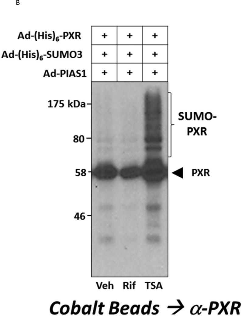
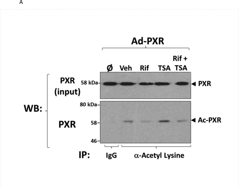
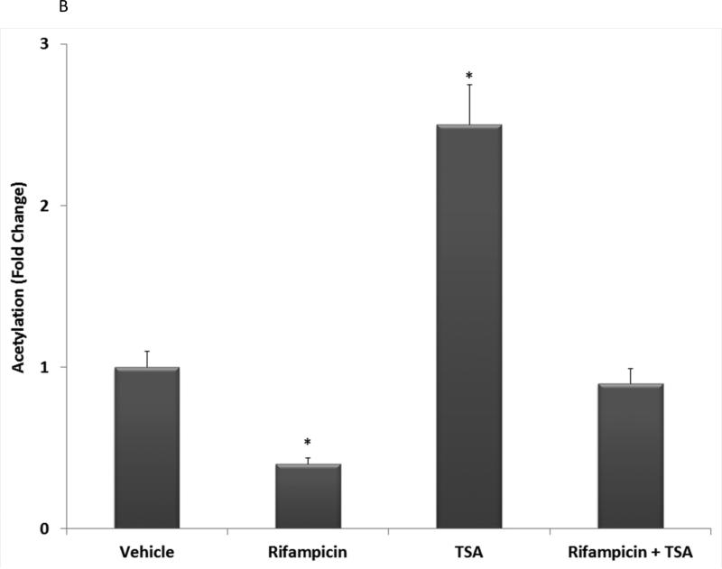
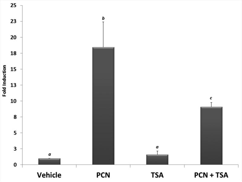
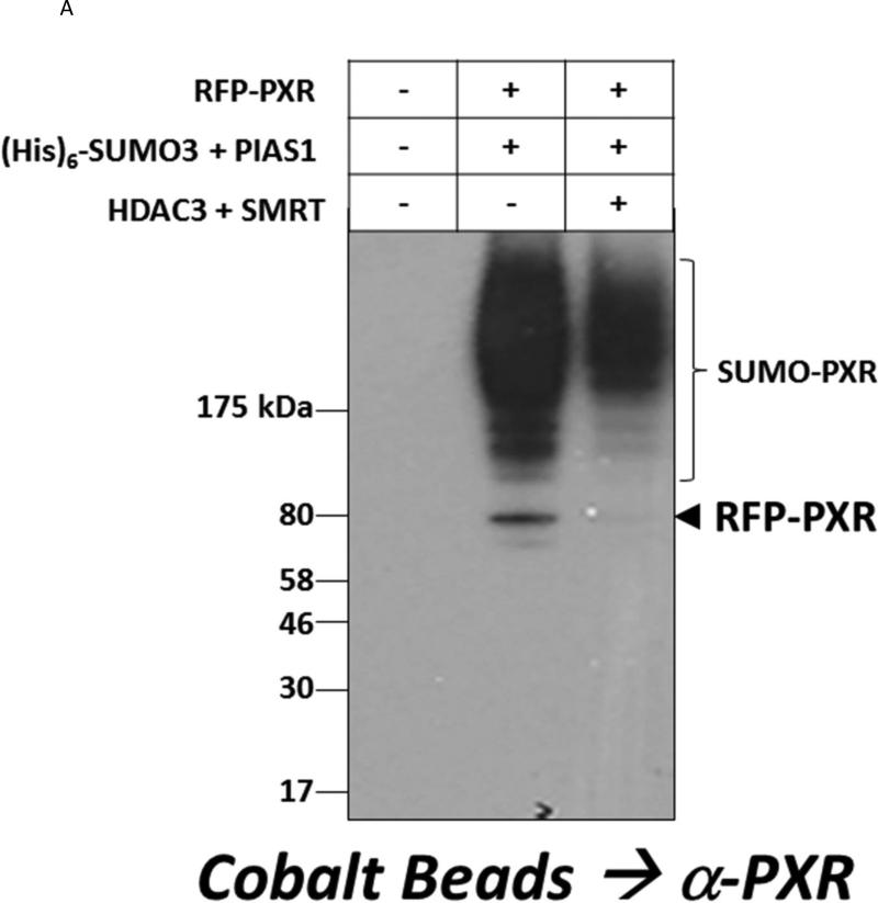
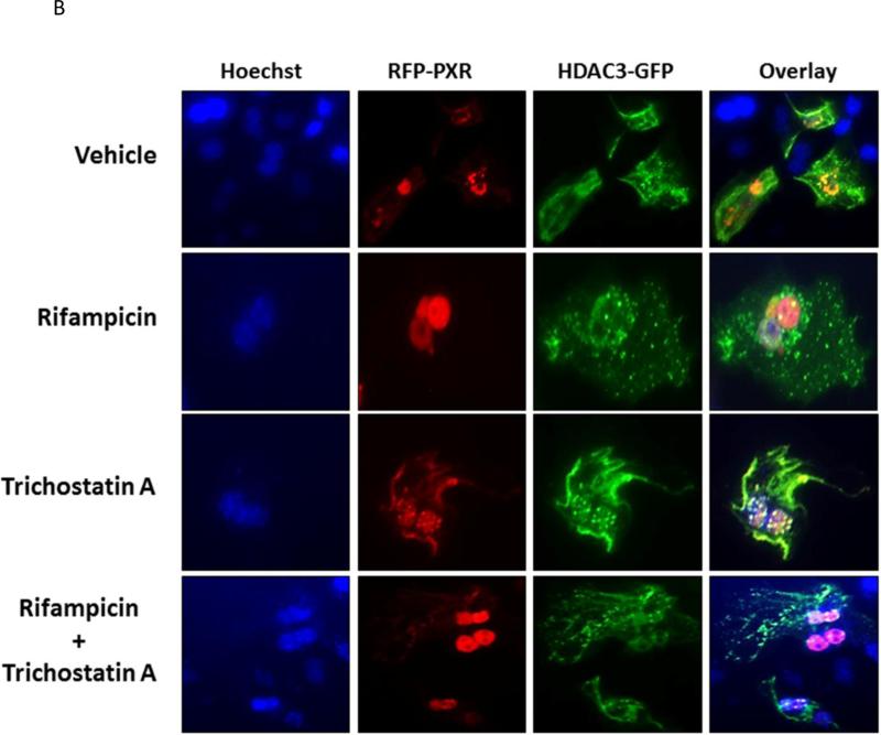
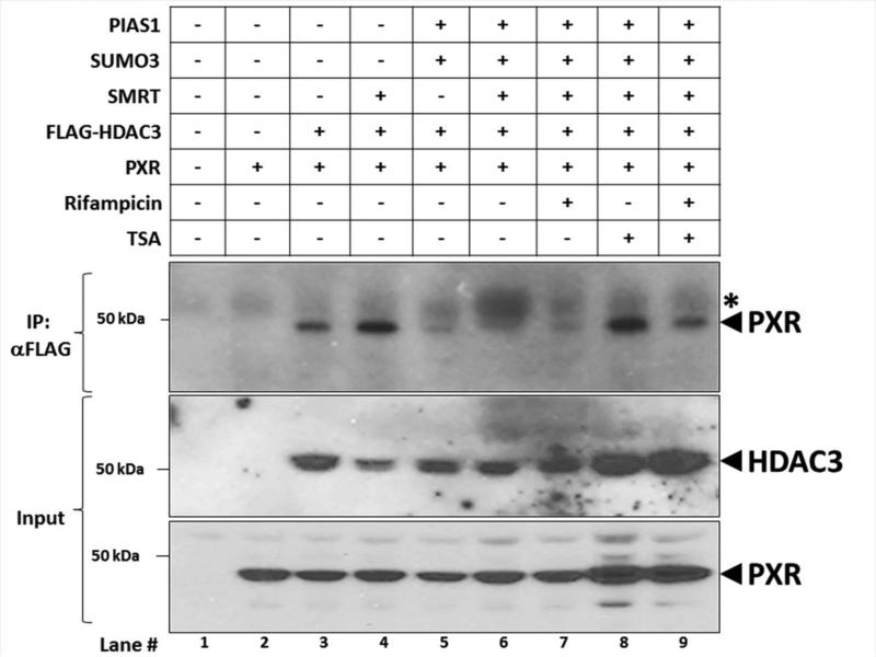
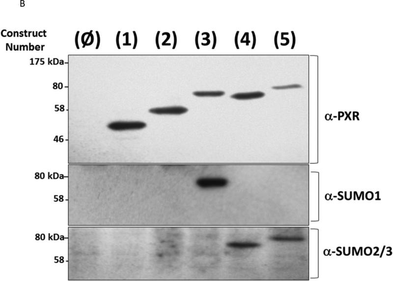
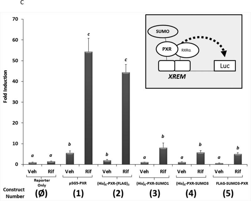
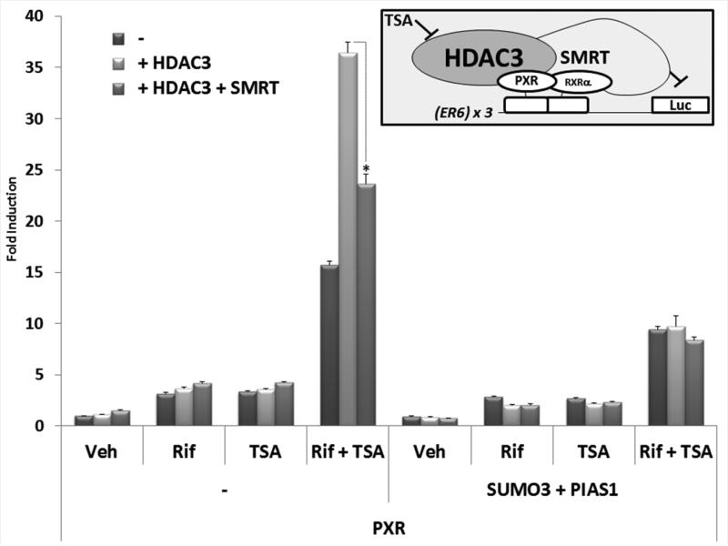
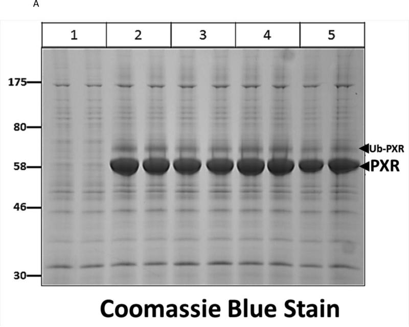
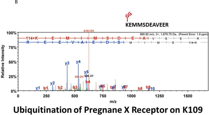
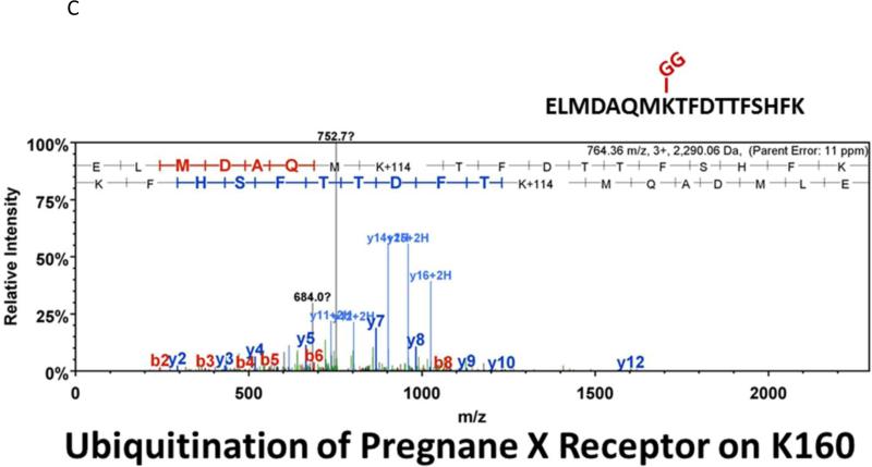
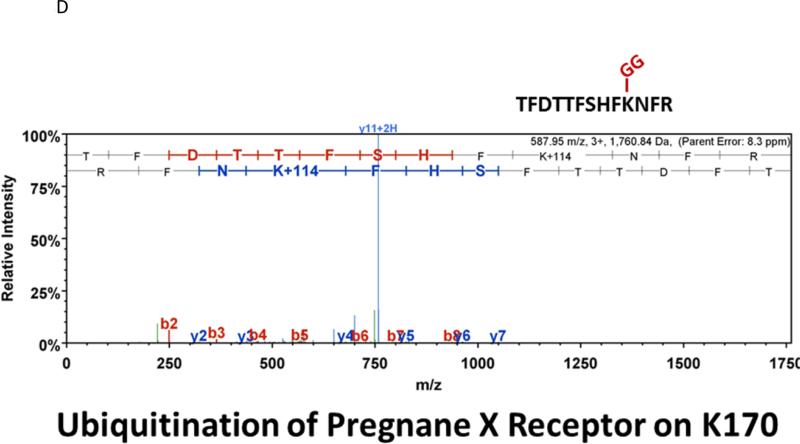
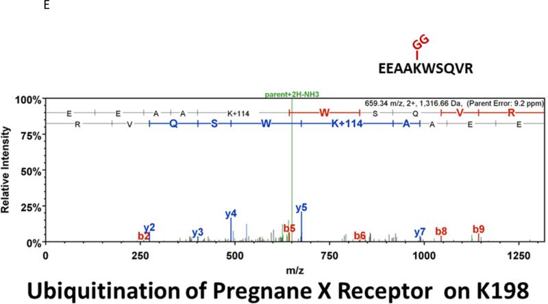
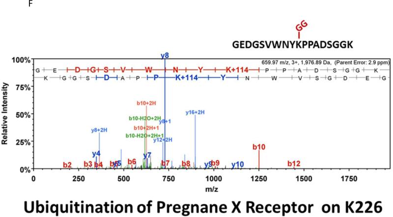
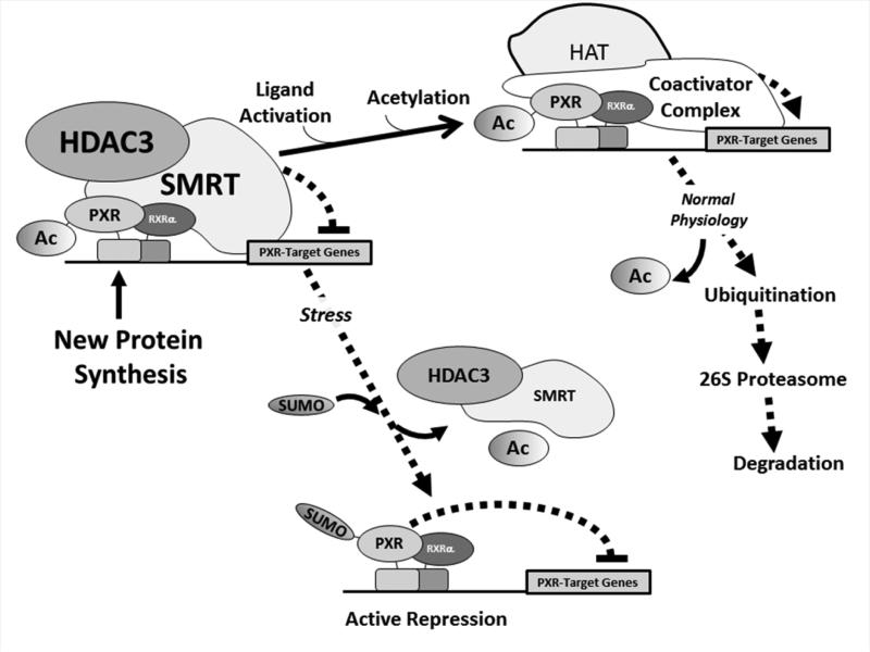
Post-translational modification (PTM) of nuclear receptor superfamily members regulates various aspects of their biology to include sub-cellular localization, the repertoire of protein-binding partners, as well as their stability and mode of degradation. The nuclear receptor pregnane X receptor (PXR, NR1I2) is a master-regulator of the drug-inducible gene expression in liver and intestine. The PXR-mediated gene activation program is primarily recognized to increase drug metabolism, drug transport, and drug efflux pathways in these tissues. The activation of PXR also has important implications in significant human diseases including inflammatory bowel disease and cancer. Our recent investigations reveal that PXR is modified by multiple PTMs to include phosphorylation, SUMOylation, and ubiquitination. Using both primary cultures of hepatocytes and cell-based assays, we show here that PXR is modified through acetylation on lysine residues. Further, we show that increased acetylation of PXR stimulates its increased SUMO-modification to support active transcriptional suppression. Pharmacologic inhibition of lysine de-acetylation using trichostatin A (TSA) alters the sub-cellular localization of PXR in cultured hepatocytes, and also has a profound impact upon PXR transactivation capacity. Both the acetylation and SUMOylation status of the PXR protein is affected by its ability to associate with the lysine de-acetylating enzyme histone de-acetylase (HDAC)3 in a complex with silencing mediator of retinoic acid and thyroid hormone receptor (SMRT). Taken together, our data support a model in which a SUMO-acetyl 'switch' occurs such that acetylation of PXR likely stimulates SUMO-modification of PXR to promote the active repression of PXR-target gene expression. This article is part of a Special Issue entitled: Xenobiotic nuclear receptors: New Tricks for An Old Dog, edited by Dr. Wen Xie.

Keywords: Acetylation; Histone deacetylase 3; Nuclear receptor; Post-translational modification; Pregnane X receptor; SUMOylation; Silencing mediator of retinoic acid and thyroid hormone receptor; Ubiquitination.


Figures

Figure 1
Figure 1. The Interface between Acetylation and SUMOylation of PXR
(A) Hepa1-6 cells were transfected with the indicated plasmid-based expression vectors. Cell lysates were produced using strong denaturing conditions to inhibit de-SUMOylation enzymes. SUMOylated proteins were enriched using cobalt beads and were washed sequentially using both guanidine-HCl and urea-based wash buffers. Proteins were eluted using 2X-Laemmli buffer and were then resolved using 10% SDS-PAGE. Western blot analysis was performed with an anti-PXR antibody that detects all modified forms of the protein (Santa Cruz, H-11 monoclonal Ab). (B) Hepa1-6 cells were transduced with the indicated adenoviral expression vectors. Twenty-four hr post-transduction, cells were treated with vehicle (0.1% DMSO) rifampicin (Rif, 10 μM) or Trichostatin A (TSA, 0.5 μM) for an additional twenty-four hr. SUMOylated proteins were gathered as in (A) and western blot with the anti-PXR antibody was used to analyze the extent of SUMO-modification.
Figure 2
Figure 2. Acetylation of PXR is Altered during Transactivation by Ligand
(A) Primary hepatocytes isolated from wild type C57BL/6 mice were isolated and transduced with an adenoviral expression vector encoding a FLAG-tagged form of human PXR (Left Panel). Total acetylated proteins were immunoprecipitated from cell extracts using a cocktail of four anti-acetylated lysine monoclonal antibodies as described in Materials and Methods. A non-immune antibody was also used as a negative control (IgG). Acetylated PXR was identified by western blotting with anti-PXR polyclonal antibody. (B) Western Blot images were quantitated by densitometric scanning of the X-ray films with the UVP Biodoc-It 220 image analysis system and 1D Gel Analysis Software. The numbers represent the relative densitometric image intensity of acetylated PXR divided by the image intensity of input levels of PXR, where vehicle treated control group was set to equal 1. Asterisks indicate a statistical difference from vehicle-treated samples (n = 3, and P < 0.05).
Figure 3
Figure 3. PXR Transactivation Capacity is the Molecular Target of Acetylation in Hepatocytes
Primary hepatocytes isolated from C57BL/6 mice were cultured overnight. Following twenty-four hr treatment with pregnenolone 16α-carbonitrile (PCN, 10 μM), trichostatin A (TSA, 0.5 μM), or both together, total RNA was isolated and the relative expression level of the Cyp3a11 gene was determined. Data are normalized to β-actin levels and are presented as fold induction. Letters different from each other indicate a statistical difference between treatment groups (n=3, and p<0.05).
Figure 4
Figure 4. The HDAC3 Deacetylase Enzyme Affects PXR-SUMOylation and Co-localizes with PXR in Hepatocytes
(A) Hepa1-6 cells were transfected with the indicated plasmid expression vectors. SUMOylated proteins were captured using cobalt beads and the extent of RFP-PXR modification was analyzed using western blotting with an anti-PXR antibody. (B) Primary cultures of mouse hepatocytes were transfected with RFP-PXR and HDAC3-GFP. Twenty-four hr post-transfection, hepatocytes were treated with rifampicin (10 μM), trichostatin A (0.5 μM), or both for an additional 24 hr. Fluorescent cells were imaged as described under Materials and Methods. To facilitate the visualization of the nucleus Hoechst 33342 (3 μM) was added to the live cells thirty min prior to imaging.
Figure 5
Figure 5. Acetylated PXR Interacts with HDAC3-SMRT Corepressor Complex
Hepa1-6 cells were transfected with the indicated expression vectors. Twenty-four hr post-transfection, cells were treated with rifampicin (10 μM), trichostatin A, or both for an additional 24 hr. Cell extracts were subjected to immunoprecipitation with an antibody that recognizes FLAG-HDAC3 (α-FLAG). A non-immune antibody was also used as a negative control (IgG). Western blot analysis was conducted using an anti-PXR polyclonal antibody to detect interaction between HDAC3-SMRT corepressor protein complex and PXR. The asterisk (*) indicates a non-specific background band.
Figure 6
Figure 6. Linear SUMO-fusion Proteins are Deficient in Transactivation Capacity
(A) A series of expression vectors were constructed (Left Panel) encoding various forms of PXR, with some fused to SUMO proteins, as described in Materials and Methods. (B) Western blot analysis was performed using the indicated antibodies that recognize PXR, SUMO1, and SUMO3 using cell extracts from CV-1 cells that were transfected with the plasmid-based expression vectors as indicated. (C) CV-1 cells were transfected with the XREM-Luc reporter gene together with the indicated construct. The reporter gene alone was used as a negative control (Reporter Only). Twenty-four hr post-transfection, cells were treated with either vehicle (0.1% DMSO) or rifampicin (Rif, 10 μM) for an additional 24 hours. Luciferase activity was normalized to β-galactosidase controls and data are presented as fold induction + SEM. Letters different from each other indicate statistically significant differences between relevant treatment groups (p<0.05).
Figure 7
Figure 7. SUMOylation of PXR Represses PXR Transactivation
CV-1 cells were transfected with the (ER6)×3-Luc reporter gene together with the indicated expression vectors. Twenty-four hr post-transfection, cells were treated with either vehicle (Veh, 0.1% DMSO), rifampicin (Rif, 10 μM), trichostatin A (TSA, 0.5 μM), or both together for an additional twenty-four hours. Luciferase activity was normalized to β-galactosidase controls and data are presented as fold induction above vehicle control + SEM. Asterisks indicate statistically significant differences between relevant treatment groups (p<0.05).
Figure 8
Figure 8. Identification of Ubiquitin-Modified Peptides of PXR Isolated from Hepatocytes using LC-MS/MS
(A) Primary hepatocytes were isolated from a male 14 week old rat and were cultured overnight in ten separate 15 cM dishes. The adenoviral expression vector encoding the six-histidine-tagged form of PXR [Ad-(His)6-PXR] was added to experimental groups 2 through 5 on the morning of day 2. On day 3, cultures were treated with vehicle (Groups 2 and 3) or 0.5 μM TSA (Groups 4 and 5) for an additional 24 hours. Following IMAC-enrichment under denaturing conditions (1 lane per 15 cM dish), the bands corresponding to PXR and Ub-PXR were excised and in-gel trypsin digestion was performed. (B-F) ESI-CID-MS/MS analysis of in-gel digested PXR resulted in a number of spectra that were assigned to tryptic peptides carrying covalently bound ubiquitin residues of Gly-Gly (ubiquitin di-glycine remnant post-trypsin digestion). The tryptic peptides of ubiquitin-modified PXR were identified with a mass addition of 114 at the lysine residues K109, K160, K170, K198, and K226 based on the assignment of multiple product ions (y and b ions) as indicated in the MS/MS scan.
Figure 9
Figure 9. Working model of the Role of the Acetyl-SUMO Switch in PXR Biology
Newly synthesized PXR protein is acetylated and poised on canonical PXR-target genes in a complex with the HDAC3-SMRT co-repressor multi-protein complex and is transcriptionally silent. Ligand activation promotes hyper-acetylation of the genomic locus, likely through the action of canonical histone/lysine acetyltransferase enzymes belonging to the E1A binding protein p300/CREB-binding protein coactivator family. Following one round of transcription the PXR-associated multi-protein complex is degraded by the 26S proteasome in an ubiquitin-dependent manner, and the promoter is thus cleared and poised to receive another round of transcriptional machinery. In the presence of specific signals, such an inflammatory stress or potentially other extra-cellular stimuli, PXR is de-acetylated and the HDAC3-SMRT co-repressor multi-protein complex is disassociated. The resulting signal-dependent action of a SUMO E3 ligase enzyme, such as PIAS1, promotes PXR-SUMOylation to inhibit PXR-target gene expression in an acetylation-dependent manner.

Similar articles

Cited by

References

    1. Kliewer SA, Moore JT, Wade L, Staudinger JL, Watson MA, Jones SA, McKee DD, Oliver BB, Willson TM, Zetterstrom RH, Perlmann T, Lehmann JM. An orphan nuclear receptor activated by pregnanes defines a novel steroid signaling pathway. Cell. 1998;92:73–82. - PubMed
    1. Lehmann JM, McKee DD, Watson MA, Willson TM, Moore JT, Kliewer SA. The human orphan nuclear receptor PXR is activated by compounds that regulate CYP3A4 gene expression and cause drug interactions. The Journal of clinical investigation. 1998;102:1016–1023. - PMC - PubMed
    1. Gotoh S, Negishi M. Statin-activated nuclear receptor PXR promotes SGK2 dephosphorylation by scaffolding PP2C to induce hepatic gluconeogenesis. Scientific reports. 2015;5:14076. - PMC - PubMed
    1. Gao J, Xie W. Pregnane X receptor and constitutive androstane receptor at the crossroads of drug metabolism and energy metabolism. Drug metabolism and disposition: the biological fate of chemicals. 2010;38:2091–2095. - PMC - PubMed
    1. Robbins D, Chen T. Tissue-specific regulation of pregnane X receptor in cancer development and therapy. Cell & bioscience. 2014;4:17. - PMC - PubMed

Publication types

MeSH terms