. 2016 Feb 17:6:21465.
doi: 10.1038/srep21465.

Tracking the immunopathological response to Pseudomonas aeruginosa during respiratory infections

Affiliations

Tracking the immunopathological response to Pseudomonas aeruginosa during respiratory infections

Cristina Cigana et al. Sci Rep. .

Abstract

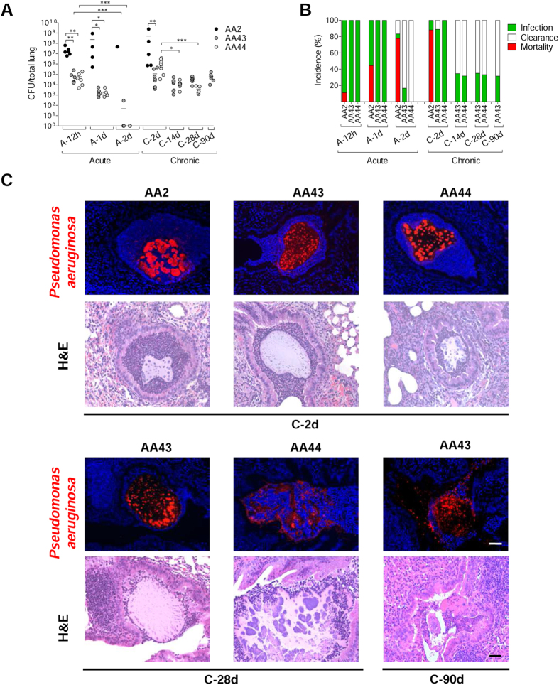
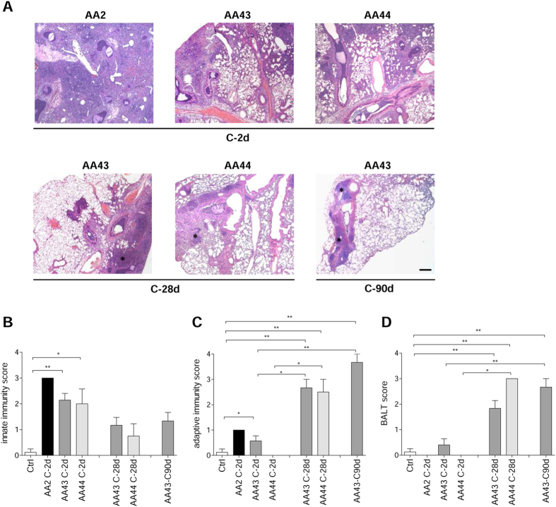
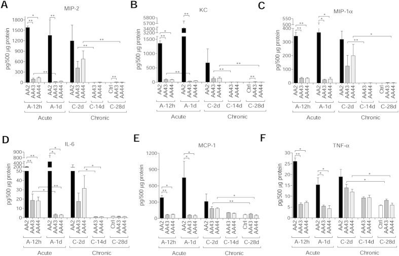
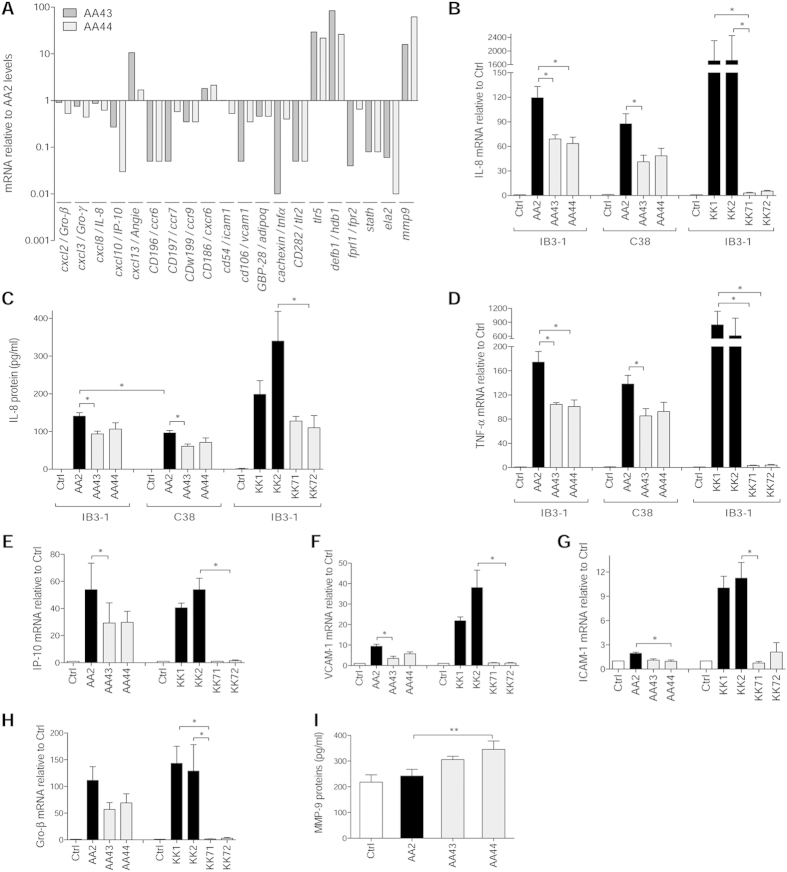
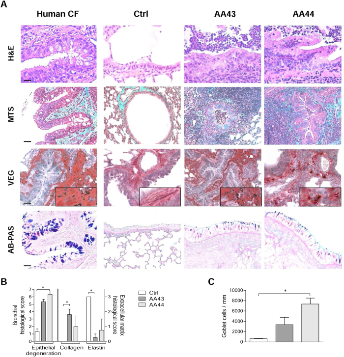
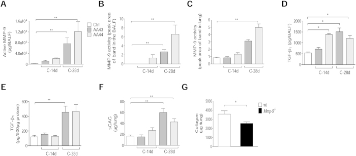
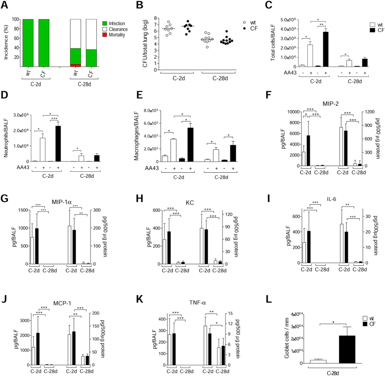
Repeated cycles of infections, caused mainly by Pseudomonas aeruginosa, combined with a robust host immune response and tissue injury, determine the course and outcome of cystic fibrosis (CF) lung disease. As the disease progresses, P. aeruginosa adapts to the host modifying dramatically its phenotype; however, it remains unclear whether and how bacterial adaptive variants and their persistence influence the pathogenesis and disease development. Using in vitro and murine models of infection, we showed that P. aeruginosa CF-adaptive variants shaped the innate immune response favoring their persistence. Next, we refined a murine model of chronic pneumonia extending P. aeruginosa infection up to three months. In this model, including CFTR-deficient mice, we unveil that the P. aeruginosa persistence lead to CF hallmarks of airway remodelling and fibrosis, including epithelial hyperplasia and structure degeneration, goblet cell metaplasia, collagen deposition, elastin degradation and several additional markers of tissue damage. This murine model of P. aeruginosa chronic infection, reproducing CF lung pathology, will be instrumental to identify novel molecular targets and test newly tailored molecules inhibiting chronic inflammation and tissue damage processes in pre-clinical studies.


Figures

Figure 1
Figure 1. Virulence of P. aeruginosa isolates and bacterial localization in murine models of airways infection.
Two groups of minimum five C57Bl/6NCrlBR mice were infected with 5 × 106 CFU/lung of planktonic bacteria for the acute infection (data modified from Fig. 5P included in Lorè NI et al. PLoS One 2012;7(4):e35648.) and with 1 to 2 × 106 CFU/lung of isolates embedded in agar beads for the chronic infection and analyzed during a time course post-infection (12 hours, 1 and 2 days of acute infection and 2, 14, 28 and 90 days of chronic lung infection). (A) CFUs were evaluated in total lung. Dots represent CFUs in individual mice and horizontal lines represent median values. The data are pooled from at least two independent experiments (n = 1–9). Statistical significance is indicated: *p < 0.05, **p < 0.01, ***p < 0.001. (B) The incidences of mortality induced by bacteremia (red), clearance (white) and airway infection (green) were determined. The data are pooled from at least two independent experiments (n = 8–24). (C) Bacterial and agar-beads localization in the lung were evaluated on challenged mice by immunofluorescence (with specific antibody against P. aeruginosa, stained in red, and with 4,6-Diamidino-2-phenylindole dihydrochloride, stained in blue) and H&E staining. Scale bar: 25 μm.
Figure 2
Figure 2. Lung histology and histopathological score of immune cells recruitment in the murine model of P. aeruginosa chronic airways infection.
C57Bl/6NCrlBR mice were infected with 1 to 2 × 106 CFU/lung of isolates embedded in agar beads for the chronic infection and analyzed during a time course post-infection (2, 28 and 90 days). (A) Lung histopathology was performed on challenged mice by H&E staining. Scale bars: 200 μm. BALT-like structures are indicated by asterisks. Innate (B) and adaptive (C) immune cells infiltration and BALT activation (D) were scored in tissue section of murine lungs stained with H&E. The data are pooled from at least two independent experiments (n = 2–7). Values represent the mean ± standard error of the mean (SEM). Statistical significance is indicated: *p < 0.05, **p < 0.01.
Figure 3
Figure 3. Cytokines/chemokines profiles in murine models after P. aeruginosa infection.
C57Bl/6NCrlBR mice were infected with 5 × 106 CFU/lung of planktonic bacteria for the acute infection or 1 to 2 × 106 CFU/lung of strains embedded in agar beads for the chronic infection and analyzed during a time course post-infection (12 hours and 1 day of acute infection and 2, 14 and 28 days of chronic lung infection). Cytokines and chemokines, including MIP-2 (A), KC (B), MIP-1α (C), IL-6 (D), MCP-1 (E) and TNF-α (F), were measured by Bioplex in lung homogenates. The data are pooled from at least two independent experiments (n = 3–6). Values represent the mean ± SEM. Statistical significance is indicated: *P < 0.05, **P < 0.01.
Figure 4
Figure 4. Expression/release of markers of inflammation and tissue damage in cell lines after infection with P. aeruginosa phenotypic variants.
Bronchial epithelial CF cells IB3-1 were infected for 4 hours with P. aeruginosa AA2-AA43-AA44 isolates, RNA extracted and retrotranscribed, and macroarray conducted. (A) Genes expression is expressed after normalization on expression induced by AA2. Validation of gene expression was performed by real time PCR in IB3-1 (B,DH) and isogenic non-CF cells C38 (B,D) after infection with AA2, AA43, AA44, KK1, KK2, KK71 and KK72. (C) Validation of IL-8 protein release was performed by ELISA in culture medium of IB3-1 and C38 after infection with isolates mentioned above. (I) Macrophagic-like cells THP-1 were infected for with P. aeruginosa AA2, AA43 and AA44 isolates (MOI 1), and MMP-9 release was measured in the culture supernatants by ELISA. Values represent the mean ± SEM. The data are pooled from at least three independent experiments. Statistical significance is indicated: *p < 0.05, **p < 0.01.
Figure 5
Figure 5. Lung histology and histopathological score of tissue damage in murine lung after infection with P. aeruginosa.
C57Bl/6NCrlBR mice were infected with 1 to 2 × 106 CFU/lung of isolates embedded in agar beads and analyzed 28 days post-infection. Sections of airways from transplanted CF patients and from C57Bl/6NCrlBR infected with CF-adapted isolates AA43 and AA44 and sterile beads were stained with H&E, MTS for collagen, VEG for elastic fibers and AB/PAS for mucopolysaccarides, according to the standard procedures (A). Scale bars: 12.5 μm for H&E, 25 μm for MTS, VEG and AB-PAS. Scorings of bronchial epithelial degeneration, collagen deposition and elastin degradation (B) were performed on slices stained with H&E, MTS and VEG, respectively. Goblet cells numbers were evaluated on slices stained with AB-PAS (C). The data are pooled from at least two independent experiments (n = 2–6). Values represent the mean ± SEM. Statistical significance is indicated: *p < 0.05.
Figure 6
Figure 6. Tissue damage markers, including MMP-9, in murine models after P. aeruginosa infection.
C57Bl/6NCrlBR mice were infected with 1 to 2 × 106 CFU/lung of CF-adapted isolates embedded in agar beads for 14 and 28 days. Levels of MMP-9 protein (A) in BALF by ELISA, MMP-9 activity (B) in BALF and (C) lung homogenate by zymography, TGF-β1 (D) in BALF and (E) lung homogenate by Bioplex and sGAG (F) in lung homogenate by a dye-binding colorimetric assay were measured after 14 and 28 days of chronic lung infection. Values represent the mean ± SEM. The data are pooled from at least two independent experiments (n = 3–12). G) B6.FVB(Cg)-Mmp9tm1Tvu/J and congenic mice were infected with 2 × 106 CFU/lung of AA43 strain embedded in agar beads. Collagen levels were evaluated by a dye-binding assay in lung homogenate after 28 days of chronic lung infection with the P. aeruginosa CF-adapted isolate AA43. Values are represented as mean ± SEM. The data derive from one experiment (n = 6–7). Statistical significance is indicated: *p < 0.05, **p < 0.01.
Figure 7
Figure 7. Bacterial virulence and lung inflammatory response in CF and congenic wt mice after P. aeruginosa chronic lung infection.
Gut-corrected CFTR-deficient C57Bl/6 Cftrtm1UNCTgN(FABPCFTR)#Jaw and congenic wt mice were infected with 2 × 106 CFU/lung of P. aeruginosa AA43 strain embedded in agar beads and analyzed after 2 and 28 Days. A) The incidences of mortality induced by bacteremia (red), clearance (white) and airway infection (green) were determined. The data are pooled from two independent experiments (n = 8–28). B) CFU were evaluated in total lung. Dots represent individual mice measurements and horizontal lines represent the median values reported in a log scale (n = 8–11). The number of total cells (C) and in particular of neutrophils (D) and macrophages (E) in the airways were analyzed in BALF of mice. Cytokines/chemokines, including MIP-2 (F), MIP-1α (G), KC (H), IL-6 (I), MCP-1 (J) and TNF-α (K), were measured by Bioplex in murine BALF (left side of the graph) and lung homogenates (right side of the graph). Sections of murine lungs infected with AA43 for 28 days were stained with AB/PAS for mucopolysaccharides, according to the standard procedure. Goblet cells numbers were evaluated on slices stained with AB-PAS (L). Values represent the mean ± SEM. The data are pooled from at least two independent experiments (n = 3–11). Statistical significance is indicated: *p < 0.05, **p < 0.01, ***p < 0.001.

References

    1. Barinaga M. Knockout mice offer first animal model for CF. Science 257, 1046–1047 (1992). - PubMed
    1. Cohen T. S. & Prince A. Cystic fibrosis: a mucosal immunodeficiency syndrome. Nat Med 18, 509–519, 10.1038/nm.2715 (2012). - DOI - PMC - PubMed
    1. Devereux G. et al. An observational study of matrix metalloproteinase (MMP)-9 in cystic fibrosis. J Cyst Fibros 13, 557–563, 10.1016/j.jcf.2014.01.010 (2014). - DOI - PubMed
    1. Gaggar A. et al. A novel proteolytic cascade generates an extracellular matrix-derived chemoattractant in chronic neutrophilic inflammation. J Immunol 180, 5662–5669, 180/8/5662 (2008). - PMC - PubMed
    1. Weathington N. M. et al. A novel peptide CXCR ligand derived from extracellular matrix degradation during airway inflammation. Nat Med 12, 317–323, nm1361 (2006). - PubMed

Publication types

MeSH terms