. 2016 Feb 17:7:10501.
doi: 10.1038/ncomms10501.

Adaptive resistance to therapeutic PD-1 blockade is associated with upregulation of alternative immune checkpoints

Affiliations

Adaptive resistance to therapeutic PD-1 blockade is associated with upregulation of alternative immune checkpoints

Shohei Koyama et al. Nat Commun. .

Abstract

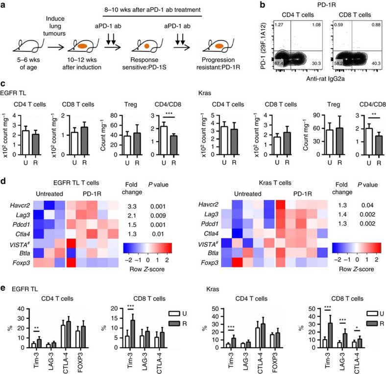
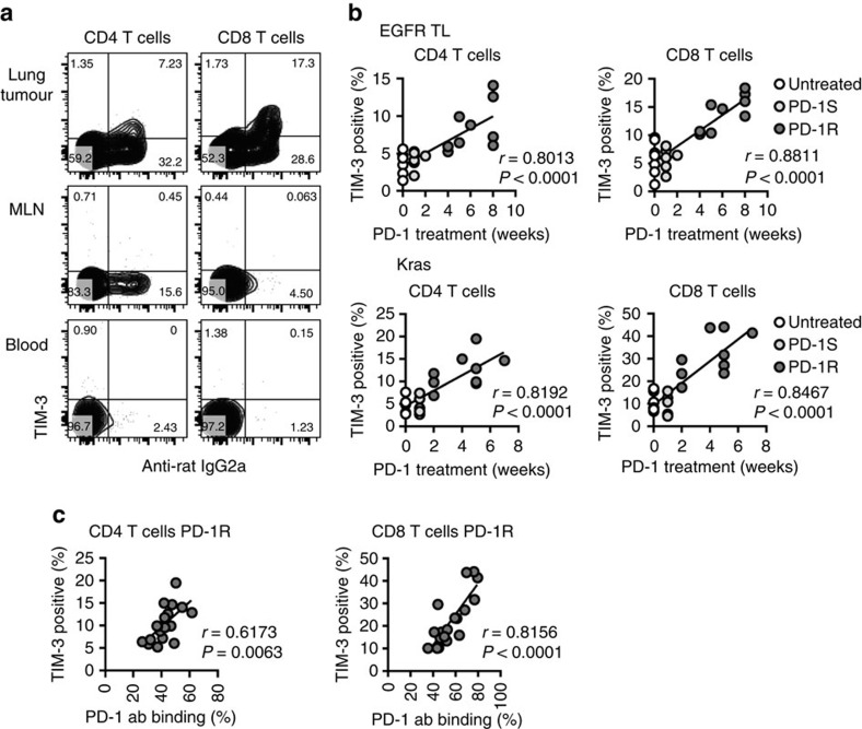
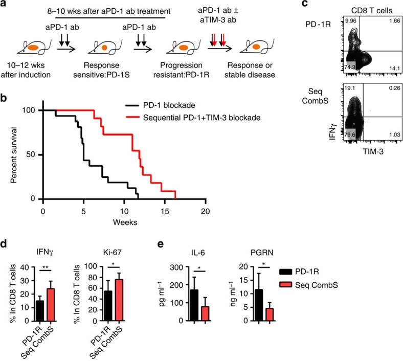
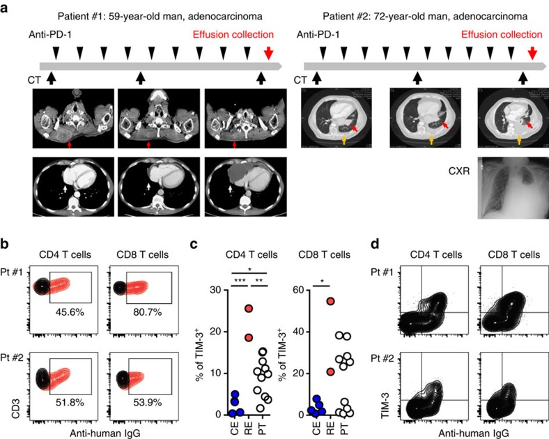
Despite compelling antitumour activity of antibodies targeting the programmed death 1 (PD-1): programmed death ligand 1 (PD-L1) immune checkpoint in lung cancer, resistance to these therapies has increasingly been observed. In this study, to elucidate mechanisms of adaptive resistance, we analyse the tumour immune microenvironment in the context of anti-PD-1 therapy in two fully immunocompetent mouse models of lung adenocarcinoma. In tumours progressing following response to anti-PD-1 therapy, we observe upregulation of alternative immune checkpoints, notably T-cell immunoglobulin mucin-3 (TIM-3), in PD-1 antibody bound T cells and demonstrate a survival advantage with addition of a TIM-3 blocking antibody following failure of PD-1 blockade. Two patients who developed adaptive resistance to anti-PD-1 treatment also show a similar TIM-3 upregulation in blocking antibody-bound T cells at treatment failure. These data suggest that upregulation of TIM-3 and other immune checkpoints may be targetable biomarkers associated with adaptive resistance to PD-1 blockade.


Conflict of interest statement

G.D. received sponsored research support from Bristol-Myers Squibb and Novartis. He is currently an employee of Novartis. F.S.H. is a Bristol-Myers Squibb nonpaid consultant, Novartis, Merck and Genentech consultant and receives clinical trial support to the institution from these companies. G.J.F. receives patent royalties on the PD-1 pathway from Bristol-Myers-Squibb, Roche, Merck, EMD-Serrono, Boehringer-Ingelheim, Amplimmune/AstraZeneca and Novartis and patent royalties on the TIM-3 pathway from Novartis. D.B.C. is a consultant for Pfizer. S.J.R. receives research support from Bristol-Myers Squibb and the Center for Immune Oncology, DFCI. The remaining authors declare no competing financial interests.

Figures

Figure 1
Figure 1. Upregulation of TIM-3 in T cells at the time of acquired resistance to anti-PD-1 blockade.
(a) Schematic of in vivo treatment with anti-PD-1 antibody until adaptive resistance (b) Representative flow cytometry data from anti-PD-1 resistant (PD-1R) EGFR TL mouse. PD-1 expression and anti-Rat IgG2a (therapeutic antibody binding) were evaluated. Fluorescent conjugated anti-PD-1 antibody is the same clone (29F.1A12) as the therapeutic antibody. (c) Cell number of T cell subsets: CD4 T cells, CD8 T cells and regulatory T cells (Treg) and CD4/CD8 ratio. Untreated (U) EGFR TL (n=7), Kras (n=7) and anti-PD-1 resistant (R) EGFR TL (n=9), Kras (n=9) were analysed (EGFR TL ***P<0.001, Kras **P=0.0028, student's t-test). Data are shown as mean±s.d. (d) Expression of six genes with an annotated role in the T-cell response in sorted T cells from EGFR models (five anti-PD-1 treated and three untreated tumours) in EGFR models and KRAS models (five anti-PD-1 treated and five untreated tumours). For each gene, expression values across the samples are plotted as log-transformed FPKM values (row-scaled and coloured on a blue-red scale to emphasize the difference between treated and untreated samples). The magnitude of change between resistant and genotype-matched untreated samples are shown as fold change and P values for differentially expressed genes (defined as having an absolute fold change greater than 1.25 and a P value <0.05 as calculated by the limma package37). Note the gene name of VISTA is 4632428N05Rik (Gene ID: 74048). (e) Surface expression of inhibitory T cell markers: TIM-3, LAG-3, CTLA-4 and FOXP3. Untreated (U) EGFR TL (n=7), Kras (n=7) and anti-PD-1 resistant (R) EGFR TL (n=9), Kras (n=9) were analysed (*P<0.05, **P<0.01, ***P<0.001). Data are shown as mean±s.d.
Figure 2
Figure 2. TIM-3 expression in tumour-infiltrating T cells correlates with treatment time and PD-1 antibody binding.
(a) TIM-3 expression in T cells from tumour-bearing lung, mediastinal lymph node and peripheral blood. Representative flow cytometry data from anti-PD-1 resistant EGFR TL mouse. (b) Significant correlation was detected between TIM-3 positivity and the duration of PD-1 blocking treatment in EGFR TL mice (untreated (0 week): n=7, anti-PD-1 sensitive (PD-1S): n=6 and resistant (PD-1R): n=9) and Kras mice (untreated: n=7, anti-PD-1 sensitive (PD-1S): n=6, resistant (PD-1R): n=9). (c) Significant correlation was detected among TIM-3 positivity and the amount of bound therapeutic PD-1 antibody in anti-PD-1 resistant (PD-1R) EGFR TL and Kras mice (both EGFR and Kras mice were combined: n=18). Correlation was evaluated using Pearson's correlation coefficient.
Figure 3
Figure 3. Sequential anti-TIM-3 blocking displays clinical efficacy in anti-PD-1 adaptive resistant tumours.
(a,b) Survival after PD-1 blockade alone (anti-PD-1 resistant) or PD-1 and sequential TIM-3 blockade combination treatment (PD-1 alone: n=16 and sequential combination treatment: n=11) (P=0.0008) after documented tumor burden. Treatment started at week 0. Median survival PD1 5 weeks vs PD-1+TIM-3 sequential treatment 11.9 weeks. (c) Representative flow cytometry data of IFN-gamma expression in CD8 T cells from anti-PD-1 resistant (PD-1R) and sequential anti-PD-1 plus anti-TIM-3 combination (Seq CombS): 2 weeks′ anti-PD-1 and anti-TIM-3 combination treatment after development of resistance to PD-1 single treatment. Fluorescent conjugated anti-TIM-3 antibody is the same clone (RMT3-23) as the therapeutic antibody. (d) IFN-gamma and Ki-67 positive CD8 T cell counts from anti-PD-1 resistant (PD-1R) (n=6) and sequential anti-PD-1 plus anti-TIM-3 combination (Seq CombS) (n=6) (*P<0.05, **P<0.01). (e) IL-6 and PGRN production in BALFs from PD-1R (n=6) and comb (Seq CombS: n=6) (*P<0.05). Data are shown as mean±s.d., P values are calculated using student's t test for all data except for the survival data.
Figure 4
Figure 4. Upregulation of TIM-3 in two resistant patient cases after anti-PD-1 treatment.
(a) Clinical course of patient #1 and patient #2, who were treated with PD-1 blocking antibodies. Both initially responded to treatment but subsequently developed treatment resistance with effusions. Arrow indicates soft tissue metastasis (red arrow) and pericardial effusion (white arrow) in patient #1 and left lower lobe tumour (red arrow) and pleural effusion (orange arrow) in patient #2. (b) Detection of therapeutic antibody (human IgG) binding in CD4 and CD8 T cells. Human IgG and isotype control are shown in red and black, respectively. (c) Percentage of TIM-3 positive CD4 and CD8 T cells in effusions from two anti-PD-1 resistant patients (resistant effusions: RE), NSCLC patients without PD-1 blocking treatment (control effusions: CE, n=5) and surgically resected primary tumours: PT (n=11). Mean % of TIM-3 in T cells from RE versus CE versus PT (CD4 T cells 22.10 versus 2.52 versus 9.06 and CD8 T cells 37.85 versus 3.19 versus 17.58. In CD4 T cells, RE versus CE ***P=0.0001, RE versus PT **P=0.0023 and CE versus PT *P=0.0247. In CD8 T cells, RE versus CE *P=0.0256. (d) TIM-3 expression and therapeutic antibody binding (human IgG) in CD4 and CD8 T cells. Data are shown as mean±s.d., P values are calculated using one-way analysis of variance with Tukey's multiple comparison test.

Comment in

References

    1. Topalian S. L. et al.. Safety, activity, and immune correlates of anti-PD-1 antibody in cancer. N. Engl. J. Med. 366, 2443–2454 (2012). - PMC - PubMed
    1. Brahmer J. R. et al.. Safety and activity of anti-PD-L1 antibody in patients with advanced cancer. N. Engl. J. Med. 366, 2455–2465 (2012). - PMC - PubMed
    1. Page D. B., Postow M. A., Callahan M. K., Allison J. P. & Wolchok J. D. Immune modulation in cancer with antibodies. Annu. Rev. Med. 65, 185–202 (2014). - PubMed
    1. Keir M. E., Butte M. J., Freeman G. J. & Sharpe A. H. PD-1 and its ligands in tolerance and immunity. Annu. Rev. Immunol. 26, 677–704 (2008). - PMC - PubMed
    1. Pardoll D. M. The blockade of immune checkpoints in cancer immunotherapy. Nat. Rev. Cancer 12, 252–264 (2012). - PMC - PubMed

Publication types