. 2016 May;151(1):193-203.
doi: 10.1093/toxsci/kfw035. Epub 2016 Feb 15.

Cardiac-Specific Deletion of the Pdha1 Gene Sensitizes Heart to Toxicological Actions of Ischemic Stress

Affiliations

Cardiac-Specific Deletion of the Pdha1 Gene Sensitizes Heart to Toxicological Actions of Ischemic Stress

Wanqing Sun et al. Toxicol Sci. 2016 May.

Erratum in

Abstract

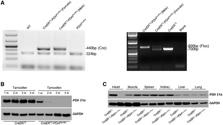
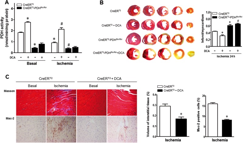
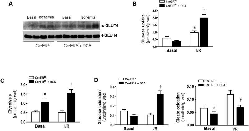
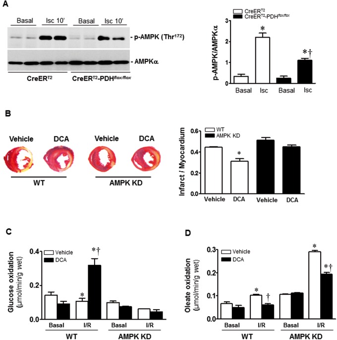
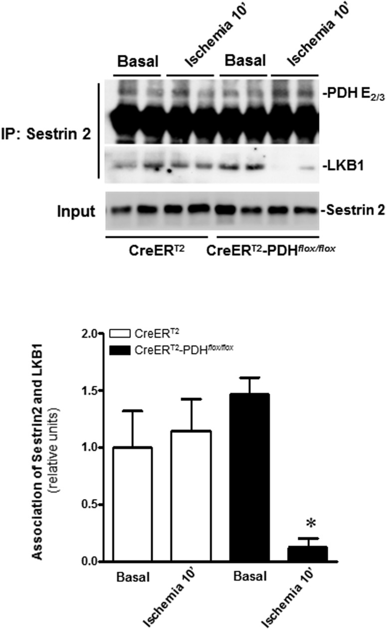
Pyruvate dehydrogenase (PDH) plays a key role in aerobic energy metabolism and occupies a central crossroad between glycolysis and the tricarboxylic acid cycle. We generated inducible cardiac-specific PDH E1α knockout (CreER(T2)-PDH(flox/flox)) mice that demonstrated a high mortality rate. It was hypothesized that PDH modulating cardiac glucose metabolism is crucial for heart functions under normal physiological and/or stress conditions. The myocardial infarction was conducted by a ligation of the left anterior descending coronary arteries. Cardiac PDH E1α deficiency caused large myocardial infarcts size and macrophage infiltration in the hearts (P < .01 vs wild-type [WT]). Wheat germ agglutinin and Masson trichrome staining revealed significantly increased hypertrophy and fibrosis in PDH E1α-deficient hearts (P < .05 vs WT). Measurements of heart substrate metabolism in an ex vivo working heart perfusion system demonstrated a significant impairment of glucose oxidation in PDH E1α-deficient hearts during ischemia/reperfusion (P < .05 vs WT). Dichloroacetate, a PDH activator, increased glucose oxidation in WT hearts during ischemia/reperfusion and reduced myocardial infarct size in WT, but not in PDH E1α-deficient hearts. Immunoblotting results demonstrated that cardiac PDH E1α deficiency leads to an impaired ischemic AMP-activated protein kinase activation through Sestrin2-liver kinase B1 interaction which is responsible for an increased susceptibility of PDH E1α-deficient heart to ischemic insults. Thus, cardiac PDH E1α deficiency impairs ischemic AMP-activated protein kinase signaling and sensitizes hearts to the toxicological actions of ischemic stress.

Keywords: AMP-activated protein kinase.; myocardial infarction; pyruvate dehydrogenase.


Figures

FIG. 1.
FIG. 1.
Cardiac-specific PDH E1α deficiency mice. A, Genotyping of CreERT2-PDHflox/flox and CreERT2(α-MHC-MerCreMer) mice by PCR analysis. B, Immunoblotting verified the level of PDH E1α in heart. The PDH E1α protein level was markedly reduced in the adult mouse heart 2 months after tamoxifen (TM) injection for 5 days. C, The immunoblotting shows the PDH E1α was specifically knock out in the adult mouse heart after TM injection.
FIG. 2.
FIG. 2.
Cardiac abnormalities in the PDH E1α iCKO mice. A, Survival analysis in PDH E1α-deficient (CreERT2-PDHflox/flox) and CreERT2 control mice. B, H&E staining of heart sections. The ratios of heart weight and body weight increased in PDH E1α-deficient (CreERT2-PDHflox/flox) mice when compared with CreERT2 control mice (upper panel). Wheat germ agglutinin (WGA) staining showed larger ventricular cardiomyocytes in cardiac PDH E1α-deficient heart than that in CreERT2 heart (the second panel). The expression of Mac-2 protein in the heart was identified by immunohistochemistry and quantified by positive staining of the cells (the third panel). Masson staining of heart sections showed the PDH E1α deficiency increased cardiac fibrosis (the fourth panel). C, Serial echocardiographic studies performed at baseline (before injection of tamoxifen), 1 month, and 2 months after tamoxifen injection. The bar graphs show the left ventricle inner diameter during diastole (LVIDd) and ejection fraction (EF). D, The mRNA levels of collagen I, IL-6 and MMP-9 in PDH E1α-deficient (CreERT2-PDHflox/flox) and CreERT2control heart were analyzed by Q-PCR. E, Glucose/oleate oxidation ih the PDH E1α-deficient (CreERT2-PDHflox/flox) and CreERT2control heart. Data are means ± SEM (n  =  4–7 per group). *P <.05 versus CreERT2 control.
FIG. 3.
FIG. 3.
Cardiac-specific PDH E1α deficiency increased susceptibility to ischemic injury. A, PDH activity was abolished in PDH E1α-deficient (CreERT2-PDHflox/flox) heart. There is reduction of PDH activity in CreERT2 heart during ischemia. Dichloroacetate (DCA) treatment increased PDH activity in CreERT2 heart under both basal and ischemic conditions. Values are means ± SEM (n  =  4 per group). *P <.05 versus CreERT2 basal, #P <.05 versus CreERT2 ischemia alone. B, Cardic PDH E1α-deficient (CreERT2-PDHflox/flox) mice and CreERT2 control mice with and without DCA administration were subjected to in vivo regional ischemia for 24 h. DCA (50 mg/kg/day) was given for 3 days before myocardial ischemia by ligation of left anterior descending coronary artery (LAD). Representative sections are shown the extent of myocardial infarction. The ratios of the infarction area to the total heart are shown in the right bar graph. Values are means ± SEM (n  =  4–6 per group). *P <.05 versus CreERT2 ischemia alone, #P <.05 versus CreERT2 ischemia + DCA. C, Masson trichrome stained to determine fibrosis and Mac-2 stained for inflammation measurement in 1 week after ischemia. Values are means ± SEM (n  =  4 per group). *P <.05 versus CreERT2 ischemia alone.
FIG. 4.
FIG. 4.
Activation of PDH by DCA modulates glucose and fatty acid oxidation. A, DCA was given at the beginning of the perfusion, after balancing 20 min, isolated CreERT2 hearts were subjected to 10 min of ischemia for GLUT4 translocation measurement. After balancing 20 min, isolated CreERT2 hearts were subjected to 10 min of ischemia followed by 20 min of reperfusion for measurement of glucose uptake (B), glycolysis (C), and (D) glucose oxidation and oleate oxidation in the heart. Values are means ± SEM (n  =  4–6 per group). *P <.05 versus CreERT2 basal, †P <.05 versus CreERT2 I/R alone.
FIG. 5.
FIG. 5.
AMPK mediates the cardioprotection of DCA. A, Cardic PDH E1α-deficient (CreERT2-PDHflox/flox) heart and CreERT2 control heart with or without DCA administration were subjected to ischemia 10 min by LAD ligation. The levels of phospho-AMPK and total AMPK in the heart were determined by immunoblotting. Values are means ± SEM (n  =  3 per group). *P <.05 versus CreERT2 basal; †P <.05 versus CreERT2 ischemia. B, AMPK kinase-dead (AMPK KD) transgenic mice and wild type (WT) mice with or without dichloroacetate (DCA) treatment were subjected to in vivo regional ischemia for 24 h. Representative sections show the extent of myocardial infarction. The ratios of the infarction area to the total heart is shown in the bar graph. Values are means ± SEM (n  =  4 per group). *P <.05 versus WT Vehicle. (C,D) AMPK KD transgenic hearts and WT hearts with or without DCA treatment were subjected to a working heart perfusion system to measure substrate metabolism. C, Glucose oxidation measurement with U-14C-glucose (20 μCi/l). Values are means ± SEM (n  =  4–6 per group). *P <.05 versus Basal vehicle; †P <.05 versus WT I/R. (D) Oleate oxidation measurement with [9, 10]-3H-oleate (50 μCi/l). Values are means ± SEM (n  =  4 per group). *P <.05 versus Basal vehicle, respectively; †P <.05 versus WT I/R vehicle or AMPK KD vehicle I/R, respectively.
FIG. 6.
FIG. 6.
Cardiac-specific PDH E1α deficiency alters LKB1-Sestrin2 interaction. Immunoprecipitation was performed using anti-Sestrin2 antibody. The precipitates were analyzed by Western blotting with antibodies recognized against PDH E2/3 and LKB1, respectively. Values are means ± SEM (n  =  4 per group). *P <.05 versus CreERT2-PDHflox/floxBasal.

Similar articles

Cited by

References

    1. Anderson J., Khou S., Nawarskas J. (2000). Ranolazine: A potential new treatment for chronic stable angina. Heart Dis. 3, 263–269. - PubMed
    1. Bainey K. R., Armstrong P. W. (2014). Clinical perspectives on reperfusion injury in acute myocardial infarction. Am. Heart J. 167, 637–645. - PubMed
    1. Bersin R. M., Stacpoole P. W. (1997). Dichloroacetate as metabolic therapy for myocardial ischemia and failure. Am. Heart J. 134, 841–855. - PubMed
    1. Budanov A. V., Karin M. (2008). p53 target genes sestrin1 and sestrin2 connect genotoxic stress and mTOR signaling. Cell 134, 451–460. - PMC - PubMed
    1. Calvert J. W., Gundewar S., Jha S., Greer J. J., Bestermann W. H., Tian R., Lefer D. J. (2008). Acute metformin therapy confers cardioprotection against myocardial infarction via AMPK-eNOS-mediated signaling. Diabetes 57, 696–705. - PubMed

MeSH terms