

Dilated cardiomyopathy with cardiogenic shock in a child with Kearns-Sayre syndrome
- PMID: 26884075
- PMCID: PMC5483557
- DOI: 10.1136/bcr-2015-213813
Dilated cardiomyopathy with cardiogenic shock in a child with Kearns-Sayre syndrome
Abstract
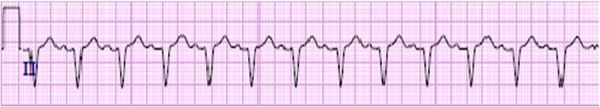
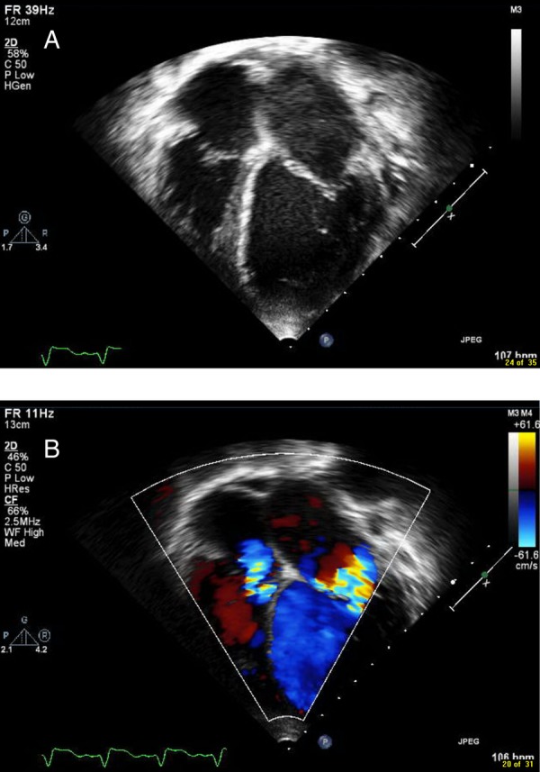
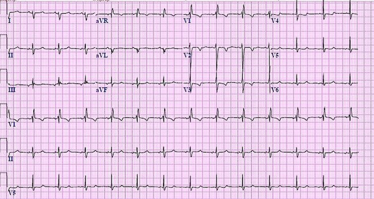
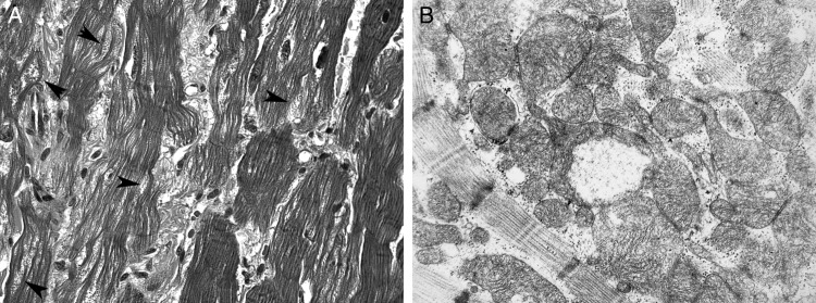
Kearns-Sayre syndrome (KSS) is a mitochondrial myopathy resulting from mitochondrial DNA deletion. This syndrome primarily involves the central nervous system, eyes, skeletal muscles and the heart. The most well-known cardiac complications involve the conduction system; however, there have been case reports describing cardiomyopathy. We describe a case of a child with KSS who presented with decompensated cardiac failure from dilated cardiomyopathy representing cardiomyocyte involvement of KSS. Our patient had a rapidly progressing course, despite maximal medical management, requiring emergent institution of extracorporeal membrane oxygenation and transition to a ventricular assist device. To the best of our knowledge, this is the youngest patient in the literature to have dilated cardiomyopathy in KSS.
2016 BMJ Publishing Group Ltd.
Figures
Similar articles
-
Cardiomyopathy in the Kearns-Sayre syndrome.Br Heart J. 1988 Apr;59(4):486-90. doi: 10.1136/hrt.59.4.486. Br Heart J. 1988. PMID: 3370184 Free PMC article.
-
Congestive heart failure due to mitochondrial cardiomyopathy in Kearns-Sayre syndrome.Klin Wochenschr. 1987 May 15;65(10):480-6. doi: 10.1007/BF01712843. Klin Wochenschr. 1987. PMID: 3599796
-
Cardiac transplantation in an incomplete Kearns-Sayre syndrome with mitochondrial DNA deletion.Neuromuscul Disord. 1993 Sep-Nov;3(5-6):561-6. doi: 10.1016/0960-8966(93)90116-2. Neuromuscul Disord. 1993. PMID: 8186712
-
Cardiac extracorporeal life support: state of the art in 2007.Cardiol Young. 2007 Sep;17 Suppl 2:104-15. doi: 10.1017/S1047951107001217. Cardiol Young. 2007. PMID: 18039404 Review.
-
Kearns-Sayre syndrome: a case report and review of cardiovascular complications.Pacing Clin Electrophysiol. 2005 May;28(5):454-7. doi: 10.1111/j.1540-8159.2005.40049.x. Pacing Clin Electrophysiol. 2005. PMID: 15869681 Review.
Cited by
-
Mitochondrial cardiomyopathies: navigating through different clinical and management pictures between adult and paediatric forms.Front Cardiovasc Med. 2025 Jul 3;12:1621096. doi: 10.3389/fcvm.2025.1621096. eCollection 2025. Front Cardiovasc Med. 2025. PMID: 40678571 Free PMC article. Review.
-
Cardiac Involvement in Mitochondrial Disorders.Curr Heart Fail Rep. 2023 Feb;20(1):76-87. doi: 10.1007/s11897-023-00592-3. Epub 2023 Feb 18. Curr Heart Fail Rep. 2023. PMID: 36802007 Free PMC article. Review.
-
Kearns Sayre syndrome: a rare etiology of complete atrioventricular block in children (case report).Pan Afr Med J. 2021 Nov 15;40:154. doi: 10.11604/pamj.2021.40.154.24281. eCollection 2021. Pan Afr Med J. 2021. PMID: 34970396 Free PMC article.
-
Uncovering the etiology of ptosis prior to blepharoplasty.Arch Plast Surg. 2020 Sep;47(5):487. doi: 10.5999/aps.2020.00409. Epub 2020 Sep 15. Arch Plast Surg. 2020. PMID: 32971602 Free PMC article. No abstract available.
References
Publication types
MeSH terms
LinkOut - more resources
Full Text Sources
Other Literature Sources
Medical