. 2016 Mar 1;113(9):E1236-45.
doi: 10.1073/pnas.1519444113. Epub 2016 Feb 16.

Deep phenotyping of 89 xeroderma pigmentosum patients reveals unexpected heterogeneity dependent on the precise molecular defect

Affiliations

Deep phenotyping of 89 xeroderma pigmentosum patients reveals unexpected heterogeneity dependent on the precise molecular defect

Hiva Fassihi et al. Proc Natl Acad Sci U S A. .

Abstract

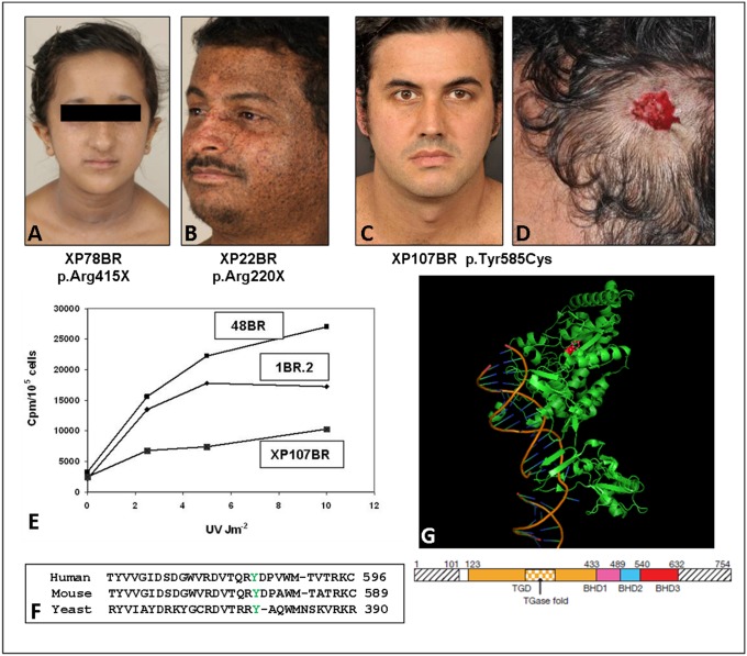
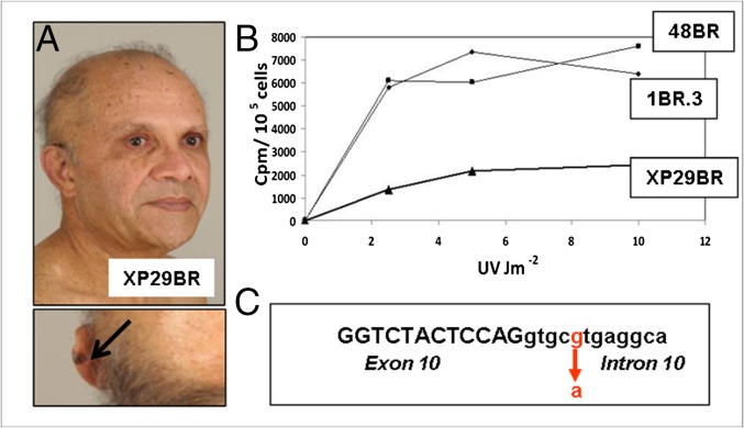
Xeroderma pigmentosum (XP) is a rare DNA repair disorder characterized by increased susceptibility to UV radiation (UVR)-induced skin pigmentation, skin cancers, ocular surface disease, and, in some patients, sunburn and neurological degeneration. Genetically, it is assigned to eight complementation groups (XP-A to -G and variant). For the last 5 y, the UK national multidisciplinary XP service has provided follow-up for 89 XP patients, representing most of the XP patients in the United Kingdom. Causative mutations, DNA repair levels, and more than 60 clinical variables relating to dermatology, ophthalmology, and neurology have been measured, using scoring systems to categorize disease severity. This deep phenotyping has revealed unanticipated heterogeneity of clinical features, between and within complementation groups. Skin cancer is most common in XP-C, XP-E, and XP-V patients, previously considered to be the milder groups based on cellular analyses. These patients have normal sunburn reactions and are therefore diagnosed later and are less likely to adhere to UVR protection. XP-C patients are specifically hypersensitive to ocular damage, and XP-F and XP-G patients appear to be much less susceptible to skin cancer than other XP groups. Within XP groups, different mutations confer susceptibility or resistance to neurological damage. Our findings on this large cohort of XP patients under long-term follow-up reveal that XP is more heterogeneous than has previously been appreciated. Our data now enable provision of personalized prognostic information and management advice for each XP patient, as well as providing new insights into the functions of the XP proteins.

Keywords: UV radiation; neurodegeneration; nucleotide excision repair; ocular disease; skin cancer.


Conflict of interest statement

The authors declare no conflict of interest.

Figures

Fig. S1.
Fig. S1.
Role of XP proteins in NER. The figure represents a scheme for NER, showing only the roles of the XP proteins. For simplicity, other proteins involved have not been included. In GG-NER, XPE and XPC are involved in damage recognition, TFIIH-containing XPB and XPD are recruited and open out the DNA structure in the vicinity of the damage. XPA verifies the damaged structure before dual incisions by XPF-ERCC1 and XPG. After the damaged section has been removed, DNA polymerases and ligases fill in the gap. In TC-NER, blockage of RNA polymerase by the damage is the recognition signal. Proteins CSB and CSA, defective in Cockayne syndrome, are recruited, and these, in turn recruit TFIIH to enable the rest of the NER process to proceed.
Fig. S2.
Fig. S2.
Translesion synthesis. DNA replication, carried out by DNA polymerases δ or ε, is blocked by a UVR lesion, and in many cases reinitiates beyond the lesion. The resulting gap is filled in by DNA polymerase ƞ, which is deficient in XP variants.
Fig. 1.
Fig. 1.
XP-C patients. (A) XP78BR, aged 12 y; note lentigines on face. (B) XP22BR, aged 38 y; note the extensive pigmentary changes. (C) XP107BR, aged 37 y; note the almost complete absence of pigmentary changes, despite not being diagnosed until 34 y of age. (D) Site of excised melanoma from XP107BR. (E) UDS, measured as incorporated 3H-thymidine cpm per 105 cells following UV irradiation of XP107BR with different UVC doses compared with two normal controls: 1BR2 and 48BR. (F) Alignment of XPC protein from human and mouse and yeast Rad4 in the region around Tyr585 (indicated in green) that is mutated in XP107BR. (G) Crystal structure of Rad4 (22) with Tyr379 (corresponding to Tyr585 in human XPC) indicated in red in the structure and the schematic below.
Fig. 2.
Fig. 2.
XP29BR. (A) Patient XP29BR, with a melanoma on his left ear (arrow). (B) UDS, measured as incorporated 3H-thymidine cpm per 105 cells following UV irradiation of XP29BR with different UVC doses compared with normal fibroblasts 1BR.3 and 48BR. (C) Sequence around the exon 10/intron 10 boundary showing the position of the mutation.
Fig. 3.
Fig. 3.
XP-D patients. (A) Severe sunburn after minimal sun exposure in XP87BR before XP diagnosis (see ref. for another photograph of this patient). (B) XP16BR and (C) XPJCLO showing minimal pigmentary skin changes due to early diagnosis and excellent UVR protection. (D) Scheme of XPD protein, showing the seven helicase domains, I, Ia, II–VI, and the positions of the mutations in the XPD patients. Below is an expanded view of aa715–740 showing the p.Ala717Gly alteration and the deletion of aa716-730 resulting from the c.C2150 > G mutation. Top line: WT protein sequence with p.Ala717Gly indicated in green and the deleted 15 aa resulting from abnormal splicing of intron 22 indicated in brown; middle and bottom lines: WT and mutant DNA with exon 22 sequence in caps, intron 22 in lowercase, with abnormally spliced-out bases in brown. (E) XP59BR, showing multiple surgical scars as a result of poor UVR protection (see ref. for another photograph of this patient). (F) Crystal structure of XPD around Arg683, showing projected hydrogen bonding with Asp681 and DNA (magenta). (Modified from ref. .)
Fig. 4.
Fig. 4.
XP-F patients. (A) XP32BR. (B) XP72BR. (C) XP24BR. Note the very mild skin changes despite only moderate UVR protection. (D) Scheme of XPF protein, showing the five motifs of the disrupted helicase domains (I, Ia, II, III, and IV), the nuclease domain (NUCL), and the two Helix-hairpin-helix (HhH) domains.
Fig. 5.
Fig. 5.
XP-G patients. (A) XP34BR. (B) Structure of the XPG protein (39), showing the N and I domains required for nuclease activity, the spacer region, nuclear localization signals (NLS), and binding sites for other proteins. Sites of the mutations in the patients are indicated in blue, and the deletion of aa16-88 in XP55BR and XP56BR is indicated by a green inverted V. (C) XP104BR. (D) XP56BR. (E) XP55BR. (F) XP101BR.

References

    1. Kleijer WJ, et al. Incidence of DNA repair deficiency disorders in western Europe: Xeroderma pigmentosum, Cockayne syndrome and trichothiodystrophy. DNA Repair (Amst) 2008;7(5):744–750. - PubMed
    1. Hirai Y, et al. Heterozygous individuals bearing a founder mutation in the XPA DNA repair gene comprise nearly 1% of the Japanese population. Mutat Res. 2006;601(1-2):171–178. - PubMed
    1. Zghal M, et al. Xeroderma pigmentosum: Manifestations cutanées, oculaires et neurologiques à partir de 49 patients tunisiens. Tunis Med. 2005;83(12):760–763. - PubMed
    1. Bradford PT, et al. Cancer and neurologic degeneration in xeroderma pigmentosum: Long term follow-up characterises the role of DNA repair. J Med Genet. 2011;48(3):168–176. - PMC - PubMed
    1. Sethi M, et al. Patients with xeroderma pigmentosum complementation groups C, E and V do not have abnormal sunburn reactions. Br J Dermatol. 2013;169(6):1279–1287. - PubMed

Publication types

LinkOut - more resources